Search results (30 results)
-
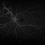
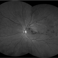
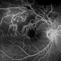 Branch Retinal Vein Occlusion- Fluorescein Angiogram, Montage
Apr 15 2016 by James B. Soque, CRA, OCT-C, COA, FOPS
A fluorescein angiogram of an 80-year-old white female with a superotemporal branch retinal vein occlusion, and retinal edema of the right eye. Currently receiving Lucentis 0.5 injection therapy.
Photographer: James Soque, CRA OCT-C COA, Island Retina, Shirley, NY
Imaging device: Topcon TRC, MERGE Imaging Software V. 11.2.0
Condition/keywords: branch retinal vein occlusion (BRVO), montage, non-perfused branch retinal vein occlusion (BRVO)
-
---thumb.jpg/image-square;max$300,300.ImageHandler) Non-perfused BRVO with macular infarction
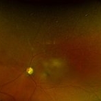
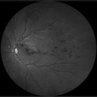
Non-perfused BRVO with macular infarction
Aug 20 2013 by Hamid Ahmadieh, MD
Late venous phase angiogram of the right eye of a 55-year-old woman with decreased vision due to BRVO. Notice capillary nonperfusion involving the macula.
Photographer: Naghmeh Nozhat, Negah Eye Center, Tehran
Condition/keywords: macular infarction, non-perfused branch retinal vein occlusion (BRVO)
-
---thumb.jpg/image-square;max$300,300.ImageHandler) Non-perfused BRVO with macular infarction
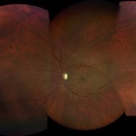
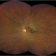
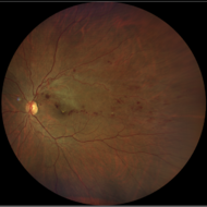
Non-perfused BRVO with macular infarction
Aug 20 2013 by Hamid Ahmadieh, MD
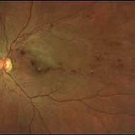
Color fundus photograph of the right eye of a 55-year-old woman with decreased vision due to BRVO. There are multiple cotton wool patches and retinal hemorrhages involving the macular center.
Photographer: Naghmeh Nozhat, Negah Eye Center, Tehran
Condition/keywords: non-perfused branch retinal vein occlusion (BRVO)
-
---thumb.jpg/image-square;max$300,300.ImageHandler) Non-perfused BRVO with macular infarction
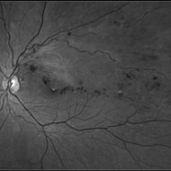
Non-perfused BRVO with macular infarction
Aug 20 2013 by Hamid Ahmadieh, MD
Mid arterio venous phase angiogram of the right eye of a 55-year-old woman with decreased vision due to BRVO. Notice capillary nonperfusion involving the macula.
Photographer: Naghmeh Nozhat, Negah Eye Center, Tehran
Condition/keywords: non-perfused branch retinal vein occlusion (BRVO)
-
 Vein Occlusion Zoom in a BRVO
Apr 29 2020 by Gabriel Castilho S Barbosa, MD
A zoomed vein occlusion in a young patient with systemic arterial hypertension.
Photographer: Gabriel Castilho, Suel Abujamra Institute, São Paulo.
Condition/keywords: branch retinal vein occlusion (BRVO), macular branch retinal vein occlusion (BRVO), non-perfused branch retinal vein occlusion (BRVO)
-
 BRVO With Non-perfusion
May 3 2014 by Mallika Goyal, MD
Late phase fluorescein angiogram in an eye with superotemporal BRVO shows delayed filling of retinal vessels and non-perfusion in the affected quadrant .
Photographer: Mallika Goyal, MD, Apollo Health City, Jubilee Hills, Hyderabad, India
Condition/keywords: non-perfused branch retinal vein occlusion (BRVO)
-
 BRVO With Non-Perfusion
May 3 2014 by Mallika Goyal, MD
Left eye superotemporal BRVO in a 53-year-old hypertensive male patient.
Photographer: Mallika Goyal, MD, Apollo Health City, Jubilee Hills, Hyderabad, India
Condition/keywords: non-perfused branch retinal vein occlusion (BRVO)
-
 BRVO With Non-perfusion
May 3 2014 by Mallika Goyal, MD
Late phase fluorescein angiogram in an eye with superotemporal BRVO shows delayed filling of retinal vessels and non-perfusion in the affected quadrant.
Photographer: Mallika Goyal, MD, Apollo Health City, Jubilee Hills, Hyderabad, India
Condition/keywords: non-perfused branch retinal vein occlusion (BRVO)
-
 BRVO With Non-Perfusion
May 3 2014 by Mallika Goyal, MD
Mid-phase fluorescein angiogram in an eye with superotemporal BRVO shows delayed filling of retinal vessels and non-perfusion in the affected quadrant.
Photographer: Mallika Goyal, MD, Apollo Health City, Jubilee Hills, Hyderabad, India
Condition/keywords: non-perfused branch retinal vein occlusion (BRVO)
-
 BRVO With Non-Perfusion
May 3 2014 by Mallika Goyal, MD
Fluorescein angiogram in an eye with superotemporal BRVO shows delayed filling of retinal vessels and non-perfusion in the affected quadrant.
Photographer: Mallika Goyal, MD, Apollo Health City, Jubilee Hills, Hyderabad, India
Condition/keywords: non-perfused branch retinal vein occlusion (BRVO)
-
 BRVO With Non-Perfusion
May 3 2014 by Mallika Goyal, MD
Early phase fluorescein angiogram in an eye with superotemporal BRVO shows delayed filling of retinal vessels in the affected quadrant.
Photographer: Mallika Goyal, MD, Apollo Health City, Jubilee Hills, Hyderabad, India
Condition/keywords: non-perfused branch retinal vein occlusion (BRVO)
-
 BRVO With Non-perfusion
May 3 2014 by Mallika Goyal, MD
Late phase fluorescein angiogram of an eye with superotemporal BRVO shows delayed filling of retinal vessels, dilation and tortuosity of the affected veins, and non-perfusion in the affected quadrant .
Photographer: Mallika Goyal, MD, Apollo Health City, Jubilee Hills, Hyderabad, India
Condition/keywords: non-perfused branch retinal vein occlusion (BRVO)
-
 BRVO With Non-perfusion
May 3 2014 by Mallika Goyal, MD
Fluorescein angiogram in an eye with superotemporal BRVO shows delayed filling of retinal vessels and non-perfusion in the affected quadrant.
Photographer: Mallika Goyal, MD, Apollo Health City, Jubilee Hills, Hyderabad, India
Condition/keywords: non-perfused branch retinal vein occlusion (BRVO)
-
 BRVO With Non-perfusion
May 3 2014 by Mallika Goyal, MD
Mid-phase fluorescein angiogram in an eye with superotemporal BRVO shows delayed filling of retinal vessels and non-perfusion in the affected quadrant.
Photographer: Mallika Goyal, MD, Apollo Health City, Jubilee Hills, Hyderabad, India
Condition/keywords: non-perfused branch retinal vein occlusion (BRVO)
-
 BRVO - Color
Apr 10 2018 by Hosam Attia, MD
56-year-old African American male with ischemic nasal BRVO with advanced cupping OS - Optos California.
Imaging device: Optos California
Condition/keywords: branch vein occlusion (BVO), non-perfused branch retinal vein occlusion (BRVO)
-
 Ischemic BRVO With Retinal Neovascularization
Apr 26 2021 by Niloofar Piri, MD
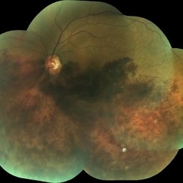
Wide field fundus photograph of the left eye demonstrating sclerotic superotemporal vein resulting from ischemic old BRVO with secondary retinal neovascularization. Note the unrelated atrophic hole within lattice degeneration in temporal periphery.
Photographer: Niloofar Piri, MD, St. louis University
Condition/keywords: branch retinal vein occlusion (BRVO), ischemia, non-perfused branch retinal vein occlusion (BRVO), retinal neovascularization
-
 BRVO
BRVO
Apr 29 2018 by mahesh aryal
Fundus photo of 46-year-old man with BRVO montage photo.
Photographer: MAHESH ARYAL LUMBINI EYE INSTITUTE
Condition/keywords: non-perfused branch retinal vein occlusion (BRVO)
-
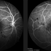
 BRVO - FA
Apr 10 2018 by Hosam Attia, MD
56-year-old African American male with ischemic nasal BRVO with advanced cupping OS - Optos California
Imaging device: Optos California
Condition/keywords: branch vein occlusion (BVO), non-perfused branch retinal vein occlusion (BRVO)
-
 Branch retinal vein occlusion - Colour & Red free image - ring shaped collaterals
Jul 18 2023 by Harsh Vardhan Singh, MS
43-year-old woman presented with left eye old STBRVO with chronic CME of duration 6month showing ring shaped collaterals more evident on red free image
Photographer: Harsh Vardhan Singh, AIIMS, Guwahati
Imaging device: Zeiss Clarus 700
Condition/keywords: branch retinal vein occlusion (BRVO), BRVO, non-perfused branch retinal vein occlusion (BRVO)
-
 Buds on Tree Appearance on FFA: Old BRVO
Mar 12 2024 by MEENAL SONI
A middle-aged man with idiopathic hypertension presented with old IT BRVO, sclerosed vein on with hemorrhages on fundus examination. FFA reveals delayed filling of vein with pruning of venules resembling buds on tree.
Photographer: Dr. Meenal Soni, Fellow VR, ASG eye Hospital Jodhpur
Imaging device: ZEISS Visucam 400
Condition/keywords: non-perfused branch retinal vein occlusion (BRVO)
-
 Branch retinal vein occlusion - Colour & Red free image - ring shaped collaterals
Jul 18 2023 by Harsh Vardhan Singh, MS
43-year-old woman presented with left eye old STBRVO with chronic CME of duration 6month showing ring shaped collaterals more evident on red free image
Photographer: Harsh Vardhan Singh, AIIMS, Guwahati
Imaging device: Zeiss Clarus 700
Condition/keywords: branch retinal vein occlusion (BRVO), BRVO, non-perfused branch retinal vein occlusion (BRVO)
-
 Branch retinal vein occlusion - Colour & Red free image - ring shaped collaterals
Jul 18 2023 by Harsh Vardhan Singh, MS
43-year-old woman presented with left eye old STBRVO with chronic CME of duration 6month showing ring shaped collaterals more evident on red free image
Photographer: Harsh Vardhan Singh, AIIMS, Guwahati
Imaging device: Zeiss Clarus 700
Condition/keywords: branch retinal vein occlusion (BRVO), BRVO, non-perfused branch retinal vein occlusion (BRVO)
 Loading…