Search results (67 results)
-
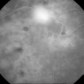
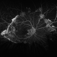
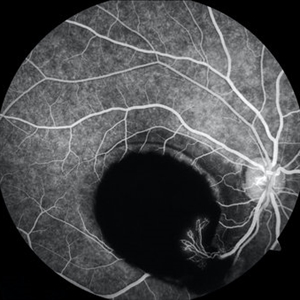 Sub-ILM Hemorrhage with Neovessels
Apr 30 2020 by Saurabh Deshmukh, MBBS, DNB, FVRS, MNAMS
Late arteriovenous phase FA showing a large sub-internal limiting membrane hemorrhage with overlying neovessels. This hypertensive patient presented with a visual acuity of counting fingers at 2 meters. The patient was advised intravitreal anti-VEGF injection, Nd: YAG Membranotomy, and systemic control of hypertension.
Photographer: Saurabh Deshmukh, Sri Sankaradeva Nethralaya, Guwahati, India
Imaging device: Topcon TRC-50 DX
Condition/keywords: hypertensive retinopathy, neovascularization elsewhere (NVE), subILM hemorrhage
-
 Venous Loop & Venous Beading
May 31 2014 by Hamid Ahmadieh, MD
Color fundus photograph of the left eye of a diabetic patient with NVD, NVE, venous loop and venous beading.
Photographer: Elham Salehi, Negah Eye Center, Tehran
Condition/keywords: color fundus photograph, neovascularization elsewhere (NVE), neovascularization of the disc (NVD), proliferative diabetic retinopathy (PDR), venous beading, venous loop
-
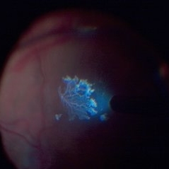
 Sea Fan Neovascularisation
Apr 27 2015 by Neha Goel, MS DNB FRCS (Glasg)
Fluorescein angiography of the left eye of a 40-year-old male.
Photographer: Neha Goel
Imaging device: Zeiss visucam
Condition/keywords: Eales disease, neovascularization elsewhere (NVE), vasculitis
-
 Marked Retinal Ischemia in Patient with Mixed Connective Tissue Disease
Feb 26 2013 by Sharon Fekrat, MD FACS FASRS
Fluorescein angiogram of the right eye of a 27-year-old female with mixed connective tissue disease and marked retinal ischemia. Panretinal laser photocoagulation (PRP) has been performed for neovascularization elsewhere (NVE).
Condition/keywords: mixed connective tissue disease, retinal ischemia
-
 NVE from BRVO
Feb 19 2015 by H. Michael Lambert, MD
Color photo of NVE after BRVO.
Condition/keywords: branch retinal vein occlusion (BRVO), neovascularization elsewhere (NVE)
-
 Preretinal Bleed in Lasered PDR
Jun 4 2014 by Neha Goel, MS DNB FRCS (Glasg)
Fundus photograph of the right eye of a 40-year-old diabetic male. Panretinal photocoagulation had been performed. There is a dense preretinal bleed overlying the macula and nasal to the disc.
Photographer: Neha Goel
Imaging device: Zeiss Visucam
Condition/keywords: laser photocoagulation, neovascularization elsewhere (NVE), preretinal hemorrhage, proliferative diabetic retinopathy (PDR)
-
 Proliferative Sickle Cell Retinopathy, Color OD
May 23 2018 by Hosam Attia, MD
45-year-old African American, male with sickle cell anemia (SC disease) with arteriolar attenuation, mild venous tortuosity, Sunburst (S) and large, partially auto-infarcted sea fan with fresh heme, OD.
Imaging device: Optos California Ultra-Wide Field Fundus Camera
Condition/keywords: neovascularization elsewhere (NVE), proliferative retinopathy, sea fan, sickle cell, sickle cell retinopathy
-
---thumb.jpg/image-square;max$300,300.ImageHandler) Binder3 P12 Slide82
Binder3 P12 Slide82
Feb 15 2013 by From the Collections of Thomas M. Aaberg, MD and Thomas M. Aaberg Jr., MD
Color fundus photograph showing peripheral retinal nonperfusion, retinal neovascularization elsewhere (NVE), venous beading and dilatation, retinal microaneurysms, and intraretinal hemorrhage.
Condition/keywords: peripheral retinal nonperfusion, proliferative retinopathy, retinal neovascularization
-
 Proliferative Diabetic Retinopathy Inverted
May 28 2015 by Matt Poe, COA
This is a young man with advanced proliferative diabetic retinopathy.
Photographer: Matt Poe, COA. Northwest Arkansas Retina Associates, Springdale, AR.
Imaging device: Heidelberg HRA
Condition/keywords: capillary dropouts, diabetes, neovascularization elsewhere (NVE), proliferative diabetic retinopathy (PDR)
-
 Detached NVE During PVD induction
Apr 27 2018 by Michael J. Koss, MD, PhD, MBA
A 73-year-old woman with macular pucker underwent a pars plana vitrectomy with membrane peeling. Additionally the patient suffers from diabetic retinopathy after being diagnosed with type 2 diabetes mellitus sixteen years ago. Prior to the procedure she was treated with a series of intravitreal Bevacizumab-injections due to diabetic macular edema. There was no history of a proliferative DRP. During the vitrectomy a branch of an obliterated NVE spontaneously detached and floated freely in the vitreous. The 3D shot was captured via Alcon’s NGENUITY® 3D Visualization System in form of photograph and video providing an outstandingly detailed image of the branched NVE.
Photographer: Michael Koss, Augenzentrum Nymphenburger Hoefe
Imaging device: Alcon’s NGENUITY® 3D Visualization System
Condition/keywords: diabetes, diabetic retinopathy, neovascularization elsewhere (NVE), pars plana vitrectomy (PPV), PVD induction
-
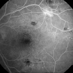 Neovascularization - RDP
Jun 29 2014 by Ratimir Lazic, MD, PhD
A FAG image of a 84-year-old female. Late venous phase of the left eye. Hyperfloercent area in upper temporal quadrant represents NVE. Many hyperflorescent dots can be seen. Few hypoflorescent areas are deep retinal hemorrhages.
Photographer: Marko Lukic, University Eye Clinic Svjetlost
Imaging device: Zeis Visucam Lite 2
Condition/keywords: neovascularization (NV), neovascularization elsewhere (NVE)
-
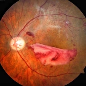 Proliferative Diabetic Retinopathy With Subhyaloid Hemorrhage
Mar 28 2018 by awaneesh m upadhyay, MBBS, DNB
56-year-old gentleman came with complaints of sudden onset painless loss of vision over 2 months, known diabetic, hypertensive with chronic kidney disease. Fundus photograph of left eye
Photographer: Dr Awaneesh Upadhyay
Imaging device: Zeiss
Condition/keywords: neovascularization elsewhere (NVE), proliferative diabetic retinopathy (PDR), subhyaloid hemorrhage
-
 BRVO With PRP laser
Feb 19 2015 by H. Michael Lambert, MD
Color photo of NVE after BRVO. Laser performed,ERM present
Condition/keywords: ischemia, neovascularization elsewhere (NVE)
-
 NVE from BRVO
Feb 19 2015 by H. Michael Lambert, MD
Color photo of NVE after BRVO.
Condition/keywords: branch retinal vein occlusion (BRVO), neovascularization elsewhere (NVE)
-
 NVE from BRVO
Feb 19 2015 by H. Michael Lambert, MD
Color photo of NVE after BRVO.
Condition/keywords: branch retinal vein occlusion (BRVO), neovascularization elsewhere (NVE)
-
 Proliferative Diabetic Retinopathy with Temporal Seafan NVE Dragging Retinal Vein
Feb 15 2018 by Kushal S Delhiwala, MBBS, MS, FMRF,FICO, FAICO
58-year-old diabetic male presenting with Bilateral Proliferative diabetic retinopathy and centre involving Diabetic macular edema.Left eye fundus photograph showing large seafan NVE temporal to macula causing upward dragging of inferotemporal retinal vein and arteriovenous anastomosis.
Photographer: Dr Kushal Delhiwala, Netralaya superspeciality eye hospital, Ahmedabad
Imaging device: Zeiss Visucam 500
Condition/keywords: neovascularization elsewhere (NVE), proliferative diabetic retinopathy (PDR), sea fan
-
 Neovascularization - RDP
Jun 29 2014 by Ratimir Lazic, MD, PhD
FAG image of a 84-year-old female. Dye leakage from the NVE can be seen.
Photographer: Marko Lukic, University Eye Clinic Svjetlost
Imaging device: Zeis Visucam Lite 2
Condition/keywords: neovascularization elsewhere (NVE)
-
 Hypertensive Retinopathy
Dec 24 2017 by Purva Patwari
52-year-old female diagnosed of hypertension by retina evaluation.
Photographer: Dr Purva Patwari, Patwari Retina Center, Ahmedabad, Gujarat , India
Imaging device: ZEISS VISU500
Condition/keywords: hypertensive retinopathy, neovascularization elsewhere (NVE), Roth spots
-
 Active neovascularization in Proliferative Diabetic Retinopathy
Jan 10 2018 by Peter H. Tang, MD, PhD
Fluorescein angiography image from a 46-year-old woman with uncontrolled proliferative diabetic retinopathy shows extensive dye leakage from active neovascularization.
Imaging device: Optos California
Condition/keywords: diabetes, diabetic retinopathy, fluorescein leakage, neovascularization elsewhere (NVE), neovascularization of the disc (NVD), pan-retinal photocoagulation (PRP), proliferative diabetic retinopathy (PDR)
-
 Proliferative Diabetic Retinopathy: Smartphone Fundus Image
Dec 15 2018 by Prithvi Chandrakanth
A 22-year-old female presented with random blood sugars of 410mg/dl and blood pressure of 170/100mmhg with OD uncorrected visual acuity of 2/60 not improving with glasses.
Photographer: Dr.Prithvi Chandrakanth, Dr.Chandrakanth Malabar Nethralaya, Kozhikode.
Imaging device: Trash To Treasure (T3) Retcam : Smartphone fundus camera
Condition/keywords: neovascularization elsewhere (NVE), neovascularization of the disc (NVD), proliferative diabetic retinopathy (PDR), smartphone fundus photography
-
 Proliferative Sickle Cell Retinopathy, Color OS
May 23 2018 by Hosam Attia, MD
45-year-old African American, male with sickle cell anemia (SC disease ) with arteriolar attenuation, mild venous tortuosity, peripheral arterio-venous anastomoses (shown better on red free), multiple small NVEs/ early sea fans (one w/ early auto-infarction) and sunburst (S) - (Not showing very well in photos) OS.
Imaging device: Optos California Ultra-Wide Field Fundus Camera
Condition/keywords: neovascularization elsewhere (NVE), proliferative retinopathy, sea fan, sickle cell, sickle cell retinopathy
-
 Proliferative Sickle Cell Retinopathy, Color OD
May 23 2018 by Hosam Attia, MD
45-year-old African American, male with sickle cell anemia (SC disease) with arteriolar attenuation, mild venous tortuosity, Sunburst (S) and large, partially auto-infarcted Seafan with fresh heme, OD.
Imaging device: Optos California Ultra-Wide Field Fundus Camera
Condition/keywords: neovascularization elsewhere (NVE), proliferative retinopathy, sea fan, sickle cell, sickle cell retinopathy
-
 Wolf Jaw
Jun 23 2019 by Veronika Yehezkeli
Wolf Jaw
Imaging device: Optos
Condition/keywords: ischemia, neovascularization (NV), neovascularization elsewhere (NVE), preretinal hemorrhage, proliferative diabetic retinopathy (PDR), wolf jaw
-
 Tractional Retinal Detachment and NVE
Dec 16 2016 by Courtney Crawford, MD, FACS
45-year-old-male with diabetes and decreased vision from a fovea-off tractional retinal detachment.
Photographer: Kristen Dunn
Imaging device: Optos
Condition/keywords: neovascularization elsewhere (NVE), tractional retinal detachment
 Loading…