Search results (80 results)
-
 Type 2 Parafoveal Telangiectasia
Aug 23 2012 by Gerardo Garcia-Aguirre, MD
Fundus photograph showing pigment migration and crystals in the temporal aspect of the fovea.
Photographer: Ricardo Montoya, Asociación para Evitar la Ceguera en México
Imaging device: Zeiss FF4
Condition/keywords: crystals, fovea, idiopathic macular telangiectasia, pigment migration
-
 Macular Telangiectasia Type 2 & CNV
Sep 22 2012 by Hamid Ahmadieh, MD
Color fundus photograph & OCT imagings of the left eye of a 70-year-old man with idiopathic macular telangiectasia type 2 and CNV.
Photographer: Hamid Ahmadieh, MD, Ophthalmic Research Center, Labbafinejad Medical Center, Shahid Beheshti University of Medical Sciences
Imaging device: Topcon Fundus Camera & Topcon OCT
Condition/keywords: choroidal neovascularization (CNV), idiopathic macular telangiectasia, optical coherence tomography (OCT)
-
 Type 2 Parafoveal Telangiectasia
Aug 23 2012 by Gerardo Garcia-Aguirre, MD
Fundus photograph showing pigment migration and crystals in the temporal aspect of the fovea.
Photographer: Noemí Hernández, Asociación para Evitar la Ceguera en México
Imaging device: Zeiss FF4
Condition/keywords: crystals, idiopathic macular telangiectasia, pigment migration
-
 Macular Telangiectasia Type 2
Sep 22 2012 by Hamid Ahmadieh, MD
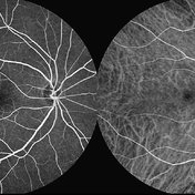
FA and ICG angiography imagings of the right eye of a 70-year-old man with idiopathic macular telangiectasia type 2.
Photographer: Hamid Ahmadieh, MD, Ophthalmic Research Center, Labbafinejad Medical Center, Shahid Beheshti University of Medical Sciences
Imaging device: HRA
Condition/keywords: idiopathic macular telangiectasia, indocyanine green (ICG) angiography
-
 Macular Telangiectasia Type 2
Sep 22 2012 by Hamid Ahmadieh, MD
Autofluorescence imagings of both eyes of a 70-year-old man with idiopathic macular telangiectasia type 2.
Photographer: Hamid Ahmadieh, MD, Ophthalmic Research Center, Labbafinejad Medical Center, Shahid Beheshti University of Medical Sciences
Imaging device: HRA
Condition/keywords: autofluorescence imaging, idiopathic macular telangiectasia
-
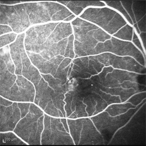
 Macular Telangiectasia (FA Early Phase)
May 16 2014 by Avris Romario Diparaja Siahaan
FA (early phase) image of a 58-year-old-man with a macular telangiectasia condition on his left eye.
Photographer: Avris Romario Diparaja Siahaan, Klinik Mata Nusantara
Imaging device: Topcon TRC 50 DX Type IA
Condition/keywords: FA early phase, macular telangiectasia
-
 Macular Telangiectasia Type 2
Sep 22 2012 by Hamid Ahmadieh, MD
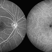
Late phase FA and ICG angiography imagings of the right eye of a 70-year-old man with idiopathic macular telangiectasia type 2.
Photographer: Hamid Ahmadieh, MD, Ophthalmic Research Center, Labbafinejad Medical Center, Shahid Beheshti University of Medical Sciences
Imaging device: HRA
Condition/keywords: idiopathic macular telangiectasia, indocyanine green (ICG) angiography
-
 Type 1A Macular Telangiectasia - Fundus photograph
Nov 11 2013 by Gerardo Garcia-Aguirre, MD
Fundus photograph of a 43-year-old male complaining of mild metamorphopsia in OS. BCVA 20/25. Some hard exudates and telangiectatic vessels are observed inferior and temporal to the fovea.
Condition/keywords: macular telangiectasia
-
 Macular Telangiectasia Type 2 & CNV
Sep 22 2012 by Hamid Ahmadieh, MD
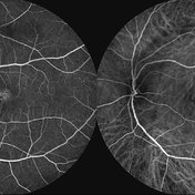
FA and ICG angiography imagings of the left eye of a 70-year-old man with idiopathic macular telangiectasia type 2 and CNV.
Photographer: Hamid Ahmadieh, MD, Ophthalmic Research Center, Labbafinejad Medical Center, Shahid Beheshti University of Medical Sciences
Imaging device: HRA
Condition/keywords: choroidal neovascularization (CNV), idiopathic macular telangiectasia, indocyanine green (ICG) angiography
-
 Macular Telangiectasia Type 2 & CNV
Sep 22 2012 by Hamid Ahmadieh, MD
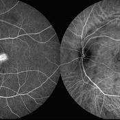
Late phase FA and ICG angiography imagings of the left eye of a 70-year-old man with idiopathic macular telangiectasia type 2 and CNV.
Photographer: Hamid Ahmadieh, MD, Ophthalmic Research Center, Labbafinejad Medical Center, Shahid Beheshti University of Medical Sciences
Imaging device: HRA
Condition/keywords: choroidal neovascularization (CNV), idiopathic macular telangiectasia, indocyanine green (ICG) angiography
-
 Type 1A Macular Telangiectasia - OCT
Nov 11 2013 by Gerardo Garcia-Aguirre, MD
SD-OCT showing intraretinal fluid in both internal and external layers of the retina. Hyper-reflective foci are also visible in the external layers of the retina.
Condition/keywords: macular telangiectasia, optical coherence tomography (OCT)
-
 Type 1A Macular Telangiectasia - Fluorescein Angiogram - Early
Nov 11 2013 by Gerardo Garcia-Aguirre, MD
Fluorescein angiogram showing hyperfluorescent spots temporal and inferior to the fovea, with mild leakage.
Condition/keywords: macular telangiectasia
-
 Telangiectasia
Sep 16 2012 by Ivan R. Batlle, MD
Mid phase fluorescein angiogram of 58-year-old female with decreased vision
Condition/keywords: idiopathic macular telangiectasia
-
 Telagiectasia
Sep 16 2012 by Ivan R. Batlle, MD
Color photograph of 58-year-old female with decreased vision
Condition/keywords: idiopathic macular telangiectasia
-
 Type 1A Macular Telangiectasia - Autofluorescence
Nov 11 2013 by Gerardo Garcia-Aguirre, MD
Autofluorescence image showing hypoautofluorescent spots corresponding to telangiectatic vessels.
Condition/keywords: macular telangiectasia
-
 Idiopathic Juxtafoveal Telangiectasia, Type 2
Nov 6 2014 by Thomas A. Ciulla, MD, MBA, FASRS
Note the telangiectactic vessels just temporal to the FAZ.
Photographer: Thomas Steele
Condition/keywords: idiopathic macular telangiectasia, juxtafoveal telangiectasis, parafoveal telangiectasia
-
 Type 1A Macular Telangiectasia - Fluorescein Angiogram - Late
Nov 11 2013 by Gerardo Garcia-Aguirre, MD
Fluorescein angiogram showing hyperfluorescent spots with diffuse leakage.
Condition/keywords: macular telangiectasia
-
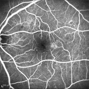
 Macular Telangiectasia (FA Late Phase)
May 16 2014 by Avris Romario Diparaja Siahaan
FA (late phase) image of a 58-year-old-man with a macular telangiectasia condition on his left eye.
Photographer: Avris Romario Diparaja Siahaan, Klinik Mata Nusantara
Imaging device: Topcon TRC 50 DX Type IA
Condition/keywords: FA late phase, macular telangiectasia
-
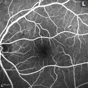
 Macular Telangiectasia (ICG Early Phase)
May 16 2014 by Avris Romario Diparaja Siahaan
ICGA (early phase) image of a 58-year-old-man with a macular telangiectasia condition on his left eye.
Photographer: Avris Romario Diparaja Siahaan, Klinik Mata Nusantara
Imaging device: Topcon TRC 50 DX Type IA
Condition/keywords: indocyanine green (ICG) angiography, macular telangiectasia
-
 Idiopathic Juxtafoveal Telangiectasia, Type 2
Nov 6 2014 by Thomas A. Ciulla, MD, MBA, FASRS
Note the telangiectactic vessels just temporal to the FAZ.
Photographer: Thomas Steele
Condition/keywords: idiopathic macular telangiectasia, juxtafoveal telangiectasis, parafoveal telangiectasia
-
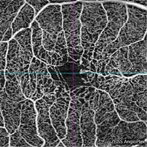 Macular Telangiectasia Type 2
Mar 8 2018 by Daniel R Agarwal, MD
OCT Angiography image in a 51-year-old male with fogging of vision and leaking on fluorescein angiography.
Photographer: Jen Welsh
Imaging device: Zeiss Angioplex OCTA
Condition/keywords: macular telangiectasia, macular telangiectasia type 2
-
 Type 1A Macular Telangiectasia - ICG Angiogram
Nov 11 2013 by Gerardo Garcia-Aguirre, MD
ICG Angiogram showing hyperfluorescent spots temporal and inferior to the fovea.
Condition/keywords: indocyanine green (ICG) angiography, macular telangiectasia
-
 Idiopathic Juxtafoveal Telangiectasia, Type 2
Nov 6 2014 by Thomas A. Ciulla, MD, MBA, FASRS
Note the telangiectactic vessels just temporal to the FAZ.
Photographer: Thomas Steele
Condition/keywords: idiopathic macular telangiectasia, juxtafoveal telangiectasis, parafoveal telangiectasia
-
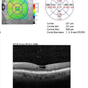 Idiopathic Juxtafoveal Telangiectasia, Type 2
Nov 6 2014 by Thomas A. Ciulla, MD, MBA, FASRS
Note the characteristic pseudocyst on OCT.
Photographer: Thomas Steele
Condition/keywords: idiopathic macular telangiectasia, juxtafoveal telangiectasis, parafoveal telangiectasia
-
 Macular Teleangiectasia
May 29 2013 by Zofia Anna Nawrocka (vel Michalewska), MD, PhD
Scanning laser ophthalmoscopy and SD-OCT of a 56-year old-woman. Visual acuity was 0.8 Snellen.
Photographer: Janusz Michalewski, MD, PhD, Ophthalmic Clinic "Jasne Blonia", Lodz, Poaldn
Imaging device: Heidelberg Spectralis
Condition/keywords: macular telangiectasia, optical coherence tomography (OCT)
 Loading…