Search results (655 results)
-
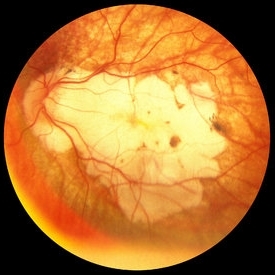
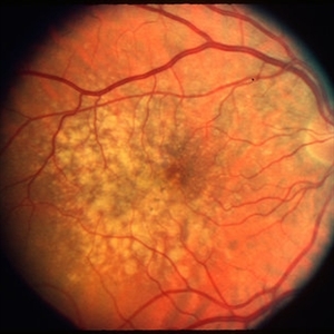
 Lacquer Cracks
Oct 13 2012 by Geoffrey G. Emerson, MD, PhD, FASRS
Lacquer cracks
Condition/keywords: lacquer cracks, myopic macular degeneration
-
---thumb.JPG/image-square;max$300,300.ImageHandler) Disciform Scar
Disciform Scar
Jul 13 2013 by Jason S. Calhoun
Poor central vision in the left eye due to macular degeneration. Disciform scar.
Photographer: Jason S. Calhoun, Department of Ophthalmology, Mayo Clinic Jacksonville, Florida
Imaging device: TOPCON TRC 50-EX
Condition/keywords: disciform scar, macular degeneration
-
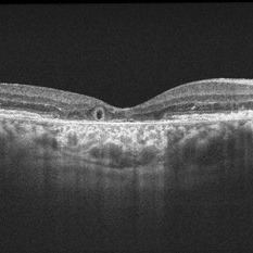
---thumb.jpg/image-square;max$300,300.ImageHandler) Geographic atrophy
Geographic atrophy
Aug 29 2012 by Young Hee Yoon, MD, PhD
OCT image of an 78-year-old woman. Her best-corrected visual acuity was counting fingers at 30cm.
Photographer: Ji Hee Kim, Asan Medical Center
Imaging device: Heidelberg spectralis
Condition/keywords: dry age-related macular degeneration (dry AMD), geographic atrophy
-
 Wet Macular Degeneration OCT
Oct 13 2012 by Geoffrey G. Emerson, MD, PhD, FASRS
Condition/keywords: optical coherence tomography (OCT)
-
 Geographic Atrophy, Fundus photograph
Aug 23 2012 by Gerardo Garcia-Aguirre, MD
Fundus photograph of an 85-year-old patient with age related macular degeneration and geographic atrophy. A large area with well-defined borders is observed, in which the choroidal vasculature is visualized.
Photographer: Noemí Hernández, Asociación para Evitar la Ceguera en México
Imaging device: Zeiss FF4
Condition/keywords: geographic atrophy
-
 Myopic CNV
Jan 11 2013 by Alex P. Hunyor, MD
Myopic macular degeneration complicated by subretinal neovascularisation, left eye.
Condition/keywords: high myopia, myopia, myopic choroidal neovascularization (CNV)
-
 Retinal Angiomatous Proliferation in Age-Related Macular Degeneration with Subretinal Neovascularization
Sep 24 2012 by James B. Soque, CRA, OCT-C, COA, FOPS
75-year-old white male with classic SRN with RAP. Lesion OD is active, and patient is receiving anti-VEGF treatment. Mid phase FA, 50 Deg, Mag 2x.
Photographer: James Soque, CRA, COA, Island Retina, Shirley, NY, USA
Imaging device: Topcon TRC 50 DX, OIS 5.0 MP Color, FA Camera, OIS Software
Condition/keywords: age-related macular degeneration (AMD), fundus autofluorescence (FAF), leakage, retinal angiomatous proliferation (RAP), subretinal neovascularization (SRNV)
-
 Fibrovascular PED
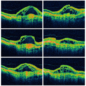
Feb 21 2014 by Roy Schwartz, MD
72-year-old female with fibrovascular PED. Upper picture - PED with sub RPE hyper-reflective substance, in a multi-layered pattern, corresponding to fibrovascular PED. CME. Lower picture - PED flattened, a denser sub RPE hyperreflective substance is seen. CME resolved.
Condition/keywords: fibrovascular pigment epithelial detachment (PED), neovascular age-related macular degeneration (AMD), optical coherence tomography (OCT), ranibizumab
-
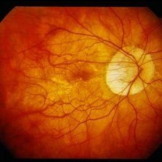
 Peripapillary Atrophy With High Myopia
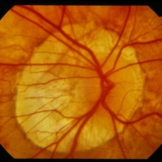
Feb 4 2015 by H. Michael Lambert, MD
Peripapillary atrophy and central macular degeneration seen in high myopia.
Condition/keywords: high myopia, peripapillary atrophy
-
 Geographic Atrophy
Aug 29 2012 by Young Hee Yoon, MD, PhD
Fundus photograph of an 78-year-old woman. Her best-corrected visual acuity was counting fingers at 30cm.
Photographer: Kyoung Woon Kim, Asan Medical Center
Imaging device: Canon CR-DGI/Version 5.1.2
Condition/keywords: dry age-related macular degeneration (dry AMD), geographic atrophy
-
 Myopic macular degeneration
Jan 11 2013 by Alex P. Hunyor, MD
Myopic macular degeneration, left eye - extensive chorioretinal atrophy.
Condition/keywords: myopic degeneration, myopic fundus, myopic macular degeneration
-
 Dry Type AMD
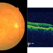
Sep 22 2012 by Hamid Ahmadieh, MD
Color fundus photograph and OCT image of a 70-year-old woman with dry type AMD.
Photographer: Hamid Ahmadieh, MD, Ophthalmic Research Center, Labbafinejad Medical Center, Shahid Beheshti University of Medical Sciences
Imaging device: Topcon Fundus Camera & Topcon OCT
Condition/keywords: dry age-related macular degeneration (dry AMD), optical coherence tomography (OCT)
-
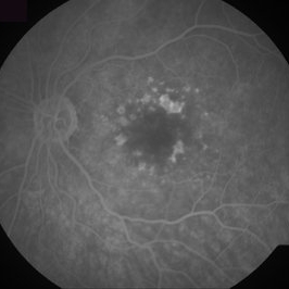
 Dry Age-Related Macular Degeneration, Fluorescein Angiogram
Aug 23 2012 by Gerardo Garcia-Aguirre, MD
Fluroescein angiogram of a 66 year-old patient with several hyperfluorescent spots corresponding to drusen.
Photographer: Noemí Hernández, Asociación para Evitar la Ceguera en México
Imaging device: FF4
Condition/keywords: age-related macular degeneration (AMD), dry age-related macular degeneration (dry AMD)
-
 Wet Age Related Macular Degeneration (WET AMD)
Sep 8 2012 by Ratimir Lazic, MD, PhD
Color fundus image of a 65 - year- old male. Drusen with suspect CNV
Photographer: Ratimir Lazic, PhD MD
Imaging device: Zeis Visucam Lite 2
Condition/keywords: fundus photograph
-
 Dry AMD
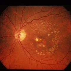
Jun 4 2014 by Henry J. Kaplan, MD
Multiple drusen with RPE changes in the macula #2.
Condition/keywords: age-related macular degeneration (AMD), dry age-related macular degeneration (dry AMD)
-
 Myopic Macular Degeneration
Oct 11 2012 by Jeffrey G. Gross, MD, FASRS
Myopic macular degeneration 20/100.
Condition/keywords: 20/100, myopic macular degeneration
-
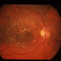
 Calcified drusen, fundus photograph
Aug 23 2012 by Gerardo Garcia-Aguirre, MD
Fundus photograph showing multiple white-yellowhish lesions corresponding to calcified drusen.
Photographer: Noemí Hernández, Asociación para Evitar la Ceguera en México
Imaging device: Zeiss FF4
Condition/keywords: calcified drusen, dry age-related macular degeneration (dry AMD)
-
 Pattern Macular Dystrophy
Oct 16 2012 by Ratimir Lazic, MD, PhD
Color fundus image of a 76-year-old female. Defect of RPE in butterfly pattern can be seen. It is easy to be misdiagnosed with dry age related macular degeneration (look at the FAG images). BCVA of that eye is 0.95.
Photographer: Marko Lukic, MD
Imaging device: Zeis Visucam Lite 2
Condition/keywords: fundus photograph, pattern macular dystrophy, retinal pigment epithelium (RPE) defect
-
 Outer-Retinal-Tubulation
Jun 27 2013 by Jason S. Calhoun
Patient with a history of wet macular degeneration and glaucoma in both eyes. VA is 20/50, right eye, 20/80, left eye. Patient is treated with Eylea in both eyes. Enhanced depth imaging OCT reveals a small like form of a cyst which in fact isn't a cyst at all. This is called outer retinal tubulation in which degenerating photo-receptors may become arranged in a circular or ovoid fashion. This is sometimes misdiagnosed as cystic changes in the retinal pigment epithelium or sub-retinal fluid.
Photographer: Jason S. Calhoun, Mayo Clinic Jacksonville, Florida
Imaging device: ZEISS OCT CIRRUS
Condition/keywords: optical coherence tomography (OCT)
-
---thumb.JPG/image-square;max$300,300.ImageHandler) "Flower" Macular Degeneration (Wet)
"Flower" Macular Degeneration (Wet)
Jul 13 2013 by Jason S. Calhoun
Patient with (wet) macular degeneration in the left eye. Notice the "flower" shape abnormal blood vessels staining.
Photographer: Jason S. Calhoun, Department of Ophthalmology, Mayo Clinic Jacksonville, Florida
Imaging device: TOPCON TRC 50-EX
Condition/keywords: choroidal neovascularization (CNV)
-
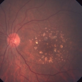
 Dry AMD
Jun 4 2014 by Henry J. Kaplan, MD
Multiple hard and calcified drusen. #1
Condition/keywords: age-related macular degeneration (AMD), calcified drusen, drusen, dry age-related macular degeneration (dry AMD)
-
 Dry Age-Related Macular Degeneration
Mar 29 2013 by Henry J. Kaplan, MD
Dry AMD with multiple confluent soft drusens
Condition/keywords: age-related macular degeneration (AMD), dry age-related macular degeneration (dry AMD)
-
 Calcified Drusen, Fluorescein Angiogram
Aug 23 2012 by Gerardo Garcia-Aguirre, MD
Fluorescein angiogram of a 72 year-old patient with dry age-related macular degeneration and calcified drusen.
Photographer: Noemí Hernández, Asociación para Evitar la Ceguera en México
Imaging device: Zeiss FF4
Condition/keywords: age-related macular degeneration (AMD), calcified drusen, dry age-related macular degeneration (dry AMD)
-
 WET Age Related Macular Degeneration (WET AMD)
Sep 8 2012 by Ratimir Lazic, MD, PhD
FAG image of a 65 - year- old male. In early venous phase hyperfluorescence due to feeling of occult CNV can be observed
Photographer: Ratimir Lazic, PhD MD
Imaging device: Zeis Visucam Lite 2
Condition/keywords: fundus photograph
-
 Peripapillary Atrophy With High Myopia
Feb 4 2015 by H. Michael Lambert, MD
Peripapillary atrophy and central macular degeneration seen in high myopia.
Condition/keywords: high myopia, peripapillary atrophy
 Loading…