Search results (119 results)
-
 PRP laser
Mar 29 2013 by Henry J. Kaplan, MD
Right after PRP laser in PDR.
Condition/keywords: laser photocoagulation, pan-retinal photocoagulation (PRP)
-
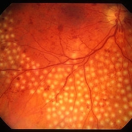
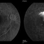
 Regressed Proliferative Diabetic Retinopathy following PRP
Sep 6 2012 by Sharon Fekrat, MD FACS FASRS
58-year-old man with regressed proliferative diabetic retinopathy in the left eye following panretinal laser photocoagulation. Note attenuated retinal vasculature.
Photographer: Sarah Enfiedjian, Ophthalmic Photographer, Durham VA Medical Center, Durham, NC
Imaging device: Zeiss
Condition/keywords: attenuated vessels, pan-retinal photocoagulation (PRP)
-
 PDR with Active NVD
Oct 8 2012 by Jeffrey G. Gross, MD, FASRS
PDR with active NVD and preretinal hemorrhage, mild VH and partial PRP.
Condition/keywords: neovascularization of the disc (NVD), preretinal hemorrhage, scatter laser photocoagulation, vitreous hemorrhage
-
 CSCR Mushroom Cloud
Feb 25 2015 by James J. Bedrick, MD
Late transit FA of a large active subfoveal CSCR leak. Focus is on peri-foveal vessels to give sense of height of large serous RD of macula. This patient presented with a BCVA of 20/200 and fluorescein and historic evidence of prior episodes of leakage. After discussion of known treatment options including observation, he was initially treated with rifampin and had partial resolution to 20/70 BCVA but this was short-lived with reaccumulation of the large serous detachment within 3 months. He then received sub-threshold micro-pulse laser photocoagulation with an 810 nm diode laser which resulted 1 month later in complete drying of the serous detachment and BCVA of 20/25.
Photographer: Diana Bodnar, COT
Imaging device: Topcon 50X with OIS capture station
Condition/keywords: CSCR subfoveal leak
-
---thumb.jpg/image-square;max$300,300.ImageHandler) Ocular Histoplasmosis Syndrome (OHS)
Ocular Histoplasmosis Syndrome (OHS)
Oct 8 2013 by Maurice F. Rabb
Thirty six year old white male stated that approximately 5 years earlier he had a blurry spot in his left eye that went away spontaneously after 3 months. Three years later the spot returned. He was seen by a local ophthalmologist who noted two "histo spots" in the left eye. Over the next 6 months his vision deteriorated from 20/30 to 20/200 in the left eye. A week prior to being seen at the UIHC he noted bulginess in his right eye. Visual acuity without correction was 20/15 OD, 20/200 OS. Color fundus photography and fluorescein angiography were performed and the patient was treated with argon laser photocoagulation. Vision decreased to 20/30 following laser surgery, but within two weeks returned to 20/15 and remained that way over the next two years. OVer the following 15 years the patient did well although he developed a recurrence in the untreated left eye and periodically he experienced vague changes in his central field.
Condition/keywords: ocular histoplasmosis syndrome (OHS)
-
 Proliferative Diabetic Retinopathy
Sep 15 2012 by Hamid Ahmadieh, MD
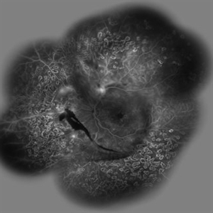
Fundus autofluorescence image of a 30-year-old woman with the history of scatter laser photocoagulation and a preretinal hemorrhage due to active PDR .
Photographer: Hamid Ahmadieh, MD, Ophthalmic Research Center, Labbafinejad Medical Center, Shahid Beheshti University of Medical Sciences
Imaging device: Heidelberg HRA
Condition/keywords: fundus autofluorescence (FAF), preretinal hemorrhage
-
 Marked Retinal Ischemia in Patient with Mixed Connective Tissue Disease
Feb 26 2013 by Sharon Fekrat, MD FACS FASRS
Fluorescein angiogram of the right eye of a 27-year-old female with mixed connective tissue disease and marked retinal ischemia. Panretinal laser photocoagulation (PRP) has been performed for neovascularization elsewhere (NVE).
Condition/keywords: mixed connective tissue disease, retinal ischemia
-
 Laser Photocoagulation
Nov 9 2012 by Norman Byer
This shows the same lesion 11 days following laser photocoagulation. Still more new hemorrhages can now be seen, and the retinal tissue in the center of the lesion is being visibly pulled forward. If you look carefully, you can see the sharp lower edge of a developing tractional horseshoe tear.
Condition/keywords: laser photocoagulation, retinal tissue, vitreous traction
-
 Laser Photocoagulation
Nov 9 2012 by Norman Byer
This is the same lesion 18 days following photocoagulation. The continuing vitreoretinal traction has now torn the retinal flap completely away from the retina and the resulting free operculum may be seen out of focus in the lower part of the photograph. The retinal tear is now easily visible with only a tiny remaining nubbin of the original flap seen above with a small hemorrhage.
Condition/keywords: free operculum, laser photocoagulation, retinal tear, vitreoretinal traction
-
 Regressed Proliferative Diabetic Retinopathy with Preretinal Hemorrhage
Oct 17 2012 by Sharon Fekrat, MD FACS FASRS
Fundus photograph of left eye of patient with regressed proliferative diabetic retinopathy following panretinal laser photocoagulation. Note fibrovascular proliferation along arcades and associated preretinal hemorrhage.
Photographer: John Reaves, Ophthalmic Photographer, Durham VA Medical Center, Durham, NC
Condition/keywords: preretinal hemorrhage, regressed
-
 Branch Retinal Artery Occlusion
Oct 2 2013 by Jerald A. Bovino, MD
There is a hollenhorst plaque causing a branch retinal artery occlusion. The patient has scars from prior panretinal laser photocoagulation.
Condition/keywords: branch retinal artery occlusion (BRAO), hollenhorst plaque, pan-retinal photocoagulation (PRP)
-
---thumb.jpg/image-square;max$300,300.ImageHandler) Chorioretinal Scarring
Chorioretinal Scarring
Feb 15 2013 by From the Collections of Thomas M. Aaberg, MD and Thomas M. Aaberg Jr., MD
Color fundus photograph showing chorioretinal scarring consistent with prior retinal laser photocoagulation to areas of peripheral retinal nonperfusion.
Condition/keywords: laser scarring, peripheral retinal nonperfusion
-
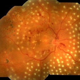 Diabetic Retinopathy With Laser Photocoagulation.
Sep 10 2017 by JEFFERSON R SOUSA, Tecg.º (Biomedical Systems Technology)
Patient patient 55-year-old, female, attended the clinic with complaint of low visual acuity. It was subjected to laser photocoagulation.
Photographer: JEFFERSON R SOUSA - Study Center and Ophthalmological Research Dr. Andre M V Gomes, Dr. Suel Abujamra Institute São Paulo-Brazil
Imaging device: Topcon TRC-50 DX, Imaginet, 50 degree field. Flash 75, mosaic with eleven images.
Condition/keywords: background diabetic retinopathy (BDR)
-
 Proliferative Diabetic Retinopathy (PDR)
Sep 11 2012 by Hamid Ahmadieh, MD
Wide- field FA image of a 55-year-old woman with active PDR and the history of scatter laser photocoagulation.
Photographer: Hamid Ahmadieh, MD, Ophthalmic Research Center, Labbafinejad Medical Center, Shahid Beheshti University of Medical Sciences
Imaging device: Heidelberg HRA
Condition/keywords: preretinal hemorrhage, retinal neovascularization, scatter laser photocoagulation
-
 Laser Photocoagulation
Nov 9 2012 by Norman Byer
This shows the same lesion four days after laser photo coagulation. The new hemorrhages seen in this photograph did not occur during the photocoagulation but developed within the next four days.
Condition/keywords: argon photocoagulation, laser photocoagulation, preretinal hemorrhage
-
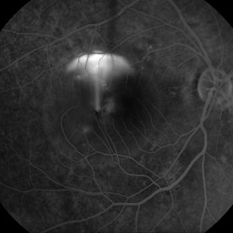
 Proliferative Diabetic Retinopathy
Sep 15 2012 by Hamid Ahmadieh, MD
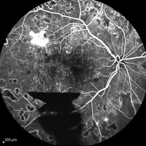
FA image of a 30-year-old woman with the history of scatter laser photocoagulation, NVE and a preretinal hemorrhage due to active PDR .
Photographer: Hamid Ahmadieh, MD, Ophthalmic Research Center, Labbafinejad Medical Center, Shahid Beheshti University of Medical Sciences
Imaging device: Heidelberg HRA
Condition/keywords: preretinal hemorrhage
-
 Brach Retinal Artery Occlusion
Oct 2 2013 by Jerald A. Bovino, MD
There is a hollenhorst plaque causing a branch retinal artery occlusion. The patient has scars from prior panretinal laser photocoagulation.
Condition/keywords: branch retinal artery occlusion (BRAO), hollenhorst plaque, pan-retinal photocoagulation (PRP)
-
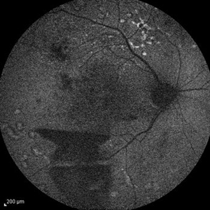
 Proliferative Diabetic Retinopathy
Sep 15 2012 by Hamid Ahmadieh, MD
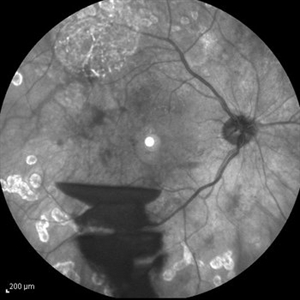
Infrared image of a 30-year-old woman with the history of scatter laser photocoagulation and a preretinal hemorrhage due to active PDR .
Photographer: Hamid Ahmadieh, MD, Ophthalmic Research Center, Labbafinejad Medical Center, Shahid Beheshti University of Medical Sciences
Imaging device: Heidelberg HRA
Condition/keywords: infrared image, preretinal hemorrhage
-
 CSCR Mushroom Cloud
Feb 23 2015 by James J. Bedrick, MD
Late transit FA of a large active sub-foveal CSCR leak. You may view this pair in stereo to appreciate the plume of leakage within this large serous RD of the macula. This patient presented with a BCVA of 20/200 and fluorescein and historic evidence of prior episodes of leakage. After discussion of known treatment options including observation, he elected to be treated initially with oral rifampin and BCVA improved to 20/40 with persistent metamorphosis and a shallower persistent macular detachment over several visits. Rifampin was discontinued and he then received sub-threshold micro-pulse laser photocoagulation with an 810 diode which resulted in the patient reporting full restoration of his vision subjectively within a month. He failed to keep his follow-up appointment.
Photographer: Diana Bodnar, COT
Imaging device: Topcon 50X with Merge capture station
Condition/keywords: CSCR subfoveal leak
-
 Eals Disease
Jan 26 2013 by Ratimir Lazic, MD, PhD
FAG image of a 28-year-old male. Staining of scars due to laser photocoagulation can be seen.
Photographer: Marko Lukic, MD
Imaging device: Zeis Visucam Lite 2
Condition/keywords: fundus photograph, laser photocoagulation
-
 CSCR Mushroom Cloud
Feb 25 2015 by James J. Bedrick, MD
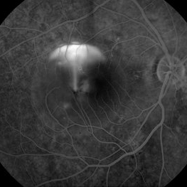
Late transit FA of a large active subfoveal CSCR leak. Focus is on peri-foveal vessels to give sense of height of large serous RD of macula. This patient presented with a BCVA of 20/200 and fluorescein and historic evidence of prior episodes of leakage. After discussion of known treatment options including observation, he was initially treated with rifampin and had partial resolution to 20/70 BCVA but this was short-lived with reaccumulation of the large serous detachment within 3 months. He then received sub-threshold micro-pulse laser photocoagulation with an 810 nm diode laser which resulted 1 month later in complete drying of the serous detachment and BCVA of 20/25.
Photographer: Diana Bodnar, COT
Imaging device: Topcon 50X with OIS capture station
Condition/keywords: CSCR subfoveal leak
-
 Exudative Macular Detachment After Intensive Laser Photocoagulation
Mar 12 2016 by Sjakon G Tahija, MD
Fundus photograph of 44-year-old man with exudative detachment of the macula after vitrectomy and ILM peeling for proliferative diabetic retinopathy combined with intensive endolaser photocagulation.
Photographer: Avris Siahaan, Klinik Mata Nusantara
Condition/keywords: exudative detachment, pan-retinal photocoagulation (PRP)
-
---thumb.jpg/image-square;max$300,300.ImageHandler) Ocular Histoplasmosis Syndrome (OHS)
Ocular Histoplasmosis Syndrome (OHS)
Oct 8 2013 by Maurice F. Rabb
Thirty six year old white male stated that approximately 5 years earlier he had a blurry spot in his left eye that went away spontaneously after 3 months. Three years later the spot returned. He was seen by a local ophthalmologist who noted two "histo spots" in the left eye. Over the next 6 months his vision deteriorated from 20/30 to 20/200 in the left eye. A week prior to being seen at the UIHC he noted bulginess in his right eye. Visual acuity without correction was 20/15 OD, 20/200 OS. Color fundus photography and fluorescein angiography were performed and the patient was treated with argon laser photocoagulation. Vision decreased to 20/30 following laser surgery, but within two weeks returned to 20/15 and remained that way over the next two years. OVer the following 15 years the patient did well although he developed a recurrence in the untreated left eye and periodically he experienced vague changes in his central field.
Condition/keywords: ocular histoplasmosis syndrome (OHS)
-
 Inactive Photocoagulated Diabetic Retinopathy
Apr 5 2017 by Linda A Cernichiaro- Espinosa, MD
Extensive pan-retinal photocoagulation in a Mexican patient that was required to inactivate the disease.
Photographer: Linda A Cernichiaro, E
Imaging device: Optos Daytona
Condition/keywords: diabetes, laser photocoagulation
-
 Retinopathy of Prematurity Under-Treated with Laser
Nov 8 2013 by Maria Ana Martinez-Castellanos, MD
Patient referred for a 2nd opinion after photocoagulation for ROP treatment. We can see under-treated avascular areas with no neovascular activity or elevated ridge
Photographer: Maria A. Martinez-Castellanos. Asociacion para Evitar la Ceguera en Mexico
Imaging device: RetCam II
Condition/keywords: laser photocoagulation, laser treatment, retinopathy of prematurity (ROP)
 Loading…