Search results (11 results)
-
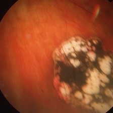
 Adenocarcinoma Arising from CHRPE
Sep 17 2015 by Marc C. Peden, MD
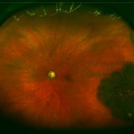
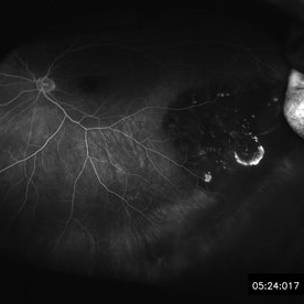
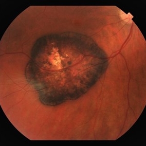
49-year-old female referred for presumed ocular melanoma. On examination was noted to have darkly pigmented lesion in the temporal retina of left eye. Lesion had characteristic scalloped edges with central lacunae, however, on ultrasonography was noted to have 1.8mm of elevation with high internal reflectivity. IVFA shows absence of dual circulation with areas of window defect. Findings were consistent with those described by Shields et al., in their April 2001 article in Archives of Ophthalmology.
Photographer: Janet Traynom
Imaging device: Optos P200MA
Condition/keywords: adenocarcinoma arising from CHRPE
-
 Adenocarcinoma Arising from CHRPE
Sep 17 2015 by Marc C. Peden, MD
49-year-old female referred for presumed ocular melanoma. On examination was noted to have darkly pigmented lesion in the temporal retina of left eye. Lesion had characteristic scalloped edges with central lacunae, however, on ultrasonography was noted to have 1.8mm of elevation with high internal reflectivity. IVFA shows absence of dual circulation with areas of window defect. Findings were consistent with those described by Shields et al., in their April 2001 article in Archives of Ophthalmology.
Photographer: Janet Traynom COT
Imaging device: Optos P200MA
Condition/keywords: adenocarcinoma arising from CHRPE
-
 Congenital Hypertrophy of the Retinal Pigment Epithelium
Nov 11 2019 by Jessica Norkus
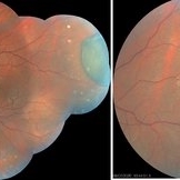
Bilateral Optos ultra wide field imaging of a 31-year-old female patient with CHRPE lesions. Lesions in OD were suspicious of Gardner Syndrome due to familial history of cancerous polyps in colon. Patient underwent colonoscopy and was deemed clear.
Photographer: Jessica Norkus, COA, Retina Specialists of Michigan
Imaging device: Optos Ultra Wide Field Camera
Condition/keywords: bear tracks, bilateral, color fundus photograph, color photo, congenital hypertrophy of the retinal pigment epithelium (CHRPE), fundus autofluorescence (FAF), fundus photograph, lacunae, macula, optic disc, Optos, pseudocolor, ultra-wide field imaging
-
 61-Year-Old Man With Large Peripheral CHRPE
Dec 9 2017 by Timothy S Fuller, MD
61-year-old man presented for evaluation of pigmented retinal lesion. Found to have a large, peripheral CHRPE with characteristic lacunae, sharp margins, and lack of elevation.
Condition/keywords: benign pigmented lesions, congenital hypertrophy of the retinal pigment epithelium (CHRPE), lacunae
-
 CHRPE
CHRPE
-
 CHRPE & Myelinated RNFL
May 21 2020 by John S. King, MD
47-year-old white female, asymptomatic, sent to evaluate a scar OD. 20/40 cc, normotensive, examination significant for a flat, solitary lesion with pigmented borders and depigmented center with early lacunae forming, along with myeliated RNFL at the temporal edge of the lesion.
Photographer: Kay Dalby
Imaging device: Topcon
Condition/keywords: congenital hypertrophy of the retinal pigment epithelium (CHRPE), myelinated nerve fiber layer
-
 CHRPE
CHRPE
Jan 12 2023 by Christopher R. Adam, M.D.
Optos color photograph of a 51 y/o F with a 9x9mm CHRPE in the nasal quadrant. The lesion is flat with a bordering halo and lacunae.
Condition/keywords: congenital hypertrophy of the retinal pigment epithelium (CHRPE)
-
 CHRPE
CHRPE
Jan 15 2021 by Priya Rasipuram Chandrasekaran, MBBS, DO, DNB, FRCS
This is the fundus photo and fundus photo montage of the left eye of a 25-year-old male showing flat, solitary, round, greyish pigmented lesion situated AT THE equator with a scalloped margin. Vessels overlying the lesion are normal and there is a clear demarcation line between this and normal retina. The margins are hypopigmented with few hypopigmented lacunae inside.
Condition/keywords: congenital hypertrophy of the retinal pigment epithelium (CHRPE)
-
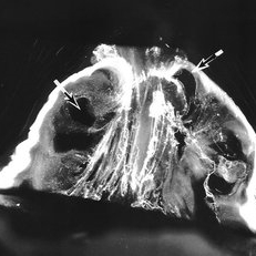 Senescent Human Vitreous Structure
Sep 1 2020 by J. Sebag, MD, FACS, FRCOphth, FARVO
Dark-field slit microscopy was performed on a fresh, unfixed, post-mortem human eye from an 88 year-old subject. The eye had dissection to peel off the sclera, choroid, and retina. The vitreous body remains attached to the anterior segment which is seen below, while the posterior pole is above in this image. Significant degenerative fibrous liquefaction is evident. Lacunae can be seen (arrows) that do not scatter light because they are primarily filled with liquid vitreous that is mostly hyaluronan and water, with little collagen to scatter light. Drug injection into a lacuna would likely have very different distribution than injection into an area of gel vitreous. [From Sebag J: The Vitreous - Structure, Function, and Pathobiology. Springer-Verlag, New York, 1989 (image © Springer Nature, reprinted with permission); reprinted in Sebag J: Vitreous in AMD therapy – the medium is the message (Guest Editorial). Retina 2015;35(9):1715-18]
Condition/keywords: vitreous
-
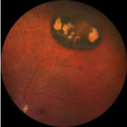
 Solitary Congenital Hypertrophy of the Retinal Pigment Epithelium with Lacunae
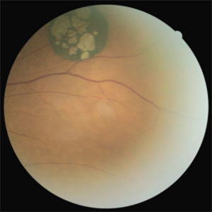
Jun 11 2023 by Ethan K Sobol, MD
Fundus photograph of a solitary CHRPE in the superotemporal quadrant with central hypopigmented lacunae.
Condition/keywords: CHRPE, congenital hypertrophy of the retinal pigment epithelium (CHRPE), lacunae
-
 Congenital Hypertrophy of RPE
Apr 2 2019 by Gary R. Cook, MD, FACS
67-year-old white male with typical CHRPE lesion demonstrating pigment hypertrophy and multiple lacunae in the inferotemporal periphery of OS. V.A. = 20/30
Imaging device: Topcon VT-50
Condition/keywords: congenital hypertrophy of the retinal pigment epithelium (CHRPE)
 Loading…