Search results (169 results)
-
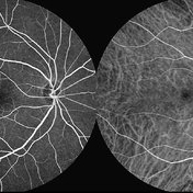
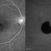
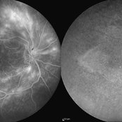
 Polypoidal Choroidal Vasculopathy
Aug 25 2012 by Hamid Ahmadieh, MD
FA & ICG angiography imagings of a 73-year-old man with a peripapillary PCV.
Photographer: Hamid Ahmadieh, Ophthalmic Research Center, Labbafinejad Medical Center
Imaging device: Heidelberg Spectralis
Condition/keywords: indocyanine green (ICG) angiography, polypoidal choroidal vasculopathy (PCV)
-
 Angioid Streaks & CNV (Fig 3)
Aug 25 2012 by Hamid Ahmadieh, MD
Early phase ICG angiography imaging of a 53-year-old woman with a juxtafoveal CNV secondary to angioid streaks.
Photographer: Hamid Ahmadieh, Ophthalmic Research Center, Labbafinejad Medical Center
Imaging device: Heidelberg Spectralis
Condition/keywords: angioid streaks, choroidal neovascularization (CNV), indocyanine green (ICG) angiography
-
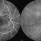
 Multifocal CSCR 2
Sep 2 2012 by Hamid Ahmadieh, MD
Early-phase FA and ICG angiograms of a 36-year-old man with an active multifocal CSCR.
Photographer: Hamid Ahmadieh, Ophthalmic Research Center, Labbafinejad Medical Center
Imaging device: Heidelberg Spectralis
Condition/keywords: central serous chorioretinopathy (CSCR), indocyanine green (ICG) angiography
-
 PED due to CSCR 4
Sep 2 2012 by Hamid Ahmadieh, MD
Early phase FA & ICG images of a 37-year-old man with a serous PED secondary to CSCR
Photographer: Hamid Ahmadieh, Ophthalmic Research Center, Labbafinejad Medical Center
Imaging device: Heidelberg Spectralis
Condition/keywords: central serous chorioretinopathy (CSCR), indocyanine green (ICG) angiography, pigment epithelial detachment (PED)
-
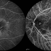
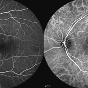
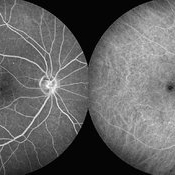
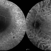 Retinitis Pigmentosa
Sep 11 2012 by Hamid Ahmadieh, MD
FA & ICG angiography images of a 40-year-old man with RP.
Photographer: Hamid Ahmadieh, MD, Ophthalmic Research Center, Labbafinejad Medical Center, Shahid Beheshti University of Medical Sciences
Imaging device: Heidelberg Spectralis
Condition/keywords: indocyanine green (ICG) angiography, retinitis pigmentosa
-
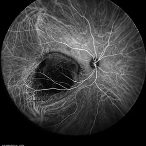
 Angioid Streaks
Sep 29 2012 by Hamid Ahmadieh, MD
Late phase ICG angiography image of the left eye of a 59-year-old man with angioid streaks.
Photographer: Hamid Ahmadieh, MD; Ophthalmic Research Center, Labbafinejad Medical Center, Shahid Beheshti University of Medical Sciences
Imaging device: Heidelberg Spectralis
Condition/keywords: angioid streaks, indocyanine green (ICG) angiography
-
 Multifocal CSCR
Sep 2 2012 by Hamid Ahmadieh, MD
Late-phase FA and ICG angiograms of a 36-year-old man with an active multifocal CSCR.
Photographer: Hamid Ahmadieh, Ophthalmic Research Center, Labbafinejad Medical Center
Imaging device: Heidelberg Spectralis
Condition/keywords: central serous chorioretinopathy (CSCR), indocyanine green (ICG) angiography
-
 Fundus Flavimaculatus and CNV
Nov 14 2013 by Hamid Ahmadieh, MD
Late phase FA and ICG angiography images of the right eye of a 35-year-old woman with subfoveal CNV secondary to fundus flavimaculatus .
Photographer: Nayereh Hadipour, Negah Eye Center, Tehran
Condition/keywords: choroidal neovascularization (CNV), fundus flavimaculatus, indocyanine green (ICG) angiography, retinal flecks
-
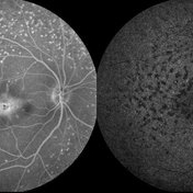
 Chronic Active Central Serous Chorioretinopathy (CSCR)
Sep 11 2012 by Hamid Ahmadieh, MD
Late phase FA & ICG angiography images of a 30-year-old man with chronic active CSCR.
Photographer: Hamid Ahmadieh, MD, Ophthalmic Research Center, Labbafinejad Medical Center, Shahid Beheshti University of Medical Sciences
Imaging device: Heidelberg Spectralis
Condition/keywords: central serous chorioretinopathy (CSCR), indocyanine green (ICG) angiography
-
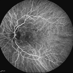
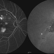
 Macular Telangiectasia Type 2
Sep 22 2012 by Hamid Ahmadieh, MD
FA and ICG angiography imagings of the right eye of a 70-year-old man with idiopathic macular telangiectasia type 2.
Photographer: Hamid Ahmadieh, MD, Ophthalmic Research Center, Labbafinejad Medical Center, Shahid Beheshti University of Medical Sciences
Imaging device: HRA
Condition/keywords: idiopathic macular telangiectasia, indocyanine green (ICG) angiography
-
 Chronic Active Central Serous Chorioretinopathy (CSCR)
Sep 11 2012 by Hamid Ahmadieh, MD
Early phase FA & ICG angiography images of a 30-year-old man with chronic active CSCR.
Photographer: Hamid Ahmadieh, MD, Ophthalmic Research Center, Labbafinejad Medical Center, Shahid Beheshti University of Medical Sciences
Imaging device: Heidelberg Spectralis
Condition/keywords: central serous chorioretinopathy (CSCR), indocyanine green (ICG) angiography
-
 Best Disease
Mar 9 2013 by Hamid Ahmadieh, MD
FA and ICG Angiography of the left eye of a 49-year-old man with advanced Best disease.
Photographer: Soodabeh Fooladin, Negah Eye Center, Tehran
Imaging device: Heidelberg Spectralis
Condition/keywords: Best disease, indocyanine green (ICG) angiography
-
 Cystoid Macular Edema (CME)
Sep 11 2012 by Hamid Ahmadieh, MD
Late phase FA & ICG angiography imagings of the left eye a 17-year-old boy with CME & retinal periphlebitis secondary to chronic intermediate uveitis.
Photographer: Hamid Ahmadieh, MD, Ophthalmic Research Center, Labbafinejad Medical Center, Shahid Beheshti University of Medical Sciences
Imaging device: Heidelberg Spectralis
Condition/keywords: cystoid macular edema (CME), indocyanine green (ICG) angiography, intermediate uveitis
-
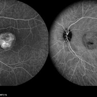
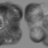
 Polypoidal Choroidal Vasculopathy: Case 1 - Image 6of 7
Oct 4 2012 by Gregg T. Kokame, MD, MMM, FASRS
OCT/Indocyanine Green Angiography image of a 57-year-old woman with treatment-naive polypoidal choroidal vasculopathy. Series of images provides an comparative view of the same condition while utilizing a variet of different imaging procedures.
Photographer: Andrew Yuen, Retina Consultants of Hawaii
Imaging device: Heidelberg Spectralis
Condition/keywords: branching vascular network (BVN), indocyanine green (ICG) angiography, optical coherence tomography (OCT), polypoidal choroidal vasculopathy (PCV)
-
 PED due to CSCR 5
Sep 2 2012 by Hamid Ahmadieh, MD
Late-phase FA and ICG images of a 37-year-old man with a serous PED secondary to CSCR.
Photographer: Hamid Ahmadieh, Ophthalmic Research Center, Labbafinejad Medical Center
Imaging device: Heidelberg Spectralis
Condition/keywords: central serous chorioretinopathy (CSCR), indocyanine green (ICG) angiography, pigment epithelial detachment (PED)
-
 Cystoid Macular Edema (CME)
Sep 11 2012 by Hamid Ahmadieh, MD
FA & ICG angiography imagings of a 17-year-old boy with CME secondary to chronic intermediate uveitis.
Photographer: Hamid Ahmadieh, MD, Ophthalmic Research Center, Labbafinejad Medical Center, Shahid Beheshti University of Medical Sciences
Imaging device: Heidelberg Spectralis
Condition/keywords: cystoid macular edema (CME), indocyanine green (ICG) angiography, intermediate uveitis
-
 Angioid Streaks
Sep 29 2012 by Hamid Ahmadieh, MD
Late phase ICG angiography image of the right eye of a 59-year-old man with angioid streaks.
Photographer: Hamid Ahmadieh, MD; Ophthalmic Research Center, Labbafinejad Medical Center, Shahid Beheshti University of Medical Sciences
Imaging device: Heidelberg Spectralis
Condition/keywords: angioid streaks, indocyanine green (ICG) angiography
-
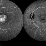
 Polypoidal Choroidal Vasculopathy: Case 1 - Image 4 of 7
Oct 4 2012 by Gregg T. Kokame, MD, MMM, FASRS
OCT/Indocyanine Green Angiography image of a 57-year-old woman with treatment-naive polypoidal choroidal vasculopathy. Series of images provides an comparative view of the same condition while utilizing a variet of different imaging procedures.
Photographer: Andrew Yuen, Retina Consultants of Hawaii
Imaging device: Heidelberg Spectralis
Condition/keywords: indocyanine green (ICG) angiography, optical coherence tomography (OCT), polypoidal choroidal vasculopathy (PCV)
-
 Cystoid Macular Edema (CME)
Sep 11 2012 by Hamid Ahmadieh, MD
Wide-field FA & ICG angiography imagings of a 17-year-old boy with CME & retinal periphlebitis secondary to chronic intermediate uveitis.
Photographer: Hamid Ahmadieh, MD, Ophthalmic Research Center, Labbafinejad Medical Center, Shahid Beheshti University of Medical Sciences
Imaging device: Heidelberg Spectralis
Condition/keywords: cystoid macular edema (CME), indocyanine green (ICG) angiography, intermediate uveitis
-
---thumb.jpg/image-square;max$300,300.ImageHandler) Polypoidal Choroidal Vasculopathy - Case 1
Polypoidal Choroidal Vasculopathy - Case 1
Oct 4 2012 by Gregg T. Kokame, MD, MMM, FASRS
Indocyanine Green Angiography image of a 57-year-old woman with treatment-naive polypoidal choroidal vasculopathy. Series of images provides an comparative view of the same condition while utilizing a variet of different imaging procedures.
Photographer: Andrew Yuen, Retina Consultants of Hawaii
Imaging device: Heidelberg Spectralis
Condition/keywords: branching vascular network (BVN), indocyanine green (ICG) angiography, polypoidal choroidal vasculopathy (PCV)
-
 Cystoid Macular Edema (CME)
Sep 11 2012 by Hamid Ahmadieh, MD
Late phase FA & ICG angiography imagings of the right eye a 17-year-old boy with CME & retinal periphlebitis secondary to chronic intermediate uveitis.
Photographer: Hamid Ahmadieh, MD, Ophthalmic Research Center, Labbafinejad Medical Center, Shahid Beheshti University of Medical Sciences
Imaging device: Heidelberg Spectralis
Condition/keywords: cystoid macular edema (CME), indocyanine green (ICG) angiography, intermediate uveitis
-
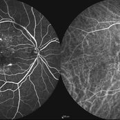
 Macular Telangiectasia Type 2
Sep 22 2012 by Hamid Ahmadieh, MD
Late phase FA and ICG angiography imagings of the right eye of a 70-year-old man with idiopathic macular telangiectasia type 2.
Photographer: Hamid Ahmadieh, MD, Ophthalmic Research Center, Labbafinejad Medical Center, Shahid Beheshti University of Medical Sciences
Imaging device: HRA
Condition/keywords: idiopathic macular telangiectasia, indocyanine green (ICG) angiography
-
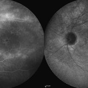
 Choroidal Tumor
Oct 17 2014 by Avris Romario Diparaja Siahaan
An ICG Angiography photography of a 27-year-old woman with a choroidal tumor in her right eye.
Photographer: Avris Romario Diparaja Siahaan, Klinik Mata Nusantara
Imaging device: Heidelberg Spectralis
Condition/keywords: choroidal tumor, indocyanine green (ICG) angiography, ultra-wide field imaging
-
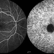
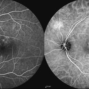
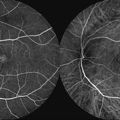 Macular Telangiectasia Type 2 & CNV
Sep 22 2012 by Hamid Ahmadieh, MD
FA and ICG angiography imagings of the left eye of a 70-year-old man with idiopathic macular telangiectasia type 2 and CNV.
Photographer: Hamid Ahmadieh, MD, Ophthalmic Research Center, Labbafinejad Medical Center, Shahid Beheshti University of Medical Sciences
Imaging device: HRA
Condition/keywords: choroidal neovascularization (CNV), idiopathic macular telangiectasia, indocyanine green (ICG) angiography
 Loading…