Search results (91 results)
-
 Diabetic Retinopathy Hard Exudates OS
Jun 30 2013 by Rogerio N Shinsato, MD, PhD
Fundus photograph with diabetic retinopathy.
Condition/keywords: diabetic macular edema, foveal hard exudates
-
 Diabetic Retinopathy, CSME, Color Fundus Photo
Mar 18 2015 by James B. Soque, CRA, OCT-C, COA, FOPS
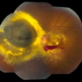
A 58-year-old diabetic male with a longstanding history of diabetic eye disease. Left eye color fundus photo shows extensive CSME, Clinically Significant Macular Edema, with deposits of hard exudates at fixation. There is extensive scattering of hard exudates and sheathing of the vessels.
Photographer: James B Soque, CRA COA
Imaging device: Topcon TRC 50 DX, OIS 5 MP Camera, MERGE software
Condition/keywords: background diabetic retinopathy (BDR), creamy yellow exudates, diabetes, exudates over the posterior pole, neovascularization of the disc (NVD), vessel sheathing
-
 Branch Retinal Vein Occlusion with Macular Edema
Aug 23 2012 by Gerardo Garcia-Aguirre, MD
Fundus photograph composition of the left eye, showing flame-shaped and blot hemorrhages in the superotemporal quadrant, with hard exudates surrounding the fovea.
Photographer: Noemí Hernández, Asociación para Evitar la Ceguera en México
Condition/keywords: branch retinal vein occlusion (BRVO), macular edema
-
---thumb.jpg/image-square;max$300,300.ImageHandler) Diabetic Retinopathy Hard Exudates OD
Diabetic Retinopathy Hard Exudates OD
Jun 30 2013 by Rogerio N Shinsato, MD, PhD
Fundus photograph with diabetic retinopathy.
Condition/keywords: diabetic macular edema, foveal hard exudates
-
 Diabetic Retinopathy, CSME, Exudates, NVD, Color Fundus Photo, Montage
Mar 18 2015 by James B. Soque, CRA, OCT-C, COA, FOPS
A 58-year-old diabetic male with a longstanding history of diabetic eye disease. Left eye color fundus photo shows extensive CSME, Clinically Significant Macular Edema, with deposits of hard exudates at fixation. There is extensive scattering of hard exudates and sheathing of the vessels.
Photographer: James B Soque, CRA COA
Imaging device: Topcon TRC 50 DX, OIS 5 MP Camera, MERGE software
Condition/keywords: background diabetic retinopathy (BDR), creamy yellow exudates, diabetes, exudates over the posterior pole, neovascularization of the disc (NVD), vessel sheathing
-
 Type 1A Macular Telangiectasia - Fundus photograph
Nov 11 2013 by Gerardo Garcia-Aguirre, MD
Fundus photograph of a 43-year-old male complaining of mild metamorphopsia in OS. BCVA 20/25. Some hard exudates and telangiectatic vessels are observed inferior and temporal to the fovea.
Condition/keywords: macular telangiectasia
-
---thumb.JPG/image-square;max$300,300.ImageHandler) diabetic macular edema
diabetic macular edema
Oct 26 2012 by Mallika Goyal, MD
Fundus photograph of left eye of 58-year-old diabetic gentleman with normal serum lipids showing foveal hard exudates.
Condition/keywords: foveal hard exudates
-
---thumb.JPG/image-square;max$300,300.ImageHandler) Diabetic Macular Edema
Diabetic Macular Edema
Oct 26 2012 by Mallika Goyal, MD
Fundus photograph of left eye of 55-year-old diabetic and hypertensive gentleman with normal serum lipids showing abundant foveal hard exudates.
Condition/keywords: diabetic macular edema
-
 Choroidal Melanoma With Radiation Retinopathy
Jul 8 2013 by Jason S. Calhoun
Patient came with follow up on choroidal melanoma. Right eye that was treated back in June of 2009 with a radioactive implant. Vein occlusion is also present with VA - hand motion. Hemorrhages visible with hard exudates from the radiation retinopathy.
Photographer: Jason S. Calhoun, Department of Ophthalmology, Mayo Clinic Jacksonville, Florida
Condition/keywords: radiation retinopathy
-
 Choroidal Melanoma
Jun 30 2013 by Jason S. Calhoun
Post-op radioactive implant with hard exudates surrounding the melanoma with hemorrhage present.
Photographer: Jason S. Calhoun, Mayo Clinic Jacksonville, Florida
-
 Leptospirosis Neuroretinitis
May 30 2014 by Mitzy E Torres Soriano, MD
50-year-old man, presented with sudden onset of reduced vision in the left eye. Visual acuity (VA) was count fingers. Fundoscopic examination revealed soft exudation adjacent the optic nerve, macular edema with hard exudates in star shape arrangement and retinal vasculitis. OCT confirmed macular edema. There were no systemic symptoms. History of alcoholism and crack cocaine addiction. Systemic work up revealed a positive leptospira. He was treated with oral doxycicline (100mg twice daily) and prednisona (1mg/kg with gradual taper) for two weeks. Follow up at six months showed an improvement of VA to 20/60 with partial resolution of clinical findings at fundoscopic exam. Leptospirosis should be ruled out in every case of neuroretinitis.
Photographer: Mitzy E. Torres Soriano, MD; Centro medico Cagua-Estado Aragua. Venezuela
Imaging device: Retinal Camera TRC-NW8, TOPCON
Condition/keywords: leptospirosis, macular star, neuroretinitis, retinal vasculitis
-
 Melanoma With Radiation Retinopathy
Jul 14 2013 by Jason S. Calhoun
Fundus photo shows treated choroidal melanoma superior, nasal in the left eye. Patient was treated with radioactive implant. Radiation retinopathy with hard exudates present.
Photographer: Jason S. Calhoun, Department of Ophthalmology, Mayo Clinic Jacksonville, Florida
Imaging device: TOPCON TRC 50-EX
Condition/keywords: radiation retinopathy
-
 Acute Necrotizing Retinal Vasculitis as Onset of Systemic Lupus Erythematosus.
Sep 3 2016 by ADRIANO FERREIRA
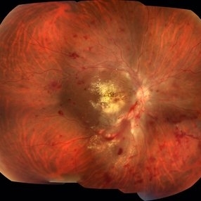
A 28-year-old white man was referred to the rheumatology clinic with gradually and rapid deterioration of the vision (both eyes). In this picture, we can observe cotton wool spots in the papillomacular area and extensive hemorrhages in posterior polo and in the middle periphery. Hard exudates are present in macular area (macular edema)
Photographer: Claudio Zett Lobo
Imaging device: TRC50DXi TOPCON
Condition/keywords: systemic lupus erythematosus (SLE) vasculitis, vasculitis
-
 Diabetic Retinopathy Hard Exudates OD
Jun 30 2013 by Rogerio N Shinsato, MD, PhD
Fundus photograph with diabetic retinopathy.
Condition/keywords: diabetic macular edema, foveal hard exudates
-
---thumb.jpg/image-square;max$300,300.ImageHandler) Coats disease
Coats disease
Jan 11 2013 by Hyung-Woo Kwak, MD
Fundus imaging shows hemorrhage and hard exudates from leaking blood vessel.
Photographer: Taegi Kim, Kyung Hee Univsersity Hospital, Seoul
Imaging device: Zeiss f 450 plus
Condition/keywords: argon photocoagulation
-
 Diabetic Retinopathy Hard Exudates OD Early Phase
Jun 30 2013 by Rogerio N Shinsato, MD, PhD
Fundus photograph with diabetic retinopathy.
Condition/keywords: diabetic macular edema, foveal hard exudates
-
---thumb.JPG/image-square;max$300,300.ImageHandler) Foveal Exudates in Diabetic Maculopathy
Foveal Exudates in Diabetic Maculopathy
Dec 13 2013 by Mallika Goyal, MD
Left eye fundus photograph of a 55-year-old lady with diabetes and dyslipidemia shows hard exudates at foveal center reducing visual acuity to 200/400.
Photographer: Mallika Goyal, MD, Apollo Health City, Hyderabad, India
Condition/keywords: diabetic maculopathy, foveal exudate
-
 Diabetic Retinopathy Hard Exudates OS
Jun 30 2013 by Rogerio N Shinsato, MD, PhD
Fundus photograph with diabetic retinopathy.
Condition/keywords: diabetic macular edema, foveal hard exudates
-
 Neovascular ARMD With Subretinal Hemorrhage, Red-Free Photos - Stereo
Nov 26 2014 by James B. Soque, CRA, OCT-C, COA, FOPS
Stereo FC, RF and FA of a 77-year-old white female with visual acuity CC 20/200-3, with left eye neovascular ARMD, drusen, and subretinal hemorrhage with hard exudates temporally. Peripheral retina reveals cobblestone degeneration.
Photographer: James Soque, CRA, COA, Island Retina, Shirley, NY
Imaging device: Topcon TRC 50 EX, with MERGE software and OIS 5 MP digital Camera
Condition/keywords: neovascular age-related macular degeneration (AMD), red-free, stereo pair
-
 Treated Melanoma
Jul 14 2013 by Jason S. Calhoun
Choroidal melanoma treated with radiation. Hard exudates formed due to radiation retinopathy.
Photographer: Jason S. Calhoun, Department of Ophthalmology, Mayo Clinic Jacksonville, Florida
Imaging device: TOPCON TRC 50-EX
Condition/keywords: radiation retinopathy
-
 Neovascular ARMD With Subretinal Hemorrhage, Fluorescein Angiography Photos - Stereo
Oct 14 2014 by James B. Soque, CRA, OCT-C, COA, FOPS
Stereo FC, RF and FA of a 77-year-old white female with visual acuity CC 20/200-3, with left eye neovascular ARMD, drusen, and subretinal hemorrhage with hard exudates temporally. Peripheral retina reveals cobblestone degeneration.
Photographer: James Soque, CRA, COA, Island Retina, Shirley, NY
Imaging device: Topcon TRC 50 EX, with MERGE software and OIS 5 MP digital Camera
Condition/keywords: neovascular age-related macular degeneration (AMD), stereo pair
-
 Adult Coats' Disease
Aug 18 2015 by Mallika Goyal, MD
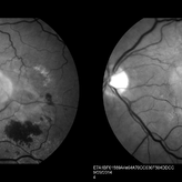
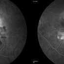
Left fundus of a 61-year-old non diabetic, non hypertensive lady complaining of vision deterioration for 1 year showed massive hard exudates at the macula. Fluorescein angiography revealed microvascular abnormalities over the posterior pole and temporal midperiphery and extensive capillary non-perfusion over the temporal retinal quadrants. OCT revealed macular edema. Fellow eye fundus and angiogram were normal.
Photographer: Mallika Goyal, MD, Apollo Health City, Jubilee Hills, Hyderabad
Condition/keywords: Coats' disease
-
 Adult Coats' Disease
Aug 18 2015 by Mallika Goyal, MD
Left fundus of a 61-year-old non diabetic, non hypertensive lady complaining of vision deterioration for 1 year showed massive hard exudates at the macula. Fluorescein angiography revealed microvascular abnormalities over the posterior pole and temporal midperiphery and extensive capillary non-perfusion over the temporal retinal quadrants. OCT revealed macular edema. Fellow eye fundus and angiogram were normal.
Photographer: Mallika Goyal, MD, Apollo Health City, Jubilee Hills, Hyderabad
Condition/keywords: Coats' disease
-
---thumb.JPG/image-square;max$300,300.ImageHandler) Exudative Diabetic Maculopathy
Exudative Diabetic Maculopathy
Nov 18 2013 by Mallika Goyal, MD
Hard exudates at macula, including at foveal centre, in an eye with diabetic retinopathy.
Photographer: Mallika Goyal, MD, Apollo Health City, Hyderabad
Condition/keywords: exudative diabetic retinopathy
-
---thumb.JPG/image-square;max$300,300.ImageHandler) Diabetic Maculopathy
Diabetic Maculopathy
Nov 18 2013 by Mallika Goyal, MD
Occasional hard exudates near foveal centre in a diabetic with mild NPDR.
Photographer: Mallika Goyal, MD, Apollo Health City, Hyderabad
Condition/keywords: diabetic maculopathy
 Loading…