Search results (60 results)
-
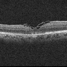
 Full-thickness Macular Hole
Aug 28 2012 by Sharon Fekrat, MD FACS FASRS
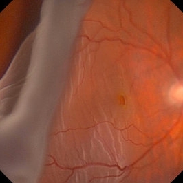
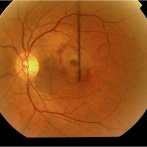
65 year old woman with a recurrent full thickness macular hole following previous 20 g pars plana vitrectomy in the right eye as well as an iatrogenic retinal hole in the papillomacular bundle. Both retinal defects are captured here in this Zeiss Stratus OCT image.
Photographer: Michael P. Kelly, FOPS Director, Duke Eye Labs, Duke University Eye Center, Durham, NC
Imaging device: Zeiss Cirrus
Condition/keywords: retinal break
-
 ILM peeling
Apr 11 2014 by Subhendu Kumar Boral, MBBS, MD(AIIMS), DNB
Brilliant blue stained ILM peeling in a case of idiopathic full thickness macular hole in a 61-year-old lady.
Photographer: Subhendu Kumar Boral
Condition/keywords: internal limiting membrane (ILM) peeling
-
 Retinal Detachment with Giant Retinal Tear and Macular Hole
Jan 6 2020 by MATTEO FORLINI, MD
A 61-year-old-male patient presented with sudden diminution of vision in the right eye due to retinal detachment with giant retinal tear and macular hole. Best corrected visual acuity (BCVA) at presentation was 20/200. A 23 G vitrectomy was performed. The edges of the tear were unrolled and complete retinal re-attachment under PFCL was achieved. A 360 degree intraoperative endolaser was performed on the peripheral retina as well as around the edges of the tears. PFCL was exchanged with silicone oil 5000cs as final tamponade. At six-months follow-up retina was attached and macular hole was repaired. Best-corrected visual acuity is 20/125 at present.
Photographer: Matteo Forlini MD, San Marino Hospital, Republic of San Marino
Condition/keywords: full thickness macular hole, giant retinal tear, silicone oil
-
---thumb.jpg/image-square;max$300,300.ImageHandler) Silicone Oil Surface
Silicone Oil Surface
Nov 14 2013 by Hamid Ahmadieh, MD
OCT image of the silicone oil surface bridging an enhanced foveal depression. There is the history of a rhegmatogenous retinal detachment associated with full thickness macular hole repair.
Photographer: Naghmeh Nozhat, Negah Eye Center, Tehran
Condition/keywords: optical coherence tomography (OCT), silicone oil
-
 Intravitreal Cysticercosis With Full Thickness Macular Hole
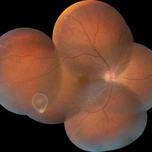
Apr 30 2018 by Vishal Agrawal, MD, FRCS,FACS,FASRS
Fundus montage picture of a 40-year-old man presenting with decreased vision in the right eye for the past 2 months. Live intravitreal cysticercosis can be seen lying on the retina. Zooming the image reveals the full thickness macular hole. The scolex invaginates with the light of the camera causing double image of the cyst because of movement .
Photographer: Vishal Agrawal MD,FRCS
Imaging device: Zeiss 524
Condition/keywords: cysticercosis, full thickness macular hole
-
 Full Thickness Macular Hole With ERM
Feb 26 2014 by Sharon Fekrat, MD FACS FASRS
Middle aged woman with a full thickness macular hole in the left eye associated with an epiretinal membrane.
Photographer: Michael P Kelly, Ophthalmic Photographer, Duke Eye Imaging, Duke Eye Center
Condition/keywords: epiretinal membrane (ERM), macular hole
-
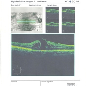
 Stage 3 Macular Hole With Operculum
Sep 25 2018 by samarth mishra
Stage 3 macular hole with operculum.
Photographer: Aditya Birla Sankara Nethralaya, Kolkata, West Bengal , India and Sankara Nethralaya, chennai , India
Condition/keywords: full thickness macular hole, macular hole, optical coherence tomography (OCT)
-
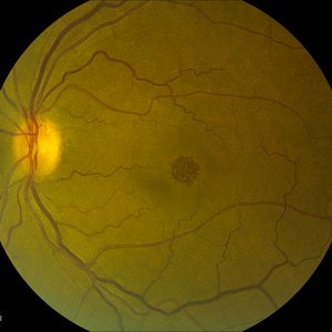
 Macular Hole and Retinoschisis in Goldmann - Favre Syndrome
May 13 2017 by ADRIANO FERREIRA
Fundus photograph of an 9-year-old child with Goldmann-Favre syndrome presenting with a macular hole and retinoschisis in the right eye.
Photographer: Jose Luiz
Condition/keywords: full thickness macular hole, Goldmann-Favre Syndrome, retinoschisis
-
 Sub-PFCL ILM Peeling
Apr 11 2014 by Subhendu Kumar Boral, MBBS, MD(AIIMS), DNB
Sub-PFCL BBG stained ILM peeling in a case of post traumatic total retinal detachment with superior large tear with full thickness macular hole.
Photographer: Subhendu Kumar Boral
Condition/keywords: internal limiting membrane (ILM) peeling
-
 Full Thickness Macular Hole
Jul 9 2012 by George W. Aylward, MD, FRCS, FRCOphth
A patient with a full thickness macular hole reducing vision to 20/200
-
 Macular hole with postoperative CNV
Dec 24 2012 by Ivan R. Batlle, MD
Pt with full thickness macular hole complicated with CNV after macular hole surgery
Condition/keywords: juxtafoveal choroidal neovascularization (CNV), macular hole
-
 Chronic Full Thickness Macular Hole
Dec 7 2016 by Jared Watson
Chronic full thickness macular hole OS, S/P attempted repair.
Condition/keywords: full thickness macular hole
-
 Macular Hole POD 1 OCT
Jun 21 2016 by John S. King, MD
POD 1 - hole closed as seen on OCT
Condition/keywords: full thickness macular hole, optical coherence tomography (OCT)
-
 Chronic Full Thickness Macular Hole
Dec 7 2016 by Jared Watson
Chronic full thickness macular hole OS, S/P attempted repair.
Condition/keywords: full thickness macular hole, IR
-
 Macular hole with postoperative CNV
Dec 24 2012 by Ivan R. Batlle, MD
Pt with full thickness macular hole complicated with CNV after macular hole surgery
Condition/keywords: juxtafoveal choroidal neovascularization (CNV)
-
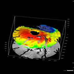 HRT of a Full Thickness Macular Hole
May 29 2013 by Zofia Anna Nawrocka (vel Michalewska), MD, PhD
3- dimensional HRT image of a full thickness macular hole.
Photographer: Zofia Michalewska, Ophthalmic Clinic "Jasne Blonia", Lodz, Poland
Imaging device: Heidelberg Retinal Tomograph
Condition/keywords: macular hole
-
 Chronic Full Thickness Macular Hole
Dec 7 2016 by Jared Watson
Chronic full thickness macular hole OS, S/P attempted repair.
Condition/keywords: full thickness macular hole
-
 Chronic Full Thickness Macular Hole
Dec 7 2016 by Jared Watson
Chronic full thickness macular hole OS, S/P attempted repair.
Imaging device: Spectralis OCT
Condition/keywords: full thickness macular hole, optical coherence tomography (OCT)
-
---thumb.jpg/image-square;max$300,300.ImageHandler) Full Thickness Macular Hole
Full Thickness Macular Hole
May 29 2013 by Zofia Anna Nawrocka (vel Michalewska), MD, PhD
3-dimensional HRT of a full thickness macular hole.
Photographer: Zofia Michalewska, Ophthalmic Clinic "Jasne Blonia", Lodz, Poland
Imaging device: Heidelberg Retinal Tomograph
Condition/keywords: macular hole
-
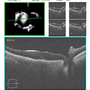
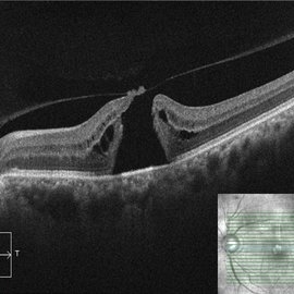 OCT Evidence of VMT Resulting in Full Thickness Macular Hole
Dec 24 2020 by Deepak Bhojwani, MS
OCT image of a patient (with past history of focal VMT ) progressing to full thickness macular hole. Note the posterior hyaloid attachment over the torn edges of fovea.
Photographer: DEEPAK BHOJWANI
Condition/keywords: full thickness macular hole, optical coherence tomography (OCT), vitreomacular traction (VMT)
-
 Full-thickness Macular Hole
Apr 8 2019 by Gary R. Cook, MD, FACS
Elderly white female with a Stage IV, full-thickness macular hole OS with whitish deposits visible at the base of the hole and a surrounding cuff of subretinal fluid
Imaging device: Topcon VT-50
Condition/keywords: full thickness macular hole, macular hole
-
 Full Thickness Macular Hole
Dec 28 2012 by Gary S. Gutow, MD, MS
Photographer: Alecia Camp, CRA - Tennessee Retina - Nashville, TN
-
 Traumatic Macular Hole
Mar 27 2019 by Gary R. Cook, MD, FACS
7-year-old white male with a traumatic macular hole and secondary epiretinal membrane formation OS; hit in the eye with a rock; V.A.= counting fingers.
Imaging device: Topcon VT-50
Condition/keywords: epiretinal membrane formation, full thickness macular hole, macular hole, traumatic macular hole
-
 7 Months Post-MH Repair Using Petalloid ILM Flap
Nov 6 2019 by John S. King, MD
68-year-old African American male with history of poor vision in the right eye, at least three weeks, was found to have a large macular hole (about 900 micron thickness), and VMT "hinge." Vision CF 2 ft with 1-2+ NSC. ILM was peeled and a petalloid type ILM flap was used along with viscoat to help keep tissue in place. 7 months later is 20/50 with a closed hole and residual, stringy like ILM remnants in the foveal region.
Imaging device: Cirrus - Macular Scan
Condition/keywords: full thickness macular hole, ILM flap
-
 OCT - Traumatic Full Thickness Macular Hole
Feb 6 2019 by awaneesh m upadhyay, MBBS, DNB
Right eye OCT image of an 8-year-old boy shows full thickness macular hole following blunt trauma of 1 week duration.
Photographer: Awaneesh Upadhyay
Imaging device: HEILDERBERG - HRA
Condition/keywords: traumatic macular hole
 Loading…