Search results (43 results)
-
 Retinal Detachment Right Eye Optomap
Mar 31 2014 by James B. Soque, CRA, OCT-C, COA, FOPS
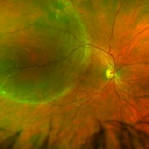
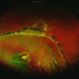
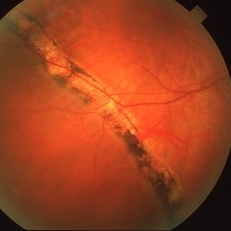
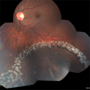
36-year-old white male presented with non traumatic retinal detachment OD, with six very distinct demarcation lines and isolated tear, and detachment parameters. Patient underwent PPV OD on 12/3/13 with 20% SF6 gas placement and face down in his first 1 month post op period.
Photographer: James Soque, CRA, COA
Imaging device: Optos Daytona
Condition/keywords: Cryopexy, demarcation line, gas pneumatic displacement, Optomap, Optos, pars plana vitrectomy (PPV), retinal tear, scanning laser ophthalmoscope
-
 Chronic Retinal Detachment: Features Slide 1
Oct 22 2012 by Ronald C. Gentile, MD
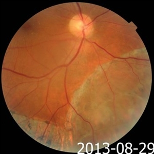
Chronic retinal detachments can be associated with demarcation lines (tidemarks), subretinal bands or sheets, and retinal cysts. Fundus photo of a chronic inferior retinal detachment reveals multiple demarcation lines inferior to the center of the fovea as a result of an inferior temporal dialysis.
Photographer: The New York Eye & Ear Infirmary Department of Medical Imaging
Condition/keywords: chronic retinal detachment, demarcation line
-
 Asymptomatic Rhegmatogenous Retinal Detachment
Sep 14 2012 by Sharon Fekrat, MD FACS FASRS
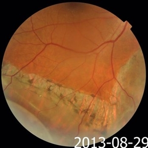
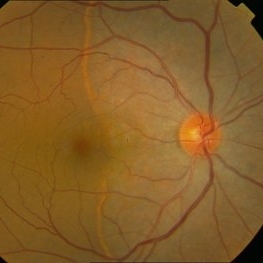
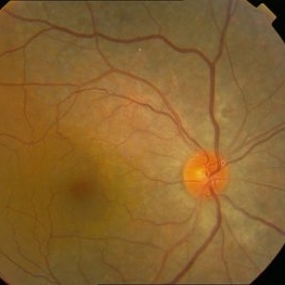
Fundus photograph of a 25-year-old emmetropic male graduate student with an inferotemporal phakic chronic asymptomatic rhegmatogenous retinal detachment with a demarcation line in the right eye. His sister who is an ophthalmology resident discovered this incidental finding. Vision 20/20.
Photographer: Brian Lutman CRA, Duke University Eye Center, Durham, NC
Condition/keywords: asymptomatic, demarcation line
-
 Acute Subhyaloid Hemorrhage CF
Oct 1 2012 by Jeffrey G. Gross, MD, FASRS
Acute subhyaloid hemorrhage CF.
Condition/keywords: demarcation line, subhyaloid hemorrhage
-
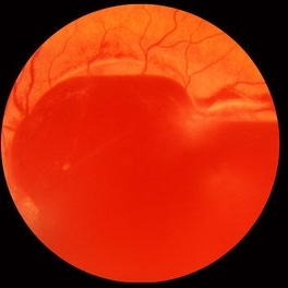
 Pigmented Demarcation Line and Retinal Macrocyst
Nov 14 2013 by Hamid Ahmadieh, MD
Color fundus photograph of the right eye of a 40-year-old man with longstanding retinal detachment showing a broad pigmented demarcation line and a retinal macrocyst.
Photographer: Elham Salehi , Negah Eye Center, Tehran
Condition/keywords: demarcation line, fundus photograph, retinal macrocyst
-
 Chronic Retinal Detachment: Features Slide 2
Oct 22 2012 by Ronald C. Gentile, MD
Chronic retinal detachments can be associated with demarcation lines (tidemarks), subretinal bands or sheets, and retinal cysts. Fundus photo of a chronic retinal detachment reveals a branching subretinal band superior nasal to the macula with a portion extending to the inferior margin of the optic disc.
Photographer: The New York Eye & Ear Infirmary Department of Medical Imaging
Condition/keywords: chronic retinal detachment, subretinal bands
-
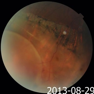 Pigmented Demarcation Line and Retinal Macrocyst
Nov 14 2013 by Hamid Ahmadieh, MD
Color fundus photograph of the right eye of a 40-year-old man with longstanding retinal detachment showing a broad pigmented demarcation line and a retinal macrocyst as well as patches of retinal hemorrhages.
Photographer: Elham Salehi , Negah Eye Center, Tehran
Condition/keywords: demarcation line, fundus photograph, retinal macrocyst
-
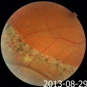
 Chronic Inferior Retinal Detachment
Mar 1 2017 by Philip J. Polkinghorne, MD
Color photograph of chronic retinal detachment with pigment demarcation line and atrophic holes visible. The vision was recorded at 20/20, and follow up is 3 years.
Photographer: Alex Fraser
Condition/keywords: atrophic retinal hole, demarcation line
-
 Pigmented Demarcation Line and Retinal Macrocyst
Nov 14 2013 by Hamid Ahmadieh, MD
Color fundus photograph of the right eye of a 40-year-old man with longstanding retinal detachment showing a broad pigmented demarcation line and a retinal macrocyst.
Photographer: Elham Salehi , Negah Eye Center, Tehran
Condition/keywords: demarcation line, fundus photograph, retinal macrocyst
-
 Scleral Buckle and Cryoptherapy Scar
Dec 29 2012 by Barbara Parolini, MD
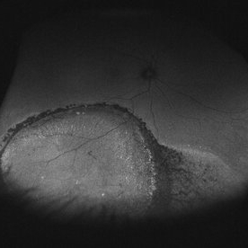
Panoramic autofluorescence photograph of a 55-year-old man after episcleral sugary for retinal detachment. An encircling scleral buckle, a superotemporal cryotherapy scar and the demarcation line of hyperautofluorescence in the previous detachment area are visible.
Photographer: Barbara Parolini, MD
Imaging device: Daytona
Condition/keywords: scleral buckle
-
 Pigmented Demarcation Line and Retinal Macrocyst
Nov 14 2013 by Hamid Ahmadieh, MD
Color fundus photograph of the right eye of a 40-year-old man with longstanding retinal detachment showing a broad pigmented demarcation line and a retinal macrocyst.
Photographer: Elham Salehi , Negah Eye Center, Tehran
Condition/keywords: demarcation line, fundus photograph, retinal macrocyst
-
 Chronic Retinal Detachment: Features Slide 3
Oct 22 2012 by Ronald C. Gentile, MD
Chronic retinal detachments can be associated with demarcation lines (tidemarks), subretinal bands or sheets, and retinal cysts. Fundus photo of a chronic retinal detachment reveals a retinal cyst within the peripherally detached temporal retina.
Condition/keywords: chronic retinal detachment
-
---thumb.jpg/image-square;max$300,300.ImageHandler) Pigmented Demarcation Line and A Blood - containing Retinal Macrocyst
Pigmented Demarcation Line and A Blood - containing Retinal Macrocyst
Nov 14 2013 by Hamid Ahmadieh, MD
Color fundus photograph of the right eye of a 40-year-old man with longstanding retinal detachment showing a broad pigmented demarcation line and a retinal macrocyst . Notice blood inside the retinal macrocyst.
Photographer: Elham Salehi , Negah Eye Center, Tehran
Condition/keywords: demarcation line, fundus photograph, retinal macrocyst
-
 Vascular loops in retinopathy of prematurity
Nov 3 2013 by Maria Ana Martinez-Castellanos, MD
Angiography of a baby with ROP treated with intravitreal anti-angiogenic therapy 1 week prior to the time this photo was taken. We can see the active vascular remodeling, vascular loops, the place where the demarcation line was at the time of the diagnosis and the growth of new vessels into the avascular zone, the leakage corresponds to immature vessels not fully covered by mural cells and not due to an inflammatory reaction.
Photographer: Maria A. Martinez-Castellanos. Asociacion para Evitar la Ceguera en Mexico
Imaging device: RetCam II
Condition/keywords: anti-VEGF, retinopathy of prematurity (ROP)
-
 Asymptomatic Chronic Retinal Detachment With Demarcation Line
Jun 11 2016 by Philip J. Polkinghorne, MD
A 65-year-old emmetrope with asymptomatic chronic retinal detachment with demarcation line.
Photographer: Alex Fraser, Greenlane Clinical Center, Auckland, New Zealand
Condition/keywords: chronic retinal detachment, fundus autofluorescence (FAF)
-
 Retinal Detachment with Demarcation Line
Apr 8 2019 by Gary R. Cook, MD, FACS
Pigmented demarcation line from a shallow, chronic retinal detachment OD
Imaging device: Topcon VT-50
Condition/keywords: demarcation line
-
 AZOOR vs. AAOOR
Mar 19 2014 by Ali Tavallali, MD, FASRS
FAF of a 47-year-old female with 20/20 VA of both eyes, note the progression of demarcation line after 4 months
Photographer: Neda Sheibani, Dr. Khodadoust Eye Hospital, Shiraz, Iran
Condition/keywords: acute zonal occult outer retinopathy (AZOOR)
-
 Bullous Retinoschisis with Outer Retinal Holes
Jun 15 2020 by Olivia Rainey
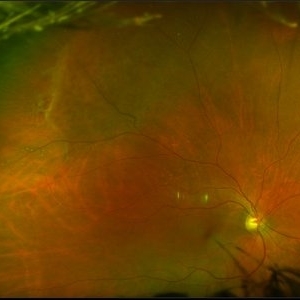
Ultra-widefield pseudocolor fundus photograph of a 56-year-old female with bullous retinoschisis with outer retinal holes affecting her right eye. The physician noted superotemporal retinoschisis in her monoculcar functioning eye. There was no demarcation line and no inner or outer layer breaks at her first appointment in February of 2020. On 6/15/20 she had a new onset outer holes and SRF tracking inferiorly. The physician recommended observation, however if this continues to progress we have discussed indications for barrier laser.
Photographer: Olivia Rainey, OCT-C, COA
Imaging device: Optos California
Condition/keywords: bullous retinoschisis, Optos, outer layer breaks, outer layer hole, pseudocolor, subretinal fluid, superior retina, ultra-wide field imaging
-
 Vascular Abnormalities in Congenital Heart Disease
Nov 3 2013 by Maria Ana Martinez-Castellanos, MD
1 month old baby born at 33 corrected age weeks, screened for ROP, we found vascular tortuosity in both eyes 4 quadrants all zones, angiography showed no demarcation line, the baby had tetralogy of Fallot and pulmonary hypertension.
Photographer: Maria A. Martinez-Castellanos. Asociacion para Evitar la Ceguera en Mexico
Imaging device: RetCam II
Condition/keywords: vascular anomaly
-
 AZOOR vs. AAOOR
Mar 19 2014 by Ali Tavallali, MD, FASRS
Color fundus photograph of a 47-year-old female with 20/20 VA of both eyes, note the demarcation line
Photographer: Neda Sheibani, Dr. Khodadoust Eye Hospital, Shiraz, Iran
Condition/keywords: acute zonal occult outer retinopathy (AZOOR)
-
 Longstanding Retinal Detachment Secondary to a Larg Retinal Tear
Dec 27 2016 by Hamid Ahmadieh, MD
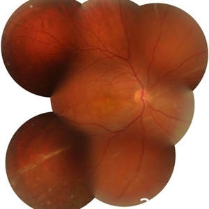
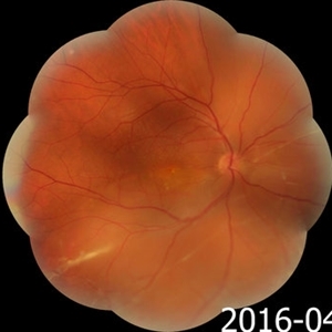
Montaged color fundus photograph of the right eye of a patient with longstanding retinal detachment. Demarcation lines are visible.
Photographer: Shabnam Poureh, Negah Eye Center, Tehran, Iran
Condition/keywords: color fundus photograph, demarcation line
-
 AZOOR vs. AAOOR
Mar 19 2014 by Ali Tavallali, MD, FASRS
Color fundus photograph of a 47-year-old female with 20/20 VA of both eyes, note the progression of demarcation line after 4 months
Photographer: Neda Sheibani, Dr. Khodadoust Eye Hospital, Shiraz, Iran
Condition/keywords: acute zonal occult outer retinopathy (AZOOR)
-
 Retinal Detachment
May 9 2016 by Nichole Lewis
Retinal detachment with partial demarcation line and same day barrier laser treatment.
Photographer: Nichole Lewis
-
 Retinal Detachment
May 13 2016 by Nichole Lewis
Inferior Retinal Detachment with some demarcation line s/p barrier laser.
Photographer: Nichole Lewis
Condition/keywords: barrier laser
-
 Longstanding Retinal Detachment Due to a Larg Retinal Tear
Dec 27 2016 by Hamid Ahmadieh, MD
Wide-field color fundus photograph of the right eye of a patient with longstanding retinal detachment. Demarcation lines are visible.
Photographer: Shabnam Poureh, Negah Eye Center, Tehran, Iran
Condition/keywords: color fundus photograph
 Loading…