Search results (94 results)
-
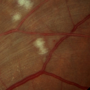
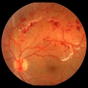 Retinal Vasculitis with Hemorrhages and Cotton Wool Spots
Oct 16 2012 by Jeffrey G. Gross, MD, FASRS
Retinal vasculitis with hemorrhages and cotton wool spots.
Condition/keywords: cotton wool spots, retinal vasculitis
-
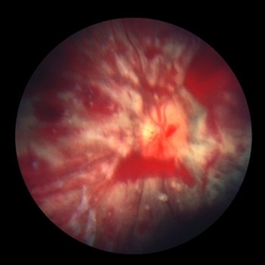 Shaken Baby Syndrome
Sep 20 2012 by Jeffrey G. Gross, MD, FASRS
Shaken Baby Syndrome with many hemorrhages and cotton wool spots
Condition/keywords: cotton wool spots, shaken baby syndrome
-
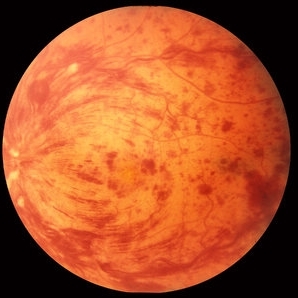
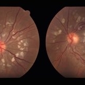
 Encephalitis with Retinal Cotton Wool Spots
Oct 15 2012 by Jeffrey G. Gross, MD, FASRS
Encephalitis, with retinal cotton wool spots, left eye, 20/30.
Condition/keywords: cotton wool spots, encephalitis, left eye
-
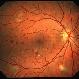
 Encephalitis with Retinal Cotton Wool Spots
Oct 15 2012 by Jeffrey G. Gross, MD, FASRS
Encephalitis with retinal cotton wool spots, right eye, 20/30.
Condition/keywords: cotton wool spots, encephalitis
-
 CRVO with Flame Hemorrhages
Oct 1 2012 by Jeffrey G. Gross, MD, FASRS
CRVO with flame hemorrhages and cotton wool spots 20/80.
Condition/keywords: 20/80, central retinal vein occlusion (CRVO), cotton wool spots
-
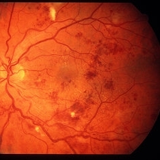
 Diabetes NPDR
Mar 29 2013 by Henry J. Kaplan, MD
NPDR.
Condition/keywords: cotton wool spots, nonproliferative diabetic retinopathy
-
 Hypertensive retinopathy
Aug 24 2012 by Geoffrey G. Emerson, MD, PhD, FASRS
A 35-year-old man has headaches and decreased vision. The right eye measures 20/25 and the left eye measures 3/200. The blood pressure measures 180/110.
Photographer: Geoffrey Emerson, MD, PhD, Retina Center, Minneapolis
Condition/keywords: cotton wool spots, hypertensive retinopathy, papilledema
-
 Purtscher's Retinopathy with Diffuse Cotton Wool Spots CF
Oct 1 2012 by Jeffrey G. Gross, MD, FASRS
Purtscher's retinopathy with diffuse cotton wool spots CF.
Condition/keywords: cotton wool spots, Purtscher's retinopathy
-
 Acute Idiopathic Occlusive Retinal Vasculitis
May 31 2014 by Hamid Ahmadieh, MD
Color fundus photograph of the right eye of a 28-year-old woman with sudden drop of vision due to acute occlusive retinal vasculitis leading to extensive nerve fiber layer infarction and retinal hemorrhages.
Photographer: Naghmeh Nozhat, Negah Eye Center, Tehran
Condition/keywords: color fundus photograph, cotton wool spots, retinal hemorrhage, retinal ischemia
-
 Proliferative Diabetic Retinopathy
Aug 23 2012 by Gerardo Garcia-Aguirre, MD
Fundus photograph of the left eye of a 45-year-old patient with proliferative diabetic retinopathy. Several microhemorrhages and cotton-wool spots are seen. A small subhyaloid hemorrhage is also observed.
Photographer: Noemí Hernández, Asociación para Evitar la Ceguera en México
Condition/keywords: cotton wool spots, microhemorrhages, subhyaloid hemorrhage
-
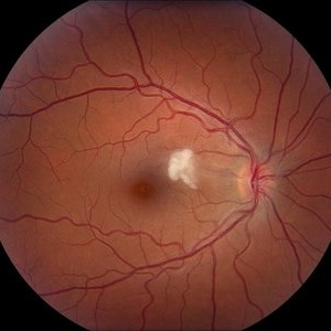 Cotton Wool Spot
Jul 10 2013 by Jason S. Calhoun
Fundus photograph shows a young male with a single cotton wool spot just nasal to the macula in the right eye.
Photographer: Jason S. Calhoun, Department of Ophthalmology, Mayo Clinic Jacksonville, Florida
Condition/keywords: cotton wool spots, hypertension
-
---thumb.jpg/image-square;max$300,300.ImageHandler) HIV Retinopathy
HIV Retinopathy
Feb 27 2013 by Henry J. Kaplan, MD
HIV retinopathy, multiple cotton wool spots, right eye. #1
Condition/keywords: cotton wool spots, HIV retinopathy
-
 HIV retinopathy with resolving CMV retinitis - left eye
Jan 11 2013 by Alex P. Hunyor, MD
HIV retinopathy and resolving CMV retinitis, left eye. 36-year-old male with HIV/AIDS. Multiple cotton wool spots due to HIV microangopathy, and an area of resolving CMV retinitis superior to the fovea (patient undergoing treatment with IV ganciclovir).
Condition/keywords: CMV retinitis, HIV retinopathy
-
 Acute Idiopathic Occlusive Retinal Vasculitis
May 31 2014 by Hamid Ahmadieh, MD
Color fundus photograph of the left eye of a 28-year-old woman with acute drop of vision due to occlusive retinal vasculitis leading to extensive nerve fiber layer infarction and retinal hemorrhages.
Photographer: Naghmeh Nozhat, Negah Eye Center, Tehran
Condition/keywords: color fundus photograph, cotton wool spots, retinal hemorrhage, retinal ischemia
-
---thumb.jpg/image-square;max$300,300.ImageHandler) SLE Retinopathy
SLE Retinopathy
Feb 26 2013 by Henry J. Kaplan, MD
SLE retinopathy,l eft eye: multiple cotton wool spots and blot hemorrhages. #2
Condition/keywords: blot hemorrhages, cotton wool spots, systemic lupus erythematosus (SLE) retinopathy
-
 Polycythemia Vera
May 2 2013 by Henry J. Kaplan, MD
CRVO in polycythemia vera; dilated and tortous retinal veins, hemorrhages and cotton wool spots.
Condition/keywords: central retinal vein occlusion (CRVO), polycythemia vera
-
 Hypertensive Retinopathy
Oct 20 2012 by Hyung-Woo Kwak, MD
The sudden appearance of cotton wool spots with hypertension retinopathy is known as accelerated hypertension. This patient has acute hypertension of rapid onset and measured systolic blood pressure was more than 200mmhg at this time.
Imaging device: Zeiss F450 plus
Condition/keywords: cotton wool spots, hypertension
-
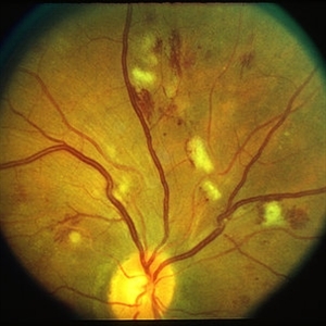 Preproliferative Diabetic Retinopathy
Mar 29 2013 by Henry J. Kaplan, MD
Multiple cotton wools in a patient with pre-proliferative diabetic retinopathy.
Condition/keywords: cotton wool spots, nonproliferative diabetic retinopathy, pre-proliferative diabetic retinopathy
-
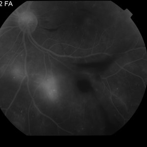
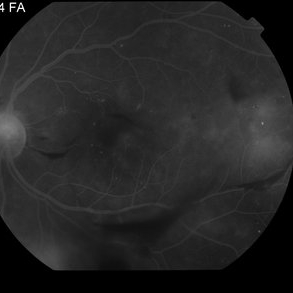 Proliferative Diabetic Retinopathy with Vitreous Hemorrhage - FA
Oct 18 2012 by Suber S. Huang, MD, MBA, FASRS
30 year old diabetic man with proliferative diabetic retinopathy and vitreous hemorrhage
Photographer: Stacie Hrvatin
Condition/keywords: cotton wool spots, neovascularization (NV), subhyaloid hemorrhage, vitreous hemorrhage
-
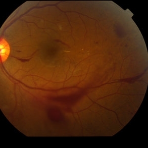 Proliferative Diabetic Retinopathy with Vitreous Hemorrhage - color fundus photo
Oct 18 2012 by Suber S. Huang, MD, MBA, FASRS
30 year old diabetic man with proliferative diabetic retinopathy and vitreous hemorrhage
Photographer: Stacie Hrvatin
Condition/keywords: cotton wool spots, neovascularization (NV), subhyaloid hemorrhage, vitreous hemorrhage
-
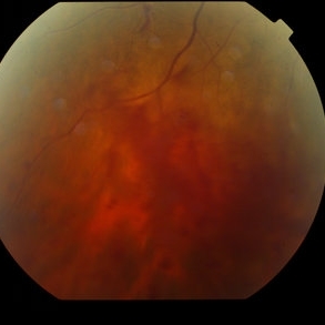 Proliferative Diabetic Retinopathy with Vitreous Hemorrhage - color fundus photo
Oct 18 2012 by Suber S. Huang, MD, MBA, FASRS
30 year old diabetic man with proliferative diabetic retinopathy and vitreous hemorrhage
Photographer: Stacie Hrvatin
Condition/keywords: cotton wool spots, neovascularization (NV), subhyaloid hemorrhage, vitreous hemorrhage
-
---thumb.jpg/image-square;max$300,300.ImageHandler) Central Retinal Vein Occlusion
Central Retinal Vein Occlusion
Oct 30 2012 by Lihteh Wu, MD
35-year-old hypertensive man with an acute CRVO. Notice the peripapillary cotton wool spots, superficial flame shaped hemorrhages and deeper dot and blot hemorrhages in all 4 quadrants. This is the typical blood and thunder appearance of a CRVO.
Condition/keywords: central retinal vein occlusion (CRVO), cotton wool spots
-
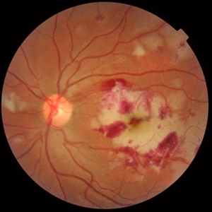
 SLE Retinopathy
Nov 14 2016 by Mitzy E Torres Soriano, MD
25-year-old female patient with systemic lupus erythematosus. Photographs show cotton wool spots, intraretinal hemorrhages and vascular tortuosity. FA demonstrated retinal vasculitis and OCT revealed cystoid macular edema. In this case diagnosis of SLE was made after ocular manifestation.
Photographer: Grupo Laser Vision, Rosario, Argentina
Condition/keywords: cotton wool spots, occlusive retinal vasculitis, occlusive vasculitis, systemic lupus erythematosus, vasculopathy
-
 Arterio-Venous Nipping, Venous Beading
Mar 1 2014 by Homayoun Tabandeh, MD, FASRS
Arterio-venous nipping, venous beading, and cotton wool spots in a patient with hypertensive and diabetic retinopathy.
Condition/keywords: arteriovenous nipping, cotton wool spots, venous beading
-
 Proliferative Diabetic Retinopathy with Vitreous Hemorrhage - FA
Oct 18 2012 by Suber S. Huang, MD, MBA, FASRS
30 year old diabetic man with proliferative diabetic retinopathy and vitreous hemorrhage
Photographer: Stacie Hrvatin
Condition/keywords: cotton wool spots, neovascularization (NV), subhyaloid hemorrhage, vitreous hemorrhage
 Loading…