Search results (19 results)
-
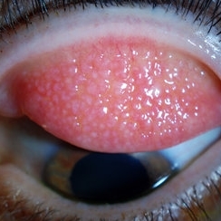 Giant Papillary Conjunctivitis, Left Upper Eyelid
Jul 22 2013 by Jason S. Calhoun
Contact lens wearer in for exam. Has rough feeling underneath both eyelids. Patient thought it was through SCL wear. Patient VA was 20/20. right eye, 20/30, left eye. Underneath the left upper eyelid, you can see papillary inflammation and redness.
Photographer: Jason S. Calhoun, Department of Ophthalmology, Mayo Clinic Jacksonville, Florida
Imaging device: TOPCON D-90 SL NIKON CAMERA
Condition/keywords: giant papillary conjunctivitis
-
 Ocular Toxocariasis slide 1
Oct 22 2012 by Ronald C. Gentile, MD
40-year-old man from South America was referred for a peripheral retinal scar in his left eye. He had a history of conjunctivitis as a child with exposure to multiple pets (cats and dogs). Fundus photo revealed a peripheral scarred sub-retinal granuloma located superior nasal with a retinal fold and traction extending to the optic nerve.
Photographer: The New York Eye & Ear Infirmary Department of Medical Imaging
Condition/keywords: toxocariasis
-
 Giant Papillary Conjunctivitis
Dec 13 2013 by Jason S. Calhoun
Patient wears soft contact lenses complained of irritation when the SCL would move. Inverted eyelid in both eyes and there was papillary +2 underneath the eyelid.
Photographer: Jason S. Calhoun, Ophthalmic Photographer, Department of Ophthalmology, Mayo Clinic Jacksonville
Imaging device: TOPCON D-90 SL NIKON CAMERA
Condition/keywords: giant papillary conjunctivitis
-
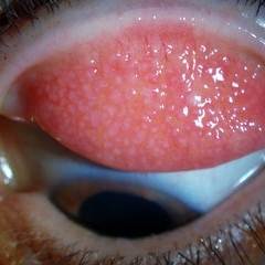 Giant Papillary Conjunctivitis, Left Upper Eyelid
Jul 22 2013 by Jason S. Calhoun
Contact lens wearer, in for exam. Has rough feeling underneath both eyelids. Patient thought it was through SCL wear. Patient VA was 20/20. right eye, 20/30, left eye. Underneath the left upper eyelid, you can see papillary inflammation and redness.
Photographer: Jason S. Calhoun, Department of Ophthalmology, Mayo Clinic Jacksonville, Florida
Imaging device: TOPCON D-90 SL NIKON CAMERA
Condition/keywords: giant papillary conjunctivitis
-
 Giant Papillary Conjunctivitis
Dec 13 2013 by Jason S. Calhoun
Patient wears soft contact lenses complained of irritation when the SCL would move. Inverted eyelid in both eyes and there was papillary +2 underneath the eyelid.
Photographer: Jason S. Calhoun, Ophthalmic Photographer, Department of Ophthalmology, Mayo Clinic Jacksonville
Imaging device: TOPCON D-90 SL NIKON CAMERA
Condition/keywords: giant papillary conjunctivitis
-
---thumb.jpg/image-square;max$300,300.ImageHandler) Superior Limbic Keratitis / Keratoconjunctivitis
Superior Limbic Keratitis / Keratoconjunctivitis
Jan 16 2014 by Gavin Thorsrud
Bilateral view of a 44-year-old female with SLK. Underwent thyroidectomy 2007, TSH currently stable on levothyroxine. Using artificial tears every 2 hours, and Restasis BID
Photographer: Gavin Thorsrud, COMT, CRA, VAMC Minneapolis
Imaging device: Zeiss 70 stereo slit lamp
Condition/keywords: keratitis, keratoconjunctivitis, superior limbic keratitis / keratoconjunctivitis (SLK)
-
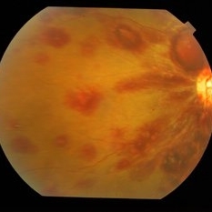
 Dengue Fever
Oct 25 2012 by Mallika Goyal, MD
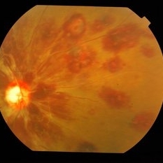
Fundus photograph of the left eye of a 32-year-old gentleman with dengue fever and thrombocytopenia. Photograph shows extensive retinal and pre-retinal haemorrhages, roth spots but no dengue retinitis. Same patient as in images 1-5.
Condition/keywords: Dengue Fever, preretinal hemorrhage, rosacea conjunctivitis
-
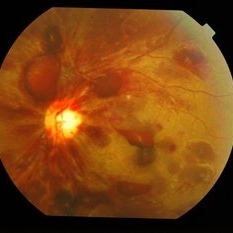
 Dengue Fever
Oct 25 2012 by Mallika Goyal, MD
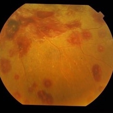
Fundus photograph of the right eye of a 32-year-old gentleman with dengue fever and thrombocytopenia. Photograph shows extensive retinal and pre-retinal haemorrhages, roth spots but no dengue retinitis. Same patient as in images 1-5.
Condition/keywords: Dengue Fever, rosacea conjunctivitis, thrombocytopenia
-
 Giant Papillary Conjunctivitis, Left Upper Eyelid
Jul 22 2013 by Jason S. Calhoun
Contact lens wearer, in for exam. Has rough feeling underneath both eyelids. Patient thought it was through SCL wear. Patient VA was 20/20. right eye, 20/30, left eye. Underneath the left upper eyelid, you can see papillary inflammation and redness.
Photographer: Jason S. Calhoun, Department of Ophthalmology, Mayo Clinic Jacksonville, Florida
Imaging device: TOPCON D-90 SL NIKON CAMERA
Condition/keywords: giant papillary conjunctivitis
-
 Dengue Fever
Oct 25 2012 by Mallika Goyal, MD
Fundus photograph of the left eye of a 32-year-old gentleman with dengue fever and thrombocytopenia. Photograph shows extensive retinal and pre-retinal haemorrhages, roth spots but no dengue retinitis. Same patient as in images 1-5
Condition/keywords: Dengue Fever, preretinal hemorrhage, rosacea conjunctivitis
-
 Dengue Fever
Oct 25 2012 by Mallika Goyal, MD
Fundus photograph of the right eye of a 32-year-old gentleman with dengue fever and thrombocytopenia. Photograph shows extensive retinal and pre-retinal haemorrhages, roth spots but no dengue retinitis. Same patient as in images 1-5.
Condition/keywords: Dengue Fever, preretinal hemorrhage, rosacea conjunctivitis
-
 Dengue Fever
Oct 25 2012 by Mallika Goyal, MD
Fundus photograph of the left eye of a 32-year-old gentleman with dengue fever and thrombocytopenia. Photograph shows extensive retinal and pre-retinal haemorrhages, roth spots but no dengue retinitis.
Condition/keywords: Dengue Fever, preretinal hemorrhage, rosacea conjunctivitis
-
---thumb.jpg/image-square;max$300,300.ImageHandler) Allergic Conjunctivitis
Allergic Conjunctivitis
Dec 27 2013 by David Callanan, MD
12-year-old male patient.
Condition/keywords: allergic conjunctivitis
-
---thumb.jpg/image-square;max$300,300.ImageHandler) Allergic Conjunctivitis
Allergic Conjunctivitis
Dec 27 2013 by David Callanan, MD
12-year-old male patient.
Condition/keywords: allergic conjunctivitis
-
---thumb.jpg/image-square;max$300,300.ImageHandler) Allergic Conjunctivitis
Allergic Conjunctivitis
Dec 27 2013 by David Callanan, MD
12-year-old male patient.
Condition/keywords: allergic conjunctivitis
-
---thumb.jpg/image-square;max$300,300.ImageHandler) Allergic Conjunctivitis
Allergic Conjunctivitis
Dec 27 2013 by David Callanan, MD
12-year-old male patient.
Condition/keywords: allergic conjunctivitis
-
 Slide 1-12
Feb 19 2019 by Lancaster Course in Ophthalmology
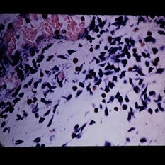
Seven small lymphocytes with a scanty rim of blue cytoplasm amid dead cells in chronic conjunctivitis. (Giemsa stain)
Condition/keywords: conjunctivitis, cytoplasm, lymphocytes
-
 Slide 1-7
Feb 19 2019 by Lancaster Course in Ophthalmology
Upper: Bilobed eosinophils. Lower: Large mast cell identified by its purple granules. Both views are from scrapings in allergic conjunctivitis. (Giemsa stain)
Condition/keywords: cell, conjunctivitis, eosinophils
-
 Slide 1-24
Feb 19 2019 by Lancaster Course in Ophthalmology
Perivascular eosinophils and PMNs in an acute conjunctivitis. (Giemsa stain)
Condition/keywords: acute conjunctivitis, perivascular eosinophils, polymorphonuclear leukocytes (PMNs)
 Loading…