Search results (85 results)
-
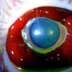
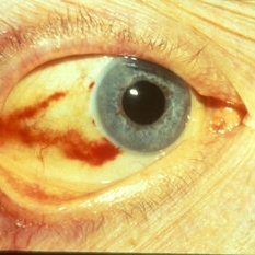
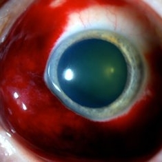 Sub-Conjunctival Hemorrhage (Chemosis)
Jul 13 2013 by Jason S. Calhoun
Chemosis or swelling of the conjunctiva with sub-conjunctival hemorrhage.
Photographer: Jason S. Calhoun, Department of Ophthalmology, Mayo Clinic Jacksonville, Florida
Imaging device: TOPCON D-90 SL NIKON CAMERA
Condition/keywords: chemosis
-
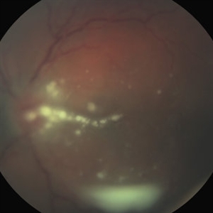 Fungal Endophthalmitis Associated With Intravenous Drug Abuse
Apr 16 2014 by Scott D. Schoenberger, MD
Fundus photograph of a 20-year-old male with pain and decreased vision OS for 3 days. His visual acuity was counting fingers and he had conjunctival injection, anterior chamber cells and vitreous cells. He admitted to intermittent use of intravenous heroin. A vitrectomy was performed and cultures were positive for candida albicans.
Condition/keywords: endogenous endophthalmitis, fungal endophthalmitis
-
 Sub-Conjunctival Hemorrhage (Chemosis)
Jul 13 2013 by Jason S. Calhoun
Chemosis or swelling of the conjunctiva with sub-conjunctival hemorrhage.
Photographer: Jason S. Calhoun, Department of Ophthalmology, Mayo Clinic Jacksonville, Florida
Imaging device: TOPCON D-90 SL NIKON CAMERA
Condition/keywords: chemosis
-
 Subconjunctival Hemorrhage
Sep 20 2012 by Jeffrey G. Gross, MD, FASRS
Subconjunctival hemorrhage, trauma in eye with choroidal rupture HM
Condition/keywords: choroidal rupture, subconjunctival hemorrhage
-
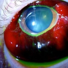
 Sub-Conjunctival Hemorrhage (Chemosis)
Jul 13 2013 by Jason S. Calhoun
Chemosis or swelling of the conjunctiva with Sub conjunctival hemorrhage.
Photographer: Jason S. Calhoun, Department of Ophthalmology, Mayo Clinic Jacksonville, Florida
Imaging device: TOPCON D-90 SL NIKON CAMERA
Condition/keywords: chemosis
-
 Sub-Conjunctival Hemorrhage (Chemosis)
Jul 13 2013 by Jason S. Calhoun
Chemosis or swelling of the conjunctiva with sub-conjunctival hemorrhage.
Photographer: Jason S. Calhoun, Department of Ophthalmology, Mayo Clinic Jacksonville, Florida
Imaging device: TOPCON D-90 SL NIKON CAMERA
Condition/keywords: chemosis
-
 Linear Nevus Sebaceous Syndrome
Feb 20 2015 by H. Michael Lambert, MD
Color photo of conjuctival lipodermoid in linear sebaceous nevus syndrome .
Condition/keywords: conjunctiva, linear nevus sebaceous syndrome, lipodermoid
-
 Sub-Conjunctival Hemorrhage (Chemosis)
Jul 13 2013 by Jason S. Calhoun
Chemosis or swelling of the conjunctiva with sub-conjunctival Hemorrhage.
Photographer: Jason S. Calhoun, Department of Ophthalmology, Mayo Clinic Jacksonville, Florida
Imaging device: TOPCON D-90 SL NIKON CAMERA
Condition/keywords: chemosis
-
 24 Hours Post Scleral Wound Closure+ Scleral Buckle+25 g Vitrectomy+Silicon Oil
Jan 23 2015 by Carlos Quezada-Ruiz, MD, FASRS
24 hours post op fundus photograph of a 43-year-old man who had perforating injury to the right eye with a small piece of plastic while he was hammering. OD LP, subconjunctival hemorrhage, clear cornea, hyphema, irido and ciclodyalisis as well as a luxated lens with traumatic cataract and a dense vitreous hemorrhage. B-US showed rhegmatogenous retinal detachment with a tear and a big inferior hemorrhagic choroidal detachment. 360 peritomy revealed 2-entry scleral wounds were found in zone II (M V and M VI) and closure was performed. 25 G PPV was performed with the infusion canal placed in the AC through the limbus. Lensectomy and removal of a dense recent vitreous hemorrhage revealed a white detached retina with an exit wound through the temporal inferior segment of the optic nerve with a nasal GRT and sub retinal hemorrhage as well as temporal inferior choroidal, PVD was induced and PFOs helped stabilizing the retina while vitrectomy and sub-retinal hemorrhage was removed through the GRT. Fluid air exchange was made and 360 endolaser over the buckle indentation was done and silicon oil was used as endotamponade. This picture was taken 24 hrs after the surgery.
Photographer: Lilibeth Rodriguez, Instituto de la Visión. Torreon, Mexico.
Condition/keywords: central retinal artery occlusion (CRAO), giant retinal tear, trauma
-
 Conjunctival Cyst
Jul 13 2013 by Jason S. Calhoun
Slit lamp exam shows conjunctival cyst in the nasal aspect. Fluorescence shows cyst in blue light.
Photographer: Jason S. Calhoun, Department of Ophthalmology, Mayo Clinic Jacksonville, Florida
Imaging device: TOPCON D-90 SL NIKON CAMERA
Condition/keywords: conjunctival cysts, cyst
-
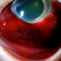
 Subconjunctival Hemorrhage
Oct 23 2012 by Larry Halperin, MD
Subconjunctival hemorrhage
Condition/keywords: subconjunctival hemorrhage
-
 Color Fundus Photograph of Macular Infarction Secondary to Subonjunctival Gentamicin Injection
May 16 2014 by Arwa Azmeh, MD, PhD
A 20-year-old male suffered from diplopia since age one. He was diagnosed to have acquired fourth nerve palsy in his left eye. VA at time of diagnosis was 20/20 in OU and Fundus exam was WNL in OU. His history revealed no other complaints. 3 days ago he underwent left superior oblique tucking for relief of his diplopia.The surgery was uneventful and at the end of surgery subconjunctival gentamicin was injected. Immediately following surgery his VA in OS decreased from 20/20 to complete loss of central vision and sensation of HM from the periphery. He was referred to us 3 days after surgery. At time of referral fundus exam of his left eye revealed macular infarction with cherry red spot appearance with few retinal hemorrhages, mild optic disc edema and CWS surrounding optic disc. Peripheral retina had normal color and appearance. The vitreous was clear. Anterior segment was quiet. IOP was WNL. Macular OCT was consistent with macular infarction. FA revealed delay in central retinal artery filling as fluorescein started to appear in the arteries at the level of the optic disc at 28 sec, and in the retinal veins at 38 sec. Macular area remained to be non-perfused throughout the whole FA. In late phases staining of blood vessels walls was noticed. The "wipe out" of large vessels and capillaries persisted in the central area. OCT through foveal area showed diffuse thickening of the retina with severe elevation in the fovea, reduced backscattering from the outer layers of the retina and enhanced reflectivity from the inner retina, due to ischemia. Complete blood count and cardiovascular study were WNL. The final diagnosis was macular infarction secondary to subconjunctival gentamicin injection.
Imaging device: OCT
Condition/keywords: macular infarction, subconjunctival gentamicin
-
---thumb.jpg/image-square;max$300,300.ImageHandler) Subconjunctival Air Bubbles
Subconjunctival Air Bubbles
Mar 21 2013 by Yusuke Oshima, MD, PhD
Slit lamp photograph demonstrates subconjunctival air bubbles, which is attributed to incomplete self-sealing of sclerotomies in a 25-gauge microincision vitrectomy surgery.
Photographer: Yusuke Takada, Osaka University Graduate School of Medicine
Condition/keywords: complication
-
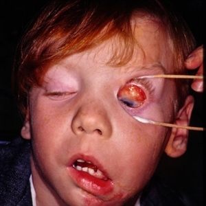
 Conjunctival Tumor
Nov 29 2013 by Jason S. Calhoun
Right conjunctival melanoma extending into anterior orbit, right eye. Temporally and nasally with pigmented masses/nodules. VA was 20/30 without correction in the right eye. Follow up to proceed with proton beam therapy.
Photographer: Jason S. Calhoun, Ophthalmic Photographer, Department of Ophthalmology, Mayo Clinic Jacksonville
Imaging device: TOPCON D-90 SL NIKON CAMERA
Condition/keywords: tumor
-
 Conjunctival Cyst
Jul 13 2013 by Jason S. Calhoun
Slit lamp exam shows conjunctival cyst in the nasal aspect. Fluorescence shows cyst in blue light.
Photographer: Jason S. Calhoun, Department of Ophthalmology, Mayo Clinic Jacksonville, Florida
Imaging device: TOPCON D-90 SL NIKON CAMERA
Condition/keywords: conjunctival cysts, cyst
-
 Chemosis
Jul 13 2013 by Jason S. Calhoun
Sub conjunctival hemorrhage with chemosis after a sub conjunctival injection of Lidocaine 2%.
Photographer: Jason S. Calhoun, Department of Ophthalmology, Mayo Clinic Jacksonville, Florida
Imaging device: TOPCON D-90 SL NIKON CAMERA
Condition/keywords: chemosis
-
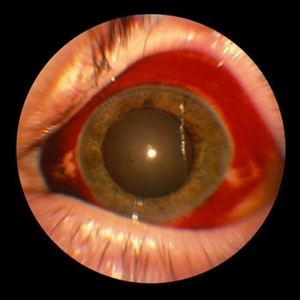
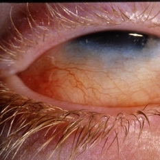
 Reticulum Cell
Feb 15 2013 by From the Collections of Thomas M. Aaberg, MD and Thomas M. Aaberg Jr., MD
Diffuse slit-lamp photograph of the left eye of a patient with systemic lymphoma showing diffuse telangiectasis, injection of conjunctival vessels and engorgement of iris vessels.
Condition/keywords: lymphoma, reticulum cell sarcoma
-
 Conjunctival Tumor
Nov 29 2013 by Jason S. Calhoun
Right conjunctival melanoma extending into anterior orbit, right eye. Temporally and nasally with pigmented masses/nodules. VA was 20/30 without correction in the right eye. Follow up to proceed with proton beam therapy.
Photographer: Jason S. Calhoun, Ophthalmic Photographer, Department of Ophthalmology, Mayo Clinic Jacksonville
Imaging device: TOPCON D-90 SL NIKON CAMERA
Condition/keywords: tumor
-
---thumb.JPG/image-square;max$300,300.ImageHandler) Posterior Scleritis Atypical
Posterior Scleritis Atypical
Dec 13 2013 by Mallika Goyal, MD
Right eye fundus of a 32-year-old male presenting with unilateral reduced quality of vision, pain and headache for 5 days; visual acuity was 20/25. There was trace RAPD, white conjunctiva, no intraocular inflammation, mild disc edema and congestion, normal retina and macula. OCT was normal. A diagnosis of optic neuritis was considered, later revised to posterior scleritis with contiguous papillitis.
Photographer: Mallika Goyal, MD, Apollo Health City, Hyderabad, India
Condition/keywords: posterior scleritis
-
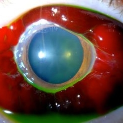
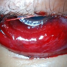
 Subconjuntival IOL After Blunt Trauma
Jun 27 2018 by Gabriel Costa Andrade, PhD
A 73-year-old male patient was referred to our ophthalmic emergency department with complaints of redness, pain, and diminution of vision in his left eye, after fall from height. The patient underwent small incision cataract surgery with polymethylmethacrylate (PMMA) IOL implantation in both the eyes 15 years back through superior sclerocorneal incision under local anesthesia. His best-corrected visual acuity was perception of light in the left eye. Ophthalmic examination using slit lamp biomicroscopy of the left eye revealed diffuse subconjunctival hemorrhage with no conjunctival laceration and inferior bulbar conjunctiva showed traumatic pseudophacocele with a sign “golden half ring,” suggesting the presence of PCIOL in subconjunctival space.There was total hyphema obscuring the view of rest of the ocular structures in his left eye.
Photographer: Gabriel Andrade, RETINA CLINIC, São Paulo, BRAZIL
Condition/keywords: dislocated intraocular lens (IOL), trauma
-
 Sub-Conjunctival Hemorrhage (Chemosis)
Jul 13 2013 by Jason S. Calhoun
Chemosis or swelling of the conjunctiva with sub-conjunctival Hemorrhage.
Photographer: Jason S. Calhoun, Department of Ophthalmology, Mayo Clinic Jacksonville, Florida
Imaging device: TOPCON D-90 SL NIKON CAMERA
Condition/keywords: chemosis
-
---thumb.JPG/image-square;max$300,300.ImageHandler) Posterior Scleritis Atypical
Posterior Scleritis Atypical
Dec 13 2013 by Mallika Goyal, MD
Right eye fundus of a 32-year-old male presenting with unilateral reduced quality of vision, pain and headache for 5 days; visual acuity was 20/25. There was trace RAPD, white conjunctiva, no intraocular inflammation, mild disc edema and congestion, normal retina and macula. OCT was normal. A diagnosis of optic neuritis was considered, later revised to posterior scleritis with contiguous papillitis.
Photographer: Mallika Goyal, MD, Apollo Health City, Hyderabad, India
Condition/keywords: posterior scleritis
-
 OCT Through Foveal Area in Macular Infarction Secondary to Subconjunctival Gentamicin Injection
May 16 2014 by Arwa Azmeh, MD, PhD
A 20-year-old male suffered from diplopia since age one. He was diagnosed to have acquired fourth nerve palsy in his left eye. VA at time of diagnosis was 20/20 in OU and fundus exam was WNL in OU. His history reaveled no other complaints. 3 days ago he underwent left superior oblique tucking for relief of his diplopia.The surgery was uneventful and at the end of surgery subconjunctival gentamicin was injected. Immediately following surgery his VA in OS decreased from 20/20 to complete loss of central vision and sensation of HM from the periphery. He was referred to us 3 days after surgery. At time of referral fundus exam of his left eye revealed macular infarction with cherry red spot appearance with few retinal hemorrhages , mild optic disc edema and CWS surrounding optic disc. Peripheral retina had normal color and appearance. The vitreous was clear. Anterior segment was quiet. IOP was WNL. Macular OCT was consistent with macular infarction. FA revealed delay in central retinal artery filling as fluorescein started to appear in the arteries at the level of the optic disc at 28 sec, and in the retinal veins at 38 sec. Macular area remained to be non-perfused throughout the whole FA. In late phases staining of blood vessels walls was noticed. The "wipe out" of large vessels and capillaries persisted in the central area. OCT through foveal area showed diffuse thickening of the retina with severe elevation in the fovea, reduced backscattering from the outer layers of the retina and enhanced reflectivity from the inner retina, due to ischemia. Complete blood count and cardiovascular study were WNL. The final diagnosis was macular infarction secondary to subconjunctival gentamicin injection.
Imaging device: OCT
Condition/keywords: macular infarction, subconjunctival gentamicin
-
 Conjunctival Melanoma
Jul 13 2013 by Jason S. Calhoun
Elderly woman with history of melanoma, shows large melanoma temporally on the conjunctiva in the left eye
Photographer: Jason S. Calhoun, Department of Ophthalmology, Mayo Clinic Jacksonville, Florida
Imaging device: TOPCON D-90 SL NIKON CAMERA
Condition/keywords: melanoma
-
 Linear Nevus Sebaceous Syndrome
Feb 20 2015 by H. Michael Lambert, MD
color photo of conjuctival lipodermoid in linear sebaceous nevus syndrome
Condition/keywords: conjunctiva, linear sebaceous nevus, linear sebaceous nevus of Jadassohn, lipodermoid
 Loading…