Search results (85 results)
-
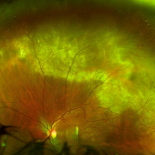
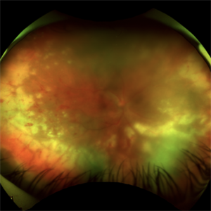 Acute Retinal Necrosis (ARN)
Jul 3 2025 by Heitor Nogueira
Fundus photograph of an 63-year-old woman who reported unilateral visual acuity loss for 10 days associated with ocular pain. He presented conjunctival hyperemia with temporal and nasal nodular scleritis, anterior chamber reaction 2+/4+, Koeppe nodules, granulomatous PKs, vitreitis 2+/4+, multiple areas of vasculitis in the arcades and periphery, associated with hemorrhages and necrotizing retinitis in the temporal, inferior and nasal periphery. Positive serology for Herpes Virus
Photographer: Heitor Nogueira, Penido Burnier Institute, Campinas, São Paulo, Brazil
Imaging device: Optos Daytona
Condition/keywords: ARN complications, Herpes, progressive outer retinal necrosis (PORN), Uveitis
-
 Acute Retinal Necrosis
Jul 3 2025 by Heitor Nogueira
Fundus photograph of an 53-year-old woman with patient who reported unilateral visual acuity loss for 10 days associated with ocular pain. She presented conjunctival hyperemia with temporal and nasal nodular scleritis, anterior chamber reaction 2+/4+, Koeppe nodules, granulomatous PKs, vitritis 2+/4+, multiple areas of vasculitis in arcades and periphery, associated with hemorrhages and necrotizing retinitis in temporal, inferior and nasal periphery. patient who reported unilateral visual acuity loss for 10 days associated with ocular pain. He presented conjunctival hyperemia with temporal and nasal nodular scleritis, anterior chamber reaction 2+/4+, Koeppe nodules, granulomatous PKs, vitreitis 2+/4+, multiple areas of vasculitis in the arcades and periphery, associated with hemorrhages and necrotizing retinitis in the temporal, inferior and nasal periphery. Positive serology for Herpes Virus.
Photographer: Heitor Nogueira, Penido Burnier Institute and CHOV, Campinas, São Paulo, Brazil
Imaging device: Optos Daytona
Condition/keywords: ARN complications, Herpes, progressive outer retinal necrosis (PORN)
-
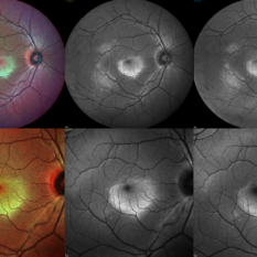 Berlin's Edema
Jun 12 2025 by Shivankar Sen, MS, FVRS
A 22 year old male came with history of sports injury to the right eye with the nose of shuttlecock while playing badminton. On examination, right eye anterior segment shows conjunctival congestion with brisk pupillary reaction and quiet anterior chamber. His best corrected visual acuity was 6/12; N6 in the right eye and 6/6; N6 in the left eye. Retinal examination revealed OD Berlin's Edema, OS within normal limits. Image Description (From Left to Right) Multicolor Reflectance (Blue-Green Enhanced) shows well defined yellowish discoloration Green reflectance and blue reflectance show corresponding whitish discoloration at the area of edema
Photographer: Dr. Shivankar Sen
Imaging device: Heidelberg Spectralis HRA+OCT
Condition/keywords: Shuttlecock Injury
-
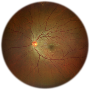 Commotio Retinae
Jun 10 2025 by CUI YUELING
The patient presented 2 hours after sustaining a left eye injury caused by a stick. Visual acuity in the left eye was 0.2 without improvement upon correction, and intraocular pressure measured 15 mmHg. Examination of the anterior segment revealed ciliary conjunctival injection accompanied by patchy subconjunctival hemorrhage. The corneal surface remained smooth, and the anterior chamber was deep with hyphema characterized by blood-tinged aqueous humor predominantly settled inferiorly. The pupil was slightly irregular, approximately 3 mm in diameter, with a superotemporal notch; pupillary light reflex was intact. The lens appeared clear. Fundus examination showed well-defined optic disc margins with normal coloration and a cup-to-disc ratio of 0.2. Retinal arteries and veins were normally distributed with an artery-to-vein ratio of 2:3. At the posterior pole, the foveal reflex exhibited concentric ripple-like changes centered on the fovea, accompanied by localized pigment attenuation and reduced reflex intensity. Irregular reflectivity was noted in the superotemporal and inferotemporal nerve fiber layers.
Photographer: Yueling Cui
Imaging device: Zeiss Clarus 500
Condition/keywords: commotio retinae
-
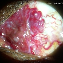
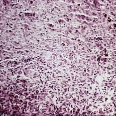
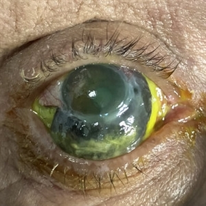 Necrotizing Scleritis
Apr 17 2025 by Gustavo Uriel Fonseca Aguirre
The clinical photograph shows necrotizing scleritis with perilimbal involvement, featuring marked scleral thinning and violaceous episcleral injection in the inferior quadrant. Focal uveal prolapse is visible at the area of maximal scleral necrosis, accompanied by peripheral ulcerative keratitis. Fluorescein staining residue is observed on the ocular surface. Associated findings include mild conjunctival chemosis and dilated episcleral vessels.
Photographer: Gustavo U. Fonseca Aguirre, Hospital Conde de Valenciana, Ciudad de México
Condition/keywords: necrotizing scleritis
-
 Rosai-Dorfman Disease
Dec 4 2024 by Virginia Gebhart
72 year old female with temporal limbal lesion that extends onto the cornea from 10:00 - 8:00 encroaching on visual axis. Possible lymphomatous process. Will refer to Emory.
Photographer: Dr Chris Bergstrom MD, OD
Condition/keywords: corneal scars and opacities, Rosai-Dorfman Disease, Subconjunctival mass
-
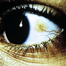
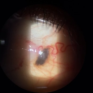 Blue Nevus
Apr 9 2024 by Hector Gabriel Moreno Solano, MD, MHA
48-year-old Hispanic male patient who comes to the clinic due to the presence of pterygium. Upon examination, a 2 mm blue nevus is found, which the patient reports having had since he was 15 years old.
Photographer: Héctor Gabriel Moreno-Solano.
Condition/keywords: conjunctiva, nevus
-
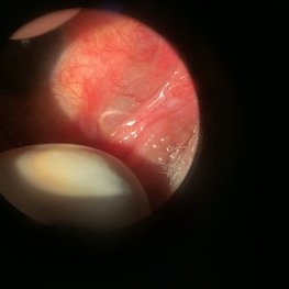
 Conjunctival AV Malformation
Dec 18 2023 by siddharth sheth
33 year old male presented with a complaint of redness since 15 years in left eye.
Photographer: Gaurav Kamble, Isha Netralaya
Imaging device: Dyanmic slit lamp imaging
Condition/keywords: conjunctival AV malformation, slit lamp photo, slit lamp photography, unilateral
-
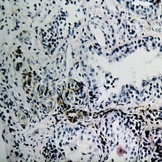
 Conjunctival involvement uveal lymphoma
Jan 20 2023 by Elaine Michele Binkley, MD
Slit lamp photograph shows characteristic "salmon-patch" conjunctival lesion in the setting of uveal marginal zone lymphoma with conjunctival involvement.
Photographer: Brice Critser, University of Iowa
Condition/keywords: Uveal Lymphoma
-
 Ocular subconjunctival worm infestation
Jun 16 2022 by Filipe Sampaio Carvalho
Ocular subconjunctival worm infestation in Manaus, Amazonas, Brazil
Photographer: Filipe Sampaio Carvalho
Imaging device: iPhone 12
Condition/keywords: worm
-
Epimacular Membrane
Oct 14 2021 by Islam bechakh
A vitrectomy is performed in our 25 G transconjunctival patient after careful decontamination of the cul-de-sacs by washing with povidone-iodine (Betadine®) 5% for 2 minutes. The panoramic system associated with the operating microscope makes it possible to control the traction on the retinal periphery and to facilitate the manipulation of the dye (Brilliant Blue G) during the surgery. The peeling of the membrane is extended to the whole macular area by trying, by a superficial grip begun in the sub-foveolar, to peel only the membrane. The internal limiting is then stained a second time and the total or partial decision is discussed on a case-by-case basis depending on the severity of the retraction and the type of diffuse or cystoid edema.
Photographer: Islam Bechakh
Condition/keywords: Epimacular membrane, vitrectomy
-
 Enucleation of an Eye with Advanced Choroidal Melanoma with Implant and Donor Sclera Replacement
Jan 11 2021 by Sophia El Hamichi, MD
Surgery of the left eye affected with advanced melanoma: Upper left image: separating the sclera from the conjunctiva and the tenon by performing a peritomy, then separating the rectus muscles that will be later sutured to the donor sclera, to preserve post-op motility. Upper right image: cutting the optic nerve. Middle left image: the globe is enucleated. Middle right image: dissection of the globe showing the melanoma. Tissue is then sent to pathology Lower left image: putting the porous polyethyline implant inside the donor sclera and marking muscles' insertion. Lower right image: reinsertion of the rectus muscles on the donor sclera, then covering with tenon and conjunctiva.
Photographer: Belinda Rodriguez, Murray Ocular Oncology and Retina, Miami
Condition/keywords: donor sclera, enucleation, implant, melanoma
-
 Massive Commotio Retinae
Oct 20 2020 by Veronika Yehezkeli
24-year-old man was injured from an explosion of a plastic bottle towards the nasal conjunctiva of his left eye. A massive commotio retinae was diagnosed superotemporally.
Photographer: Veronika Yehezkeli, Meir medical center, Israel
Condition/keywords: blunt trauma, commotio retinae, preretinal hemorrhage
-
 Swollen MIRAgel Exoplant
Apr 2 2019 by Gary R. Cook, MD, FACS
Buckle swelling of a MIRAgel exoplant and its anchoring suture visible beneath the conjunctiva in the superotemporal quadrant OD
Imaging device: Topcon VT-50
Condition/keywords: MIRAgel exoplant, retina surgery complications
-
 Slide 14-25
Mar 4 2019 by Lancaster Course in Ophthalmology
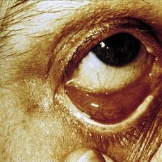
In advanced cases, melanomas may present as an orbital or subconjunctival mass.
Condition/keywords: melanoma
-
 Slide 14-24
Mar 4 2019 by Lancaster Course in Ophthalmology
In advanced cases, melanomas may present as an orbital or subconjunctival mass.
Condition/keywords: melanoma
-
 Slide 7-29
Feb 25 2019 by Lancaster Course in Ophthalmology
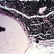
Benign lymphoid hyperplasia of the conjunctiva may resemble a normal lymph node.
Condition/keywords: conjunctiva, hyperplasia, lymph node
-
 Slide 7-28
Feb 25 2019 by Lancaster Course in Ophthalmology
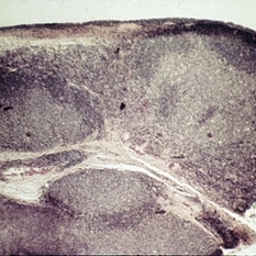
Lymphoid tumor of the conjunctiva.
Condition/keywords: conjunctiva, tumor
-
 Slide 7-27
Feb 25 2019 by Lancaster Course in Ophthalmology
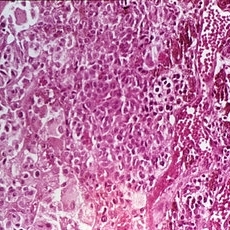
Melanoma of the conjunctiva arising from acquired melanosis.
Condition/keywords: conjunctiva, melanoma, melanosis
-
 Slide 7-26
Feb 25 2019 by Lancaster Course in Ophthalmology
Acquired melanosis of the conjunctiva showing hyperpigmentation of the basal layer of the epithelium.
Condition/keywords: conjunctiva, epithelium, melanosis
-
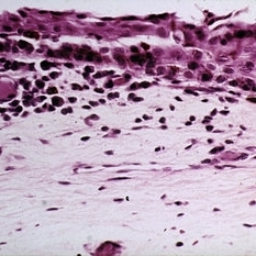
 Slide 7-24
Feb 25 2019 by Lancaster Course in Ophthalmology
Nevus of the conjunctiva. Clusters of benign nevus cells surround an epithelial inclusion cyst.
Condition/keywords: conjunctiva, cyst, epithelial, nevus
-
 Slide 7-23
Feb 25 2019 by Lancaster Course in Ophthalmology
Benign nevus of the conjunctiva.
Condition/keywords: conjunctiva, nevus
-
 Slide 7-22
Feb 25 2019 by Lancaster Course in Ophthalmology
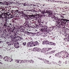
Squamous cell carcinoma of the conjunctiva with invasion into the substantia propria.
Condition/keywords: conjunctiva, substantia propia
-
 Slide 7-21
Feb 25 2019 by Lancaster Course in Ophthalmology
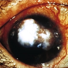
Squamous cell carcinoma of the conjunctiva may present as an exuberant tumor mass.
Condition/keywords: conjunctiva, tumor
-
 Slide 7-19
Feb 25 2019 by Lancaster Course in Ophthalmology
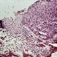
Carcinoma-in-situ. There is an abrupt change from normal conjunctiva (left) to carcinoma-in-situ.
Condition/keywords: conjunctiva
 Loading…