Search results (544 results)
-
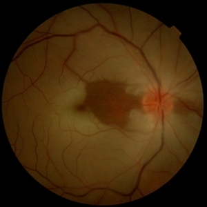
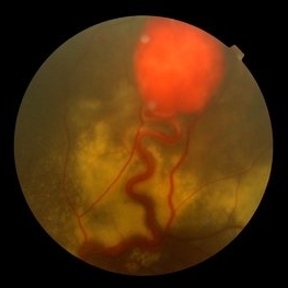
 Lacquer Cracks in Pathologic Myopia
Apr 19 2013 by Theodore Leng, MD, MS, FASRS
Color fundus photograph of a 48-year-old woman with lacquer cracks in the setting of pathologic myopia.
Condition/keywords: lacquer cracks, pathologic myopia
-
---thumb.jpg/image-square;max$300,300.ImageHandler) Presumed Ocular Histoplasmosis Syndrome
Presumed Ocular Histoplasmosis Syndrome
Feb 26 2013 by Henry J. Kaplan, MD
Color fundus photograph of the right eye of a patient with POHS shows typical punched out scars and peripapillary atrophy.
Condition/keywords: presumed ocular histoplasmosis syndrome (POHS)
-
 "Boat-Shaped" Preretinal Hemorrhage
Feb 21 2019 by Mitzy E Torres Soriano, MD
Color fundus photograph showing preretinal (subhyaloid) hemorrhage in a diabetic patient with proliferative diabetic retinopathy.
Photographer: Andrea Vitale, MD
Condition/keywords: preretinal hemorrhage, proliferative diabetic retinopathy (PDR), subhyaloid hemorrhage
-
 Venous Loop & Venous Beading
May 31 2014 by Hamid Ahmadieh, MD
Color fundus photograph of the left eye of a diabetic patient with NVD, NVE, venous loop and venous beading.
Photographer: Elham Salehi, Negah Eye Center, Tehran
Condition/keywords: color fundus photograph, neovascularization elsewhere (NVE), neovascularization of the disc (NVD), proliferative diabetic retinopathy (PDR), venous beading, venous loop
-
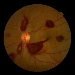 Ocular Manifestation of Acute Leukemia
Sep 8 2012 by Hamid Ahmadieh, MD
Color fundus photograph of a 26-year-old man with acute leukemia.
Photographer: Hamid Ahmadieh, MD, Ophthalmic Research Center, Labbafinejad Medical Center, Shahid Beheshti University of Medical Sciences , Tehran
Imaging device: Topcon Fundus Camera
Condition/keywords: acute leukemia, white centered retinal hemorrhage (Roth Spot)
-
---thumb.jpg/image-square;max$300,300.ImageHandler) Bergmeister's Papilla
Bergmeister's Papilla
Mar 22 2014 by Hamid Ahmadieh, MD
Color fundus photograph of the right eye of a 50-year-old man with Bergmeister's papilla.
Photographer: Naghmeh Nozhat, Negah Eye Center, Tehran
Imaging device: Topcon Fundus Camera
Condition/keywords: Bergmeister's Papillae, color photo
-
---thumb.jpg/image-square;max$300,300.ImageHandler) Reticular Pattern Dystrophy
Reticular Pattern Dystrophy
Aug 7 2013 by From the Collections of Thomas M. Aaberg, MD and Thomas M. Aaberg Jr., MD
Color fundus photograph reveals typical reticular - type pattern dystrophy, OS.
Condition/keywords: pattern macular dystrophy, reticular dystrophy
-
---thumb.jpg/image-square;max$300,300.ImageHandler) CMV Retinitis in a Patient with the Diagnosis of AIDS
CMV Retinitis in a Patient with the Diagnosis of AIDS
Feb 27 2013 by Henry J. Kaplan, MD
Color fundus photograph, right eye: CMV neuroretinitis (retinitis which begins from the arcades and is accompanied by hemorrhage and also has involved the optic nerve).
Condition/keywords: AIDS
-
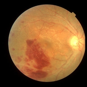
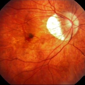 Myopic Choroidal Neovascular Membrane
Mar 25 2013 by Ratimir Lazic, MD, PhD
Color fundus photography of a 33-year-old female. In macular area subretinal hemorrhage can be seen. Area of atrophy temporal from PNO. Myopic changes of posterior pole and mid periphery can be noticed. The patient has been treated with 2 consecutive ranibizumab intravitreal injections. BCVA at baseline was 0,05 (Snellen lines) and 0,3 (Snellen lines) 2 months after.
Photographer: Marko Lukic, MD
Imaging device: Zeis Visucam Lite 2
Condition/keywords: high myopia, myopic choroidal neovascularization (CNV), ranibizumab
-
 Lattice Degeneration and Choroidal Nevus
Oct 10 2015 by Hamid Ahmadieh, MD
Color fundus photograph of the right eye of a 46-year-old woman with a typical lattice degeneration and an adjacent choroidal nevus.
Photographer: Solmaz Shahmohammad, Negah Eye Center, Tehran, Iran
Condition/keywords: choroidal nevus, color fundus photograph, lattice degeneration
-
 Choroidal Osteoma Plus CNV
Sep 2 2012 by Hamid Ahmadieh, MD
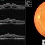
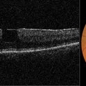
Color fundus photograph and OCT imaging of a 47-year-old man with a juxtafoveal CNV superimposed on a choroidal osteoma.
Photographer: Hamid Ahmadieh, Ophthalmic Research Center, Labbafinejad Medical Center
Imaging device: Topcon
Condition/keywords: choroidal neovascularization (CNV), choroidal osteoma, optical coherence tomography (OCT)
-
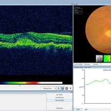
 Chronic Active Central Serous Chorioretinopathy (CSCR)
Sep 11 2012 by Hamid Ahmadieh, MD
Color fundus photograph and OCT image of a 30-year-old man with chronic active CSCR.
Photographer: Hamid Ahmadieh, MD, Ophthalmic Research Center, Labbafinejad Medical Center, Shahid Beheshti University of Medical Sciences
Imaging device: Topcon
Condition/keywords: central serous chorioretinopathy (CSCR), optical coherence tomography (OCT)
-
 White Without Pressure
Jan 31 2018 by Olivia Rainey
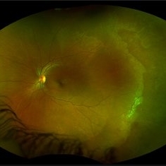
Ultra-wide field pseudocolor photograph of a 57-year-old female with white without pressure affecting her left eye. Patient will be having bloodwork done to rule out possible sarcoidosis or sickle cell.
Photographer: Olivia Rainey
Imaging device: Optos
Condition/keywords: blot hemorrhages, color fundus photograph, left eye, Optos, ultra-wide field imaging, white without pressure
-
 Central Areolar Choroidal Dystrophy
Jul 7 2015 by Hamid Ahmadieh, MD
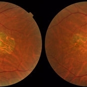
Color fundus photograph of both eyes of a 58-year-old man with progressive loss of vision. VA OD is 20/60 and VA OS is 20/400.
Photographer: Soulmaz Shahmohammad, Negah Eye Center, Tehran, Iran
Imaging device: Topcon
Condition/keywords: central areolar choroidal dystrophy (CACD), color fundus photograph
-
 Sickle Cell Retinopathy with Sea Fans
Aug 24 2012 by Geoffrey G. Emerson, MD, PhD, FASRS
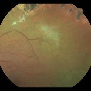
Color fundus photograph of a 40-year-old man with African heritage and sickle SC disease. A sea fan (white) is present along the superotemporal arcade adjacent to an area of ischemia.
Photographer: Geoffrey Emerson, MD, PhD, Retina Center, Minneapolis
Condition/keywords: sea fan, sickle cell retinopathy
-
 Acute Idiopathic Occlusive Retinal Vasculitis
May 31 2014 by Hamid Ahmadieh, MD
Color fundus photograph of the right eye of a 28-year-old woman with sudden drop of vision due to acute occlusive retinal vasculitis leading to extensive nerve fiber layer infarction and retinal hemorrhages.
Photographer: Naghmeh Nozhat, Negah Eye Center, Tehran
Condition/keywords: color fundus photograph, cotton wool spots, retinal hemorrhage, retinal ischemia
-
 Central Retinal Artery Occlusion & Cilioretinal Artery Sparing
Dec 22 2012 by Hamid Ahmadieh, MD
Color fundus photograph of the right eye of a 34-year-old man with sudden drop of vision due to CRAO. Please notice cherry red spot despite cilioretinal artery sparing .
Photographer: Zohre Salimi; Labbafinejad Medical Center, Shahid Beheshti University of Medical Sciences, Tehran
Imaging device: Topcon Fundus Camera
Condition/keywords: central retinal artery occlusion (CRAO), cherry red spot, cilioretinal sparing
-
 Sebaceous Cell Carcinoma of the eyelid
Dec 19 2012 by Eric A. Postel, MD
Color fundus photograph of an eyelid lesion in a 65-year-old man found to be sebaceous cell carcinoma
Condition/keywords: sebaceous cell carcinoma
-
---thumb.jpg/image-square;max$300,300.ImageHandler) Macular CHRPE
Macular CHRPE
Aug 11 2013 by Eric M. Shrier, DO
This a color fundus photograph of a 74-year-old black male with longstanding poor vision os, 20/200. He exhibits mild NPDR additionally.
Photographer: Christopher Bunce
Condition/keywords: congenital hypertrophy of the retinal pigment epithelium (CHRPE)
-
---thumb.jpg/image-square;max$300,300.ImageHandler) Progressive Outer Retinal Necrosis
Progressive Outer Retinal Necrosis
Feb 15 2013 by From the Collections of Thomas M. Aaberg, MD and Thomas M. Aaberg Jr., MD
Color fundus photograph showing extensive confluent retinal whitening, retinal exudation, intraretinal hemorrhage, and sheathing of retinal vessels consistent with infectious retinitis such as progressive outer retinal necrosis (PORN).
Condition/keywords: occlusive retinitis, retinal necrosis
-
 Macular Pseudohole
Jul 7 2015 by Hamid Ahmadieh, MD
Color fundus photograph and optical coherence tomography of the left eye of a 72-year-old woman with blurred vision due to epiretinal membrane. VA OS is 20/40 . Macular pseudohole is visible.
Photographer: Shabnam Poureh, Negah Eye Center, Tehran, Iran
Imaging device: Topcn
Condition/keywords: color fundus photograph, macular pseudohole, optical coherence tomography (OCT)
-
 Behcet's Disease
Mar 13 2013 by Hamid Ahmadieh, MD
Color fundus photograph of the right eye of a 23-year-old man with retinal vasculitis and branch retinal vein occlusion (BRVO) due to Behcet's disease .
Photographer: Solmaz Shahmohammad, Negah Eye Center, Tehran
Imaging device: Heidelberg Spectralis
Condition/keywords: branch retinal vein occlusion (BRVO), retinal vasculitis
-
---thumb.jpg/image-square;max$300,300.ImageHandler) Peripheral retinal nonperfusion, capillary abnormalities, retinal microaneurysms, and intraretinal hemorrhage
Peripheral retinal nonperfusion, capillary abnormalities, retinal microaneurysms, and intraretinal hemorrhage
Feb 15 2013 by From the Collections of Thomas M. Aaberg, MD and Thomas M. Aaberg Jr., MD
Color fundus photograph showing peripheral retinal nonperfusion, capillary abnormalities, retinal microaneurysms, and intraretinal hemorrhage.
Condition/keywords: peripheral retinal nonperfusion, proliferative retinopathy
-
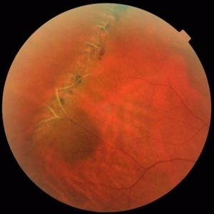
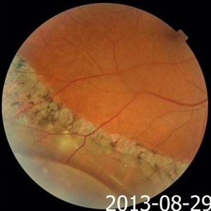 Pigmented Demarcation Line and Retinal Macrocyst
Nov 14 2013 by Hamid Ahmadieh, MD
Color fundus photograph of the right eye of a 40-year-old man with longstanding retinal detachment showing a broad pigmented demarcation line and a retinal macrocyst.
Photographer: Elham Salehi , Negah Eye Center, Tehran
Condition/keywords: demarcation line, fundus photograph, retinal macrocyst
-
 Von Hippel-Lindau
Sep 3 2012 by Hamid Ahmadieh, MD
Color fundus photograph of a 35-year-old woman with retinal angiomatosis.
Photographer: Hamid Ahmadieh, MD, Ophthalmic Research Center, Labbafinejad Medical Center, Shahid Beheshti University of Medical Sciences , Tehran
Imaging device: Topcon Fundus Camera
Condition/keywords: retinal angiomatous proliferation (RAP), Von Hippel-Lindau
 Loading…