Search results (65 results)
-
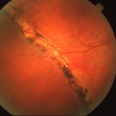
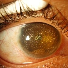 Synchysis Scintillans
Sep 17 2015 by Jessica G Lee, MD
24-year-old male with history of chronic retinal detachment.
Photographer: Bob Masini
Condition/keywords: cholesterol crystals, refractile bodies, synchysis scintillans, trauma, vitreous hemorrhage
-
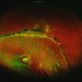
 Chronic Retinal Detachment: Features Slide 1
Oct 22 2012 by Ronald C. Gentile, MD
Chronic retinal detachments can be associated with demarcation lines (tidemarks), subretinal bands or sheets, and retinal cysts. Fundus photo of a chronic inferior retinal detachment reveals multiple demarcation lines inferior to the center of the fovea as a result of an inferior temporal dialysis.
Photographer: The New York Eye & Ear Infirmary Department of Medical Imaging
Condition/keywords: chronic retinal detachment, demarcation line
-
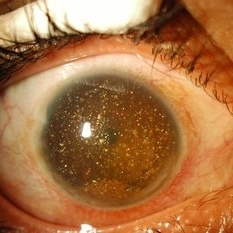 Synchisis Scintillans
Sep 17 2015 by Jessica G Lee, MD
24-year-old male with history of chronic retinal detachment.
Condition/keywords: cholesterol crystals, refractile bodies, synchysis scintillans, trauma, vitreous hemorrhage
-
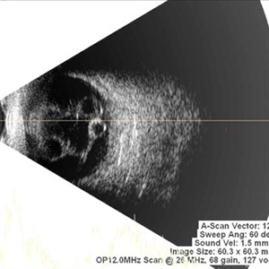
 Chronic Retinal Detachment
Oct 12 2012 by Jeffrey G. Gross, MD, FASRS
Chronic RD with multiple retinal cysts, B scan ultrasound.
Condition/keywords: B scan ultrasound, chronic retinal detachment, retinal cyst
-
 Chronic Retinal Detachment: Features Slide 2
Oct 22 2012 by Ronald C. Gentile, MD
Chronic retinal detachments can be associated with demarcation lines (tidemarks), subretinal bands or sheets, and retinal cysts. Fundus photo of a chronic retinal detachment reveals a branching subretinal band superior nasal to the macula with a portion extending to the inferior margin of the optic disc.
Photographer: The New York Eye & Ear Infirmary Department of Medical Imaging
Condition/keywords: chronic retinal detachment, subretinal bands
-
 Chronic Inferior Retinal Detachment
Mar 1 2017 by Philip J. Polkinghorne, MD
Color photograph of chronic retinal detachment with pigment demarcation line and atrophic holes visible. The vision was recorded at 20/20, and follow up is 3 years.
Photographer: Alex Fraser
Condition/keywords: atrophic retinal hole, demarcation line
-
 Chronic Retinal Detachment
Oct 12 2012 by Jeffrey G. Gross, MD, FASRS
Chronic retinal detachment, with precipitates.
Condition/keywords: chronic retinal detachment, precipitates
-
 Chronic Retinal Detachment: Features Slide 3
Oct 22 2012 by Ronald C. Gentile, MD
Chronic retinal detachments can be associated with demarcation lines (tidemarks), subretinal bands or sheets, and retinal cysts. Fundus photo of a chronic retinal detachment reveals a retinal cyst within the peripherally detached temporal retina.
Condition/keywords: chronic retinal detachment
-
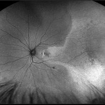
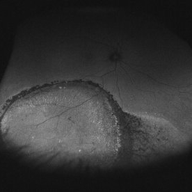 Asymptomatic Chronic Retinal Detachment With Demarcation Line
Jun 11 2016 by Philip J. Polkinghorne, MD
A 65-year-old emmetrope with asymptomatic chronic retinal detachment with demarcation line.
Photographer: Alex Fraser, Greenlane Clinical Center, Auckland, New Zealand
Condition/keywords: chronic retinal detachment, fundus autofluorescence (FAF)
-
 Retinal Detachment with Demarcation Line
Apr 8 2019 by Gary R. Cook, MD, FACS
Pigmented demarcation line from a shallow, chronic retinal detachment OD
Imaging device: Topcon VT-50
Condition/keywords: demarcation line
-
 Chronic Retinal Detachment
May 11 2016 by Andrea Arriola-Lopez, MD MSc
56-year-old man, BCVA 20/25. Incidental finding. Laser was given.
Photographer: Andrea E. Arriola-López MD MSc
Imaging device: OPTOS Dakota
Condition/keywords: chronic
-
 Multiple Retinal Cysts Associated With Chronic Retinal Detachment
Sep 24 2018 by samarth mishra
Patient presented with a diminution of vision in left eye since few months. On B-scan ultrasonography multiple retinal cysts with a total retinal detachment were noted.
Photographer: Aditya Birla Sankara Nethralaya, West Bengal , Kolkata , India
Condition/keywords: B scan ultrasound, chronic retinal detachment, intraretinal cyst, retinal cyst
-
 Angioma from Chronic Retinal Detachment
Sep 19 2014 by David Callanan, MD
36-year-old female, angioma from chronic retinal detachment.
Condition/keywords: angioma
-
 IOL
IOL
Jan 17 2018 by Emily Cooper
Optos image of 47-year-old man with a now worsening retinal detachment that had been treated by pneumatic retinopexy.
Photographer: Emily Cooper, Retina Specialists of Michigan
Imaging device: Optos Ultra Wide Field
Condition/keywords: chronic retinal detachment, intraocular lens (IOL)
-
 Iris Bombé in Late-Stage Pediatric Retinal Detachments
Oct 29 2018 by Linda A Cernichiaro- Espinosa, MD
Different iris pigmentation from two infants with late stage retinal detachment. Left- iris bombé from a white infant. Right- iris bombé from a latin american infant.
Photographer: Brenda Fallas, BS
Imaging device: RetCam III and iPhone 8s
Condition/keywords: bilateral retinal detachment, chronic retinal detachment, iris bombe, pediatric retina
-
 Angioma from Chronic Retinal Detachment
Sep 19 2014 by David Callanan, MD
36-year-old female, angioma from chronic retinal detachment.
Condition/keywords: angioma
-
 Chorioretinal Scars with Subretinal Fibrosis and an old Retinal Detachment
May 3 2018 by Nichole Lewis
Chorioretinal scars with subretinal fibrosis and an old retinal detachment.
Photographer: Nichole Lewis
Condition/keywords: chorioretinal scar, chronic retinal detachment, subretinal fibrosis
-
 Angioma from Chronic Retinal Detachment
Sep 19 2014 by David Callanan, MD
36-year-old female, angioma from chronic retinal detachment.
Condition/keywords: angioma
-
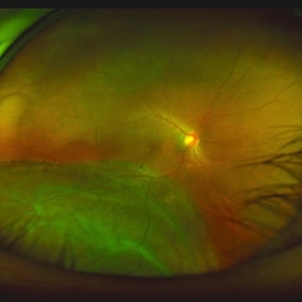
 Macula Off Retinal Detachment with CNV
Nov 11 2019 by Olivia Rainey
Ultra-wide field pseudocolor photograph of a 42-year-old female with a long-standing, macula-off retinal detachment affecting her left eye. Patient was unaware of vision loss until testing her visual acuity and she denied seeing flashing lights. Patient decided to proceed with scleral buckling. The CNV is potentially secondary the retinal detachment, but may be myopic related or idiopathic. The CNV appears fibrotic and inactive. The patient was warned that this will absolutely limit how much vision she recovers once the retina is reattached.
Photographer: Olivia Rainey
Imaging device: Optos California
Condition/keywords: choroidal neovascularization (CNV), chronic retinal detachment, fundus autofluorescence (FAF), left eye, montage, Optos, retinal detachment of the macula, ultra-wide field imaging
-
 Angioma from Chronic Retinal Detachment
Sep 19 2014 by David Callanan, MD
36-year-old female, angioma from chronic retinal detachment.
Condition/keywords: angioma
-
 Large Retinal Detachment
Sep 17 2015 by Jason S. Calhoun
Large retinal detachment in the right eye with the macula detached.
Photographer: Jason Calhoun, Mayo Clinic Jacksonville, Department of Opthalmolgy
Imaging device: OPTOS 200TX
Condition/keywords: chronic retinal detachment
-
 Angioma from Chronic Retinal Detachment
Sep 19 2014 by David Callanan, MD
36-year-old female, angioma from chronic retinal detachment.
Condition/keywords: angioma
-
 Multiple Retinal Cysts Associated With Chronic Retinal Detachment
Sep 24 2018 by samarth mishra
Patient presented with a diminution of vision in left eye since few months. On B-scan ultrasonography multiple retinal cysts with a total retinal detachment were noted.
Photographer: Aditya Birla Sankara Nethralaya, West Bengal , Kolkata , India
Condition/keywords: B scan ultrasound, chronic retinal detachment, intraretinal cyst, retinal cyst
-
 Angioma from Chronic Retinal Detachment
Sep 19 2014 by David Callanan, MD
36-year-old female, angioma from chronic retinal detachment.
Condition/keywords: angioma
-
 Angioma from Chronic Retinal Detachment
Sep 19 2014 by David Callanan, MD
36-year-old female, angioma from chronic retinal detachment.
Condition/keywords: angioma
 Loading…