Search results (345 results)
-
 Myopic CNV
Jan 11 2013 by Alex P. Hunyor, MD
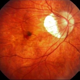
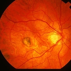
Myopic macular degeneration complicated by subretinal neovascularisation, left eye.
Condition/keywords: high myopia, myopia, myopic choroidal neovascularization (CNV)
-
 Myopic Choroidal Neovascular Membrane
Mar 25 2013 by Ratimir Lazic, MD, PhD
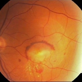
Color fundus photography of a 33-year-old female. In macular area subretinal hemorrhage can be seen. Area of atrophy temporal from PNO. Myopic changes of posterior pole and mid periphery can be noticed. The patient has been treated with 2 consecutive ranibizumab intravitreal injections. BCVA at baseline was 0,05 (Snellen lines) and 0,3 (Snellen lines) 2 months after.
Photographer: Marko Lukic, MD
Imaging device: Zeis Visucam Lite 2
Condition/keywords: high myopia, myopic choroidal neovascularization (CNV), ranibizumab
-
 Choroidal Osteoma Plus CNV
Sep 2 2012 by Hamid Ahmadieh, MD
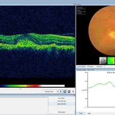
Color fundus photograph and OCT imaging of a 47-year-old man with a juxtafoveal CNV superimposed on a choroidal osteoma.
Photographer: Hamid Ahmadieh, Ophthalmic Research Center, Labbafinejad Medical Center
Imaging device: Topcon
Condition/keywords: choroidal neovascularization (CNV), choroidal osteoma, optical coherence tomography (OCT)
-
 Punctate Inner Choroidopathy with CNV Treated with Bevacizumab # 6 of 7
Feb 28 2013 by Gregory R. Blaha, MD, PhD
Fundus photo following treatment with bevacizumab in a 31-year-old female with vision loss from a choroidal neovascular membrane (CNV) from punctate inner choroidopathy. The vision improved and was stable following a single injection.
Photographer: Gerard Gauthier, Spindel Eye Assoc., Derry, NH
Imaging device: Zeiss FF 450 Plus
Condition/keywords: bevacizumab, choroidal neovascularization (CNV), punctate inner choroidopathy (PIC)
-
 Pigment Epithelial Detachment late FA with small occult CNV
Jul 6 2012 by Tarek S. Hassan, MD, FASRS
72-year-old man with VA loss and metamorphopsia of 2 months duration. PED found, testing done to rule out CNV. Very suspicious for CNV in superonasal fovea/parafovea.
Condition/keywords: choroidal neovascularization (CNV), pigment epithelial detachment (PED)
-
 Optic Nerve Head Drusen With Idiopathic CNV
Feb 17 2017 by Kristen Wagner
22-year-old female fundus photograph of a right eye with Optic Nerve Drusen with Idiopathic CNV.
Photographer: Kristen Wagner, COT, OSC Ophthalmic Photographer, Tennessee Retina, Nashville TN
Condition/keywords: choroidal neovascularization (CNV), drusen of optic disc, optic disc drusen
-
 Secondary Choroidal Neovascularization Due to Toxoplasmosis
Feb 25 2013 by Henry J. Kaplan, MD
Left eye: secondary choroidal neovascularization and subretinal hemorrhage in a patient with old macular scar of toxoplasma.
Condition/keywords: choroidal neovascularization (CNV), toxoplasmosis, toxoplasmosis chorioretinitis
-
 Extra Macular CNV
Aug 24 2012 by John S. King, MD
P/C c mild VFD
Photographer: Kristin Konecki, OcuSight Eye Care Center, Rochester, NY
Condition/keywords: choroidal neovascularization (CNV)
-
 Angioid Streaks & CNV (Fig 3)
Aug 25 2012 by Hamid Ahmadieh, MD
Early phase ICG angiography imaging of a 53-year-old woman with a juxtafoveal CNV secondary to angioid streaks.
Photographer: Hamid Ahmadieh, Ophthalmic Research Center, Labbafinejad Medical Center
Imaging device: Heidelberg Spectralis
Condition/keywords: angioid streaks, choroidal neovascularization (CNV), indocyanine green (ICG) angiography
-
 Angioid Streaks & CNV (Fig 5)
Sep 2 2012 by Hamid Ahmadieh, MD
OCT imaging of a 53-year-old woman with a juxtafoveal CNV secondary to angioid streaks.
Photographer: Hamid Ahmadieh, Ophthalmic Research Center, Labbafinejad Medical Center
Imaging device: Topcon
Condition/keywords: angioid streaks, choroidal neovascularization (CNV), optical coherence tomography (OCT)
-
---thumb.JPG/image-square;max$300,300.ImageHandler) "Flower" Macular Degeneration (Wet)
"Flower" Macular Degeneration (Wet)
Jul 13 2013 by Jason S. Calhoun
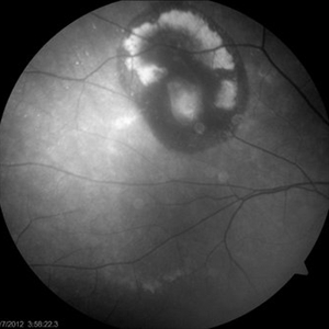
Patient with (wet) macular degeneration in the left eye. Notice the "flower" shape abnormal blood vessels staining.
Photographer: Jason S. Calhoun, Department of Ophthalmology, Mayo Clinic Jacksonville, Florida
Imaging device: TOPCON TRC 50-EX
Condition/keywords: choroidal neovascularization (CNV)
-
 Choroidal Osteoma with Choroidal Neovascularization
Aug 23 2012 by Gerardo Garcia-Aguirre, MD
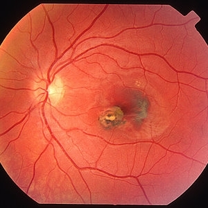
Fundus photograph of the right eye of a 32 year-old male patient with choroidal osteoma and choroidal neovascularization. The left eye also has a choroidal osteoma.
Photographer: Ricardo Montoya, Asociación para Evitar la Ceguera en México
Condition/keywords: choroidal neovascularization (CNV), macular choroidal osteoma
-
Angioid Streaks & CNV (Fig 1)
Aug 25 2012 by Hamid Ahmadieh, MD
Fundus autofluorescence (FAF) of a 53-year-old woman with a juxtafoveal CNV secondary to angioid streaks.
Photographer: Hamid Ahmadieh, Ophthalmic Research Center, Labbafinejad Medical Center
Imaging device: Heidelberg Spectralis
Condition/keywords: angioid streaks, choroidal neovascularization (CNV), fundus autofluorescence (FAF)
-
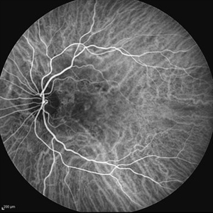
 Choroidal Neovascularization, Idiopathic
Aug 23 2012 by Gerardo Garcia-Aguirre, MD
Fluoresein Angiogram of a 40 year-old patient showing a hyperfluorescent lesion with irregular margins corresponding to a choroidal neovascularization, surrounded by hypofluorescence corresponding to subretinal hemorrhage.
Photographer: Noemí Hernández, Asociación para Evitar la Ceguera en México
Imaging device: Zeiss FF4
Condition/keywords: choroidal neovascularization (CNV)
-
 Juxtafoveal Choroidal Neovascularization Secondary to Choroidal Rupture
Aug 30 2012 by Young Hee Yoon, MD, PhD
Fundus photograph of a 14-year-old boy with a history of blunt trauma to his left eye 9 months ago. Best-corrected visual acuity remained at 20/30.
Photographer: Heon Eui Hong, Asan Medical Center
Imaging device: Canon CR-DGI / Version 5.1.2
Condition/keywords: choroidal rupture, juxtafoveal choroidal neovascularization (CNV)
-
 Juxtafoveal Choroidal Neovascularization Secondary to Choroidal Rupture
Aug 30 2012 by Young Hee Yoon, MD, PhD
SD-OCT image of a 14-year-old boy with a history of blunt trauma to his left eye 9 months ago. Best-corrected visual acuity remained at 20/30.
Photographer: Soo Hyun Cho, Asan Medical Center
Imaging device: HHeidelberg Spectralis OCTI/ version 1.7.0.0
Condition/keywords: choroidal rupture, juxtafoveal choroidal neovascularization (CNV)
-
 Punctate Inner Choroidopathy with CNV Treated with Bevacizumab # 1 of 7
Feb 28 2013 by Gregory R. Blaha, MD, PhD
Fundus photograph in a 31-year-old female with vision loss from a choroidal neovascular membrane (CNV) from punctate inner choroidopathy. Note the CNV and hemorrhage superotemporal to the fovea.
Photographer: Gerard Gauthier, Spindel Eye Associates, Derry, NH
Imaging device: Zeiss FF 450 Plus
Condition/keywords: bevacizumab, choroidal neovascularization (CNV), punctate inner choroidopathy (PIC)
-
 Presumed Ocular Histoplasmosis Syndrome
Aug 24 2012 by John S. King, MD
c histo spots
Photographer: Kristin Konecki, OcuSight Eye Care Center, Rochester, NY
Condition/keywords: choroidal neovascularization (CNV), presumed ocular histoplasmosis syndrome (POHS)
-
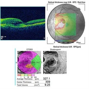
 Myopic Choroidal Neovascularization
Aug 23 2012 by Gabriela Lopezcarasa Hernandez, MD
19-year-old male who complains of scotoma and metamorphopsias.
Photographer: Gabriela Lopezcarasa Hernandez, Macular Retina Consultores
Imaging device: Heidelberg Spectralis
Condition/keywords: choroidal neovascularization (CNV), myopia
-
 CNV Due to Chronic Central Serous Retinopathy
Apr 6 2014 by Ratimir Lazic, MD, PhD
A color fundus image of a 66-year-old male with previously diagnosed chronic CSR. Few weeks ago the patient noticed rapidly worsening of the visual acuity on the left eye. Subretinal hemorrhage with big PED and subretinal exudation can be noticed. The image presents baseline clinical picture of the left eye. The antiVEGF intravitreal treatment have been started.
Photographer: Marko Lukic, University Eye Clinic Svjetlost
Imaging device: Zeis Visucam Lite 2
Condition/keywords: central serous chorioretinopathy (CSCR), choroidal neovascularization (CNV), subretinal hemorrhage
-
 Myopic CNV
May 2 2013 by Henry J. Kaplan, MD
Subretinal membrane in high myopia.
Condition/keywords: myopic choroidal neovascularization (CNV)
-
 Bruch’s membrane rupture
Jan 11 2013 by Hyung-Woo Kwak, MD
An area of Bruch’s membrane rupture involving the fovea is seen on color photograph (left).
Photographer: Misook Lee, Kyung Hee Univsersity Hospital, Seoul
Imaging device: Zeiss f 450 plus
Condition/keywords: myopic choroidal neovascularization (CNV)
-
 Choroidal Neovascularization
Oct 20 2012 by Hyung-Woo Kwak, MD
This 35-year-old young female patient has drusen-like lesions under the macular in both eyes. Such patients have a considerable risk of developing choroidal neovascular lesions.
Condition/keywords: choroidal neovascularization (CNV)
-
 Astrocytic Hamartoma
Oct 10 2012 by K. Bailey Freund, MD
Fundus photograph of an 85-year-old woman with an astrocytic hamartoma and type 2 choroidal neovascular membrane.
Condition/keywords: choroidal neovascularization (CNV)
-
---thumb.jpg/image-square;max$300,300.ImageHandler) POHS Complicated by CNV
POHS Complicated by CNV
Feb 26 2013 by Henry J. Kaplan, MD
POHS complicated by CNV: subretinal hemorrhage in the fovea adjacent to POHS scar.
Condition/keywords: choroidal neovascularization (CNV), presumed ocular histoplasmosis syndrome (POHS), subretinal hemorrhage
 Loading…