Search results (93 results)
-
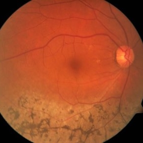
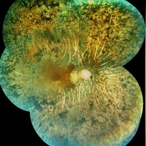
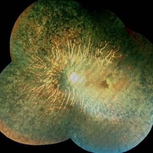
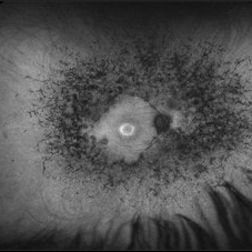
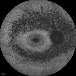 Retinitis Pigmentosa - Fundus Autofluorescence
Sep 20 2014 by Rameez N Hussain, MD
Fundus autofluorescence of retinitis pigmentosa showing hyperautofluorescent rings or foveal hyperautofluorescence.
Photographer: Dr.Rameez N Hussain, MD, Central Imaging Center, Vitreo Retinal Services, Giridhar Eye Institute, Cochin, India
Imaging device: Heidelberg Blue Peak Autofluorescence imaging.
Condition/keywords: bone spicule, cystoid macular edema (CME), fundus autofluorescence (FAF), retinitis pigmentosa
-
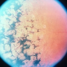
 Bone Spicules
Aug 1 2013 by From the Collections of Thomas M. Aaberg, MD and Thomas M. Aaberg Jr., MD
Bone spicules.
Condition/keywords: bone spicule
-
 Retinitis Pigmentosa
May 26 2017 by Olivia Rainey
Ultra-wide-field pseudocolor image of the right eye of an 39-year-old female with Retinitis Pigmentosa. She had slightly atypical appearance due to asymmetry: sectoral atrophy in left eye, compared to 360 degree bone spicule formation in right eye. Ddx: Pigmentary degeneration vs infection vs X-linked RP carrier due to asymmetry. Recommended genetic testing through My Retina Tracker, as well as visual field and ERG testing. Patient's vision was sc20/100 PH 20/70 in the right eye and sc20/80 PH 20/40 in the left.
Photographer: Olivia Rainey
Imaging device: Optos California
Condition/keywords: bone spicule, fundus photograph, Optos, peripheral bone spicules, pseudocolor, retinitis pigmentosa, ultra-wide field imaging
-
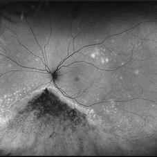
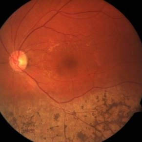 Sector Retinitis Pigmentosa
Mar 13 2014 by Hyung-Woo Kwak, MD
Fundus photograph of an 57-year-old woman with a sector retinitis pigmentosa. Regionalized areas of bone spicule pigmentation is in the inferior quadrants of the retina.
Photographer: Missok Lee, Kyung Hee University Hospital, Seoul, Korea
Imaging device: Zeiss F450 Plus
Condition/keywords: sector retinitis pigmentosa
-
 Retinitis Pigmentosa - Autofluorescence OD
Jun 18 2018 by Hosam Attia, MD
Ultra-wide fundus auto-fluorescence photograph of a 38-year-old African, American female with degenerative myopia, unilateral RP variant, depicting extensive mid-peripheral bone spicules hypo-autofluorescence, extending further into the periphery w/ relative sparing of the macula OD VF 30-V showed severe peripheral constriction OD, enlarged BS OS and OCT showed severe ellipsoid zone degeneration with saucerization and cystoid macular degeneration with no obvious late macular leakage on FA (Both, not shown)
Imaging device: Optos California
Condition/keywords: bone spicule, peripheral bone spicules, retinitis pigmentosa
-
---thumb.jpg/image-square;max$300,300.ImageHandler) Retinitis Pigmentosa
Retinitis Pigmentosa
Oct 13 2012 by Geoffrey G. Emerson, MD, PhD, FASRS
Condition/keywords: bone spicule, retinitis pigmentosa
-
 Sector Retinitis Pigmentosa
Mar 13 2014 by Hyung-Woo Kwak, MD
Fundus photograph of an 57-year-old woman with a sector retinitis pigmentosa. Regionalized areas of bone spicule pigmentation is in the inferior quadrants of the retina.
Photographer: Missok Lee, Kyung Hee University Hospital, Seoul, Korea
Imaging device: Zeiss F450 Plus
Condition/keywords: sector retinitis pigmentosa
-
 Retinitis pigmentosa AD Slide 2
Oct 22 2012 by Ronald C. Gentile, MD
Fundus photo out side the fovea reveals predominantly depigmentation of the retinal pigment epithelium with some white dots and occasional bone spicule-shaped pigment deposit peripherally.
Photographer: The New York Eye & Ear Infirmary Department of Medical Imaging
Condition/keywords: retinitis pigmentosa
-
 Peripheral Bone Spicules
Jul 31 2013 by From the Collections of Thomas M. Aaberg, MD and Thomas M. Aaberg Jr., MD
Peripheral bone spicules.
Condition/keywords: bone spicule, peripheral bone spicules
-
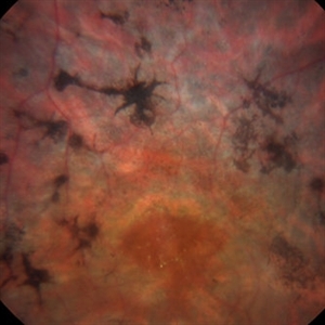 Bone Spicules
Mar 1 2014 by Homayoun Tabandeh, MD, FASRS
Bone spicule pigmentary retinopathy in a patient with retinitis pigmentosa.
Condition/keywords: bone spicule, retinitis pigmentosa
-
 Retinitis Pigmentosa
May 26 2017 by Olivia Rainey
Ultra-wide-field pseudocolor image of the left eye of an 39-year-old female with Retinitis Pigmentosa. She had slightly atypical appearance due to asymmetry: sectoral atrophy in left eye, compared to 360 degree bone spicule formation in right eye. Ddx: Pigmentary degeneration vs infection vs X-linked RP carrier due to asymmetry. Recommended genetic testing through My Retina Tracker, as well as visual field and ERG testing. Patient's vision was sc20/100 PH 20/70 in the right eye and sc20/80 PH 20/40 in the left eye.
Photographer: Olivia Rainey
Imaging device: Optos California
Condition/keywords: bone spicule, fundus photograph, left eye, Optos, peripheral bone spicules, pseudocolor, retinitis pigmentosa, ultra-wide field imaging
-
---thumb.jpg/image-square;max$300,300.ImageHandler) Retinitis Pigmentosa with peripheral arteriolar narrowing
Retinitis Pigmentosa with peripheral arteriolar narrowing
Feb 20 2013 by From the Collections of Thomas M. Aaberg, MD and Thomas M. Aaberg Jr., MD
Retinitis pigmentosa with peripheral arteriolar narrowing small sector of bone spicule
Condition/keywords: arteriolar narrowing, bone spicule, retinitis pigmentosa
-
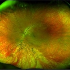
 Retinitis Pigmentosa
Aug 25 2015 by René Hernán Parada Vásquez
Fundus photograph of both eyes of a 38-year-old female with retinitis pigmentosa, bone spicule-shaped pigment deposits are present in the mid periphery, and macula with a peripheral ring of depigmentation.
Photographer: Parada René, ESO, Guatemala.
Imaging device: Canon CR-2
Condition/keywords: bilateral pigmentary retinopathy, retinitis pigmentosa, retinitis pigmentosa (RP) dystrophy
-
 Retinitis Pigmentosa
Feb 26 2020 by Manuel Ángel Alcántara Delgado, MD
Merged color fundus photograph of a 68-year-old woman with advanced retinitis pigmentosa. It is appreciated bone spicule-shaped pigment deposits, optic disc pallor, retinal atrophy, attenuated retinal vessels and surface wrinkling retinopathy.
Photographer: Manuel Ángel Alcántara Delgado
Condition/keywords: chorioretinal atrophy, choroidal circulation, optic disc pallor, pericentral retinitis pigmentosa, retina, retinitis pigmentosa, retinitis pigmentosa (RP) dystrophy, sector retinitis pigmentosa
-
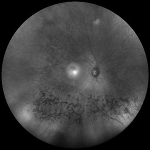 Sector Retinitis Pigmentosa
Mar 13 2014 by Hyung-Woo Kwak, MD
Wide field infrared image of an 57-year-old woman with a sector retinitis pigmentosa. Regionalized areas of bone spicule pigmentation is in the inferior quadrants of the retina.
Photographer: Missok Lee, Kyung Hee University Hospital, Seoul, Korea
Imaging device: Heidelberg Spectralis
Condition/keywords: sector retinitis pigmentosa
-
 Retinitis pigmentosa
Feb 26 2020 by Manuel Ángel Alcántara Delgado, MD
Merged color fundus photograph of a 68-year-old woman with advanced retinitis pigmentosa. It is appreciated bone spicule-shaped pigment deposits, optic disc pallor, retinal atrophy and attenuated retinal vessels.
Photographer: Manuel Ángel Alcántara Delgado
Condition/keywords: choroidal circulation, optic disc pallor, pericentral retinitis pigmentosa, retina, retinitis pigmentosa, retinitis pigmentosa (RP) dystrophy, sector retinitis pigmentosa
-
 Retinitis Pigmentosa
Apr 30 2015 by Mitzy E Torres Soriano, MD
Fundus of patient with retinitis pigments, bone spicule-shaped pigment deposits are present with retinal atrophy, while the macula is preserved . Retinal vessels are attenuated.
Photographer: Mitzy E. Torres Soriano, MD; Centro medico Cagua-Estado Aragua. Venezuela
Imaging device: TRC-NW8
Condition/keywords: pigmentary retinal dystrophy, retinal dystrophy, retinitis pigmentosa, retinitis pigmentosa (RP) dystrophy
-
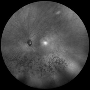 Sector Retinitis Pigmentosa
Mar 13 2014 by Hyung-Woo Kwak, MD
Wide field infrared image of an 57-year-old woman with a sector retinitis pigmentosa. Regionalized areas of bone spicule pigmentation is in the inferior quadrants of the retina.
Photographer: Missok Lee, Kyung Hee University Hospital, Seoul, Korea
Imaging device: Heidelberg Spectralis
Condition/keywords: sector retinitis pigmentosa
-
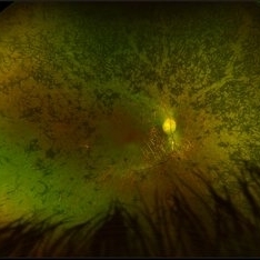
 Retinitis Pigmentosa
May 26 2017 by Olivia Rainey
Ultra-wide-field pseudocolor image of the left eye of an 39-year-old female with Retinitis Pigmentosa. She had slightly atypical appearance due to asymmetry: sectoral atrophy in left eye, compared to 360 degree bone spicule formation in right eye. Ddx: Pigmentary degeneration vs infection vs X-linked RP carrier due to asymmetry. Recommended genetic testing through My Retina Tracker, as well as visual field and ERG testing. Patient's vision was sc20/100 PH 20/70 in the right eye and sc20/80 PH 20/40 in the left eye.
Photographer: Olivia Rainey
Imaging device: Optos California
Condition/keywords: autofluorescence imaging, bone spicule, hyperautofluorescent ring, hypoautofluorescence, Optos, peripheral bone spicules, retinitis pigmentosa, ultra-wide field imaging
-
 Retinitis Pigmentosa
May 26 2017 by Olivia Rainey
Ultra-wide-field fundus autofluorescence image of the left eye of an 39-year-old female with Retinitis Pigmentosa. She had slightly atypical appearance due to asymmetry: sectoral atrophy in left eye, compared to 360 degree bone spicule formation in right eye. Ddx: Pigmentary degeneration vs infection vs X-linked RP carrier due to asymmetry. Recommended genetic testing through My Retina Tracker, as well as visual field and ERG testing. Patient's vision was sc20/100 PH 20/70 in the right eye and sc20/80 PH 20/40 in the left eye.
Photographer: Olivia Rainey
Imaging device: Optos
Condition/keywords: autofluorescence imaging, hyperautofluorescence, hypoautofluorescence, left eye, Optos, peripheral bone spicules, retinitis pigmentosa, ultra-wide field imaging
-
---thumb.jpg/image-square;max$300,300.ImageHandler) RP/RPE Bone Spicules
RP/RPE Bone Spicules
Dec 27 2013 by David Callanan, MD
54-year-old female, CF; HM; followed for some time.
Condition/keywords: bone spicule, retinal pigment epithelium
-
---thumb.jpg/image-square;max$300,300.ImageHandler) Retinitis Pigmentosa
Retinitis Pigmentosa
Feb 20 2013 by From the Collections of Thomas M. Aaberg, MD and Thomas M. Aaberg Jr., MD
bone spicules
Condition/keywords: bone spicule, retinitis pigmentosa
-
---thumb.jpg/image-square;max$300,300.ImageHandler) retinitis pigmentosa
retinitis pigmentosa
Feb 20 2013 by From the Collections of Thomas M. Aaberg, MD and Thomas M. Aaberg Jr., MD
peripheral bone spicules
Condition/keywords: bone spicule, retinitis pigmentosa
-
 Pigmentary Retinal Dystrophy
Mar 29 2019 by Jessica Norkus
Optos ultra wide field image of 41-year-old male patient with pigmentary retinal dystrophy. Atypical findings due to unilateral presentation. Patient has been experiencing symptoms for 15 years, notes significant nyctalopia.
Photographer: Jessica Norkus
Imaging device: Optos Ultra Wide Field Camera
Condition/keywords: abnormal fundus, bone spicule, color fundus photograph, color photo, fundus photograph, Optos, peripheral bone spicules, pigment changes, ultra-wide field imaging, unilateral blindness
-
---thumb.jpg/image-square;max$300,300.ImageHandler) Retinitis Pigmentosa
Retinitis Pigmentosa
Feb 20 2013 by From the Collections of Thomas M. Aaberg, MD and Thomas M. Aaberg Jr., MD
360 degree bone spicules
Condition/keywords: bone spicule, retinitis pigmentosa
 Loading…