 Initializing download.
-
 By Hosam Attia, MD
- Uploaded on Jun 18, 2018.
- Last modified by Caroline Bozell on Jun 19, 2018.
- Rating
- Appears in
- Retinitis Pigmentosa
- Condition/keywords
- bone spicule, peripheral bone spicules, retinitis pigmentosa
- Imaging device
-
Scanning laser ophthalmoscope
Optos California - Description
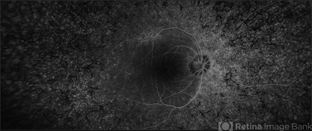
- Ultra-wide fluorescein angiogram of a 38-year-old African, American female with degenerative myopia, Unilateral RP variant, depicting abnormal fluorescence pattern with extensive mid-peripheral bone spicules hypofluorescence, extending further into the periphery w/ relative sparing of the macula OD. VF 30-V showed severe peripheral constriction OD, enlarged BS OS & OCT showed severe ellipsoid zone degeneration with saucerization and cystoid macular degeneration w/ No obvious late macular leakage on FA (Both, not shown)


---thumb.jpg/image-square;max$79,0.ImageHandler "Retinitis Pigmentosa")


