Search results (129 results)
-
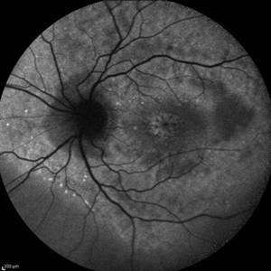
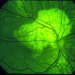
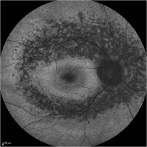 Retinitis Pigmentosa - Fundus Autofluorescence
Sep 20 2014 by Rameez N Hussain, MD
Fundus autofluorescence of retinitis pigmentosa showing hyperautofluorescent rings or foveal hyperautofluorescence.
Photographer: Dr.Rameez N Hussain, MD, Central Imaging Center, Vitreo Retinal Services, Giridhar Eye Institute, Cochin, India
Imaging device: Heidelberg Blue Peak Autofluorescence imaging.
Condition/keywords: bone spicule, cystoid macular edema (CME), fundus autofluorescence (FAF), retinitis pigmentosa
-
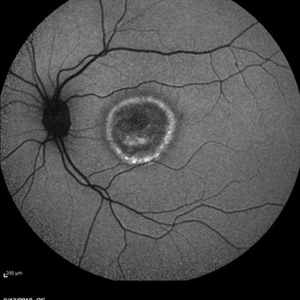
 Late Stage Stargardt's Disease
Mar 13 2013 by Hamid Ahmadieh, MD
Autofluorescence imaging of the left eye of a 46-year-old man with decreased VA due to advanced Stargardt's disease.
Photographer: Nayereh Hadipoor, Negah Eye Center, Tehran
Imaging device: Heidelberg Spectralis
Condition/keywords: autofluorescence imaging, Stargardt disease
-
 Serpiginous like Choroiditis
Aug 24 2012 by S. Natarajan, MD, FASRS, FRCS (GLASGOW) , FICO, D.Sc, FELA
Fundus photograph of a 32-year-old male with serpiginous like lesion in the posterior pole followed up with serial autofluorescence imaging.
Photographer: Prof.Dr.S. Natarajan
Imaging device: Zeiss FF 450 plus IR
Condition/keywords: autofluorescence imaging, choroiditis, serpiginous choroiditis
-
 PED due to CSCR 2
Sep 2 2012 by Hamid Ahmadieh, MD
Autofluorescence imaging of a 37-year-old man with a serous PED secondary to CSCR.
Photographer: Hamid Ahmadieh, Ophthalmic Research Center, Labbafinejad Medical Center
Imaging device: Heidelberg Spectralis
Condition/keywords: autofluorescence imaging, central serous chorioretinopathy (CSCR), pigment epithelial detachment (PED)
-
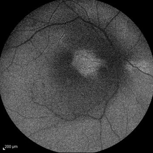
 Cystoid Macular Edema (CME)
Sep 11 2012 by Hamid Ahmadieh, MD
Autofluorescence imaging of the left eye of a 17-year-old boy with chronic intermediate uveitis showing CME.
Photographer: Hamid Ahmadieh, MD, Ophthalmic Research Center, Labbafinejad Medical Center, Shahid Beheshti University of Medical Sciences
Imaging device: Heidelberg Spectralis
Condition/keywords: autofluorescence imaging, cystoid macular edema (CME), intermediate uveitis
-
 Best Disease
Mar 9 2013 by Hamid Ahmadieh, MD
Autofluorescence Imaging of the left eye of a 49-year-old man with decreased VA due to advanced Best disease.
Photographer: Soodabeh Fooladin, Negah Eye Center, Tehran
Imaging device: Heidelberg Spectralis
Condition/keywords: autofluorescence imaging, Best disease
-
 Chronic Central Serous Chorioretinopathy
Sep 26 2012 by Hamid Ahmadieh, MD
Autofluorescence imaging of the right eye of a 50-year-old man with active chronic CSCR and BCVA of 20/100.
Photographer: Hamid Ahmadieh, MD, Ophthalmic Research Center, Labbafinejad Medical Center, Shahid Beheshti University of Medical Sciences
Imaging device: Heidelberg Spectralis
Condition/keywords: autofluorescence imaging, chronic central serous chorioretinopathy (CSCR)
-
 Central Retinal Artery Occlusion & Cilioretinal Artery Sparing
Dec 22 2012 by Hamid Ahmadieh, MD
Autofluorescence imaging of the right eye of a 34-year-old man with sudden drop of vision due to CRAO. The macula is involved despite cilioretinal artery sparing .
Photographer: Zohre Salimi; Labbafinejad Medical Center, Shahid Beheshti University of Medical Sciences
Imaging device: Heidelberg HRA
Condition/keywords: autofluorescence imaging, central retinal artery occlusion (CRAO), cilioretinal sparing
-
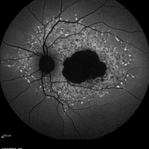
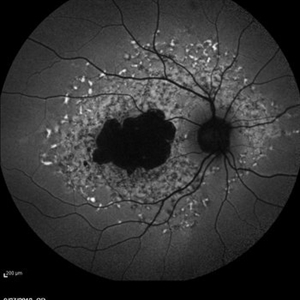
 Geographic Atrophy - Case 1: Photo 2 of 6
Oct 4 2012 by Gregg T. Kokame, MD, MMM, FASRS
Blue-peak Autofuorescence Image of patient with Geographic Atrophy
Photographer: Jaclyn Pisano, Retina Consultants of Hawaii
Imaging device: Heidelberg Spectralis
Condition/keywords: autofluorescence imaging, geographic atrophy
-
 RIP 2 FAF
Oct 7 2015 by Roberto Gallego-Pinazo, MD, PhD, DiSSO
Multicolor and autofluorescence sequence of a retinal pigment epithelium tear following intravitreal anti-VEGF injection.
Photographer: Rosa Dolz-Marco, University and Polytechnic Hospital La Fe, Valencia, Spain
Condition/keywords: age-related macular degeneration (AMD), autofluorescence imaging, choroidal neovascularization (CNV), multicolor, retinal pigment epithelium (RPE) tear
-
 Macular Telangiectasia Type 2
Sep 22 2012 by Hamid Ahmadieh, MD
Autofluorescence imagings of both eyes of a 70-year-old man with idiopathic macular telangiectasia type 2.
Photographer: Hamid Ahmadieh, MD, Ophthalmic Research Center, Labbafinejad Medical Center, Shahid Beheshti University of Medical Sciences
Imaging device: HRA
Condition/keywords: autofluorescence imaging, idiopathic macular telangiectasia
-
 Geographic Atrophy - Case 1: Photo 3 of 6
Oct 4 2012 by Gregg T. Kokame, MD, MMM, FASRS
Infrared Image of patient with Geographic Atrophy
Photographer: Jaclyn Pisano, Retina Consultants of Hawaii
Imaging device: Heidelberg Spectralis
Condition/keywords: autofluorescence imaging, geographic atrophy
-
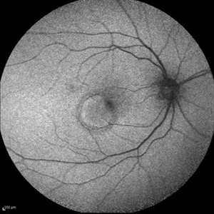
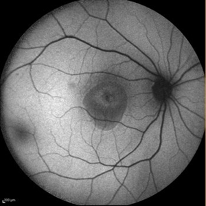
 Serpiginous Choroiditis - Fundus Autofluorescence
Sep 20 2014 by Rameez N Hussain, MD
Fundus autofluorescence of serpiginous choroiditis showing decreased autofluorescence area corresponding to the inactive lesion (RPE atrophy) and increased autofluorescence area corresponding to active lesion.
Photographer: Dr.Rameez N Hussain, MD, Central Imaging Center, Vitreo Retinal Services, Giridhar Eye Institute, Cochin, India
Imaging device: Heidelberg Blue Peak Autofluorescence imaging.
Condition/keywords: serpiginous choroiditis
-
 Geographic Atrophy - Case 1: Photo 4 of 6
Oct 4 2012 by Gregg T. Kokame, MD, MMM, FASRS
NIRAF (near infrared autofluorescence) Image of patient with Geographic Atrophy
Photographer: Jaclyn Pisano, Retina Consultants of Hawaii
Imaging device: Heidelberg Spectralis
Condition/keywords: autofluorescence imaging, geographic atrophy, near infrared autofluorescence (NIRAF)
-
 Geographic Atrophy - Case 1: Photo 1 of 6
Oct 4 2012 by Gregg T. Kokame, MD, MMM, FASRS
NIRAF (near infrared autofluorescence) Image of patient with Geographic Atrophy
Photographer: Jaclyn Pisano, Retina Consultants of Hawaii
Imaging device: Heidelberg Spectralis
Condition/keywords: autofluorescence imaging, geographic atrophy, near infrared autofluorescence (NIRAF)
-
 Geographic Atrophy - Case 1: Photo 5 of 6
Oct 4 2012 by Gregg T. Kokame, MD, MMM, FASRS
Blue-peak Autofuorescence Image of patient with Geographic Atrophy
Photographer: Jaclyn Pisano, Retina Consultants of Hawaii
Imaging device: Heidelberg Spectralis
Condition/keywords: autofluorescence imaging, geographic atrophy
-
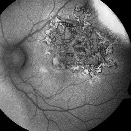
 Choriodal Rupture - AW 001 - Initial Presentation
Mar 11 2013 by Suber S. Huang, MD, MBA, FASRS
40-year-old male sustained blunt trauma OD with orbital fracture and choroidal rupture subjacent to inferior arcade with blood, subretinal fluid, and exudate extending to fovea with CF 2 feet at presentation 9-10-12. On followup 3-11-12, vision improved to 20/400 with resolution of hemorrhage, normal OCT, but speckling of foveal RPE and PMB consistent with damage.
Photographer: Mark Harrod
Condition/keywords: autofluorescence imaging, blunt trauma, choroidal hemorrhage, choroidal rupture, orbital fracture, retinal hemorrhage, submacular hemorrhage
-
 Autofluorescence 10-14-13 AZOOR
Dec 14 2013 by Robert T. Wendel, MD
Autofluorescence 10-14-13 AZOOR
Condition/keywords: acute zonal occult outer retinopathy (AZOOR), autofluorescence imaging
-
---thumb.jpg/image-square;max$300,300.ImageHandler) Polypoidal Choroidal Vasculopathy: Case 1 - Image 2 of 7
Polypoidal Choroidal Vasculopathy: Case 1 - Image 2 of 7
Oct 4 2012 by Gregg T. Kokame, MD, MMM, FASRS
Bluepeak Autofluorescence image of a 57-year-old woman with treatment-naive polypoidal choroidal vasculopathy. Series of images provides an comparative view of the same condition while utilizing a variet of different imaging procedures.
Photographer: Andrew Yuen, Retina Consultants of Hawaii
Imaging device: Heidelberg Spectralis
Condition/keywords: autofluorescence imaging, fundus autofluorescence (FAF), polypoidal choroidal vasculopathy (PCV)
-
---thumb.jpg/image-square;max$300,300.ImageHandler) Optic Disc Drusen
Optic Disc Drusen
Mar 27 2013 by Henry J. Kaplan, MD
Autofluorescence imaging shows heper AF on the optic nerve head specially superiorly due to drusen in the same patient #2.
Imaging device: Heidelberg spectralis
Condition/keywords: drusen of optic disc, optic disc drusen, optic nerve drusen
-
 Choroidal Osteoma
Mar 29 2013 by Henry J. Kaplan, MD
Autofluorescence in choroidal osteoma.
Condition/keywords: autofluorescence imaging, choroidal osteoma
-
 Choriodal Rupture - AW 002 - 6 Month F/U
Mar 11 2013 by Suber S. Huang, MD, MBA, FASRS
40-year-old male sustained blunt trauma OD with orbital fracture and choroidal rupture subjacent to inferior arcade with blood, subretinal fluid, and exudate extending to fovea with CF 2 feet at presentation 9-10-12. On follow up 3-11-12, vision improved to 20/400 with resolutionof hemorrhage, normal OCT, but speckling of foveal RPE and PMB consistent with damage.
Photographer: Mark Harrod
Condition/keywords: autofluorescence imaging, blunt trauma, choroidal hemorrhage, choroidal rupture, orbital fracture, retinal hemorrhage, submacular hemorrhage
-
 Elmiron Toxicity
Jan 12 2018 by Jessica Norkus
Bilateral ultra-wide field pseudo-color and autofluorescent images of a 46-year-old female with Elmiron Toxicity.
Photographer: Jessica Norkus
Imaging device: Optos
Condition/keywords: autofluorescence imaging, bilateral, color fundus photograph, drug toxicity, Optos, toxic maculopathy, toxic retinopathy, ultra-wide field imaging
-
 Late Stage Stargardt's Disease
Mar 13 2013 by Hamid Ahmadieh, MD
Autofluorescence imaging of the right eye of a 46-year-old man with decreased VA due to advanced Stargardt's disease.
Photographer: Nayereh Hadipoor, Negah Eye Center, Tehran
Imaging device: Heidelberg Spectralis
Condition/keywords: autofluorescence imaging, Stargardt disease
-
 Choriodal Rupture 004 - Fundus Autoflurescence - 6 Month Follow Up
Mar 11 2013 by Suber S. Huang, MD, MBA, FASRS
40-year-old male sustained blunt trauma OD with orbital fracture and choroidal rupture subjacent to inferior arcade with blood, subretinal fluid, and exudate extending to fovea with CF 2 feet at presentation 9-10-12. On follow up 3-11-12, vision improved to 20/400 with resolutionof hemorrhage, normal OCT, but speckling of foveal RPE and PMB consistent with damage.
Photographer: Mark Harrod
Condition/keywords: autofluorescence imaging, blunt trauma, choroidal hemorrhage, choroidal rupture, orbital fracture, retinal hemorrhage, submacular hemorrhage
 Loading…