Search results (226 results)
-
 Retinal capillary hemangiomas 3
Jan 11 2013 by Alex P. Hunyor, MD
Retinal capillary haemangiomas, left superior periphery, in a 20 year old female with von Hippel-Lindau disease.
Condition/keywords: hemangioma, retinal capillary hemangioma, Von Hippel-Lindau
-
 Retinal capillary hemangioma 2
Jan 11 2013 by Alex P. Hunyor, MD
Retinal capillary haemangioma, right inferior periphery, in a 20-year-old female with von Hippel-Lindau disease.
Condition/keywords: hemangioma, retinal capillary hemangioma, Von Hippel-Lindau
-
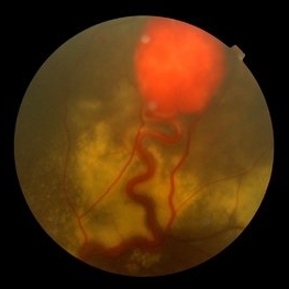
 Retinal Angiomas In VHL
Dec 24 2012 by Roy D. Brod, MD
Fundus photograph of 16 year old male with recent diagnosis of Von Hippel-Lindau disease showing typical appearance of a retinal angioma in superior mid periphery OD. Note unrelated choroidal nevus above superior arcade.
Photographer: Julia Walker
Condition/keywords: hemangioma, Von Hippel-Lindau
-
 Von Hippel-Lindau
Sep 3 2012 by Hamid Ahmadieh, MD
Color fundus photograph of a 35-year-old woman with retinal angiomatosis.
Photographer: Hamid Ahmadieh, MD, Ophthalmic Research Center, Labbafinejad Medical Center, Shahid Beheshti University of Medical Sciences , Tehran
Imaging device: Topcon Fundus Camera
Condition/keywords: retinal angiomatous proliferation (RAP), Von Hippel-Lindau
-
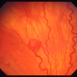
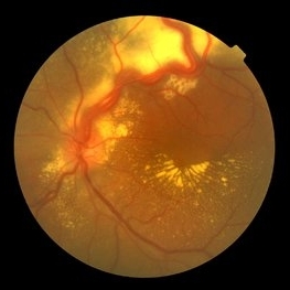
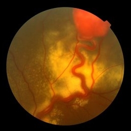
 Retinal capillary hemangioma
Jan 11 2013 by Alex P. Hunyor, MD
Retinal capillary haemangioma nasal to optic disc, right eye.
Condition/keywords: retinal capillary hemangioma, Von Hippel-Lindau
-
 Von Hippel-Lindau
Sep 3 2012 by Hamid Ahmadieh, MD
Color fundus photograph of a 35-year-old woman with retinal angiomatosis.
Photographer: Hamid Ahmadieh, MD, Ophthalmic Research Center, Labbafinejad Medical Center, Shahid Beheshti University of Medical Sciences
Imaging device: Topcon Fundus Camera
Condition/keywords: retinal angiomatous proliferation (RAP), Von Hippel-Lindau
-
---thumb.jpg/image-square;max$300,300.ImageHandler) Retinal capillary hemangioma 4 image 1
Retinal capillary hemangioma 4 image 1
Jan 11 2013 by Alex P. Hunyor, MD
Retinal capillary haemangioma, left eye, in a young female with von Hippel-Lindau disease. Color image 1 showing extensive lipid deposition in the macula.
Condition/keywords: retinal capillary hemangioma, Von Hippel-Lindau
-
 Von Hippel-Lindau
Sep 5 2012 by Hamid Ahmadieh, MD
Color fundus photograph of a 32-year-old man with retinal angiomatosis.
Photographer: Hamid Ahmadieh, MD, Ophthalmic Research Center, Labbafinejad Medical Center, Shahid Beheshti University of Medical Sciences
Imaging device: Topcon Fundus Camera
Condition/keywords: retinal angiomatous proliferation (RAP), Von Hippel-Lindau
-
 Retinal Angiomas In VHL
Dec 24 2012 by Roy D. Brod, MD
Fundus photograph of 16 year old male with recent diagnosis of Von Hippel-Lindau disease showing 2 retinal angiomas in inferior mid periphery OD.
Photographer: Julia Walker
Condition/keywords: hemangioma, Von Hippel-Lindau
-
 Von Hippel-Lindau
Sep 8 2012 by Hamid Ahmadieh, MD
Color fundus photograph of a 32-year-old man with retinal angiomatosis.
Photographer: Hamid Ahmadieh, MD, Ophthalmic Research Center, Labbafinejad Medical Center, Shahid Beheshti University of Medical Sciences
Imaging device: Topcon Fundus Camera
Condition/keywords: retinal angiomatous proliferation (RAP), Von Hippel-Lindau
-
 Retinal Angiomas In VHL
Dec 24 2012 by Roy D. Brod, MD
Mid phase fluorescein angiogram of 16 year old male with recent diagnosis of Von Hippel-Lindau disease showing hyperfluorescent angioma in superior mid periphery OD.
Photographer: Julia Walker
Condition/keywords: hemangioma, Von Hippel-Lindau
-
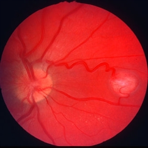
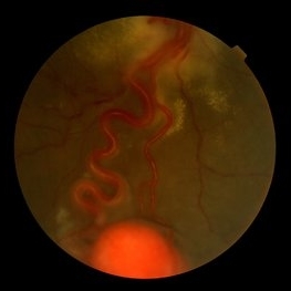
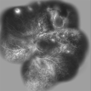
 Capillary Hemongima, Coat's Response
May 2 2013 by Henry J. Kaplan, MD
Coat's response as exudation in the macula in the same patient with retinal capillary hemangioma. Notice the dilated feeder vessles from the optic nerve infriorly; #2.
Condition/keywords: Coats' disease, retinal capillary hemangioma, Von Hippel-Lindau
-
 Von Hippel-Lindau
Sep 3 2012 by Hamid Ahmadieh, MD
Color fundus photograph of a 35-year-old woman with retinal angiomatosis.
Photographer: Hamid Ahmadieh, MD, Ophthalmic Research Center, Labbafinejad Medical Center, Shahid Beheshti University of Medical Sciences
Imaging device: Topcon Fundus Camera
Condition/keywords: retinal angiomatous proliferation (RAP)
-
 Retinal Angiomas In VHL
Dec 24 2012 by Roy D. Brod, MD
Mid phase fluorescein angiogram of 16 year old male with recent diagnosis of Von Hippel-Lindau disease showing hyperfluorescent angiomas in inferior mid periphery OD.
Photographer: Julia Walker
Condition/keywords: hemangioma, Von Hippel-Lindau
-
---thumb.jpg/image-square;max$300,300.ImageHandler) Von Hippel Lindau (VHL) Disease
Von Hippel Lindau (VHL) Disease
Oct 14 2013 by Maurice F. Rabb
16 year old was screened at age 12 for clinical manifestations of Von Hippel Lindau (VHL) disease because of positive genetic testing. His father was also affected with VHL, with ocular and renal involvement. He had spinal cord lesions and a solid renal mass. Ocular exams were: Visual acuity 20/20 OD, 20/20 OS.
Condition/keywords: Von Hippel-Lindau
-
 Von Hippel-Lindau 1
Oct 13 2012 by Hamid Ahmadieh, MD
Color fundus photograph of the left eye of a 25-year-old woman with exudative retinal detachment secondary to retinal angiomatosis (Von Hippel-Lindau).
Photographer: Hamid Ahmadieh, MD, Ophthalmic Research Center, Labbafinejad Medical Center, Shahid Beheshti University of Medical Sciences
Imaging device: Topcon Fundus Camera
Condition/keywords: exudative retinal detachment, retinal angiomatous proliferation (RAP), Von Hippel-Lindau
-
 Von Hippel-Lindau
Aug 23 2012 by Gabriela Lopezcarasa Hernandez, MD
29-year-old woman with decrease in visual acuity secondary to serous retinal detachment in Von Hippel-Lindau.
Photographer: Gabriela Lopezcarasa Hernandez, Hospital Angeles Lomas
Imaging device: FF4
Condition/keywords: serous retinal detachment
-
 Von Hippel-Lindau
Oct 13 2012 by Hamid Ahmadieh, MD
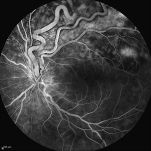
Wide field FA image of the right eye of a 25-year-old woman with retinal angiomatosis (Von Hippel-Lindau). Fundus of the right eye seemed to be normal in ophthalmoscopy.
Photographer: Soodabeh Fooladin, Negah Eye Center, Tehran
Imaging device: Heidelberg Spectralis
Condition/keywords: exudative retinal detachment, retinal angiomatous proliferation (RAP), Von Hippel-Lindau
-
 Von Hippel-Lindau
Oct 25 2012 by Gabriela Lopezcarasa Hernandez, MD
22 -year-old female with serous and tractional retinal detachment secondary to Von Hippel-Lindau dissease
Photographer: Araceli Rojas, Hospital Angeles Lomas Mexico
Imaging device: Zeiss FF4
Condition/keywords: serous retinal detachment, Von Hippel-Lindau
-
 Von Hippel Lindau
Mar 13 2013 by Carl C. Awh, MD, FASRS
TRD under silicone oil due to hemangiomas due to Von Hippel Lindau.
Photographer: Alecia Camp, CRA - Tennessee Retina - Nashville, TN
Condition/keywords: silicone oil, tractional retinal detachment, Von Hippel-Lindau
-
 Von Hippel-Lindau
Oct 13 2012 by Hamid Ahmadieh, MD
Wide field FA image of the left eye of a 25-year-old woman with exudative retinal detachment secondary to retinal angiomatosis (Von Hippel-Lindau).
Photographer: Soodabeh Fooladin, Negah Eye Center, Tehran
Imaging device: Heidelberg Spectralis
Condition/keywords: exudative retinal detachment, retinal angiomatous proliferation (RAP), Von Hippel-Lindau
-
 Von Hippel-Lindau
Oct 13 2012 by Hamid Ahmadieh, MD
Late FA image of the left eye of a 25-year-old woman with exudative retinal detachment secondary to retinal angiomatosis (Von Hippel-Lindau).
Photographer: Soodabeh Fooladin, Negah Eye Center, Tehran
Imaging device: Heidelberg Spectralis
Condition/keywords: exudative retinal detachment, retinal angiomatous proliferation (RAP)
-
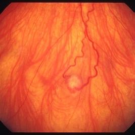
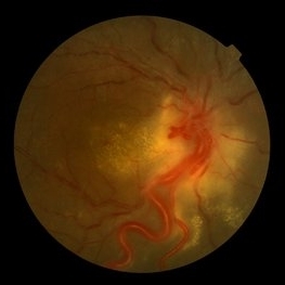
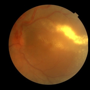
---thumb.jpg/image-square;max$300,300.ImageHandler) Capillay Hemangioma Von Hippel
Capillay Hemangioma Von Hippel
Mar 29 2013 by Henry J. Kaplan, MD
Capillary hemangioma in the temporal macular area, notice the dilated feeder vessels which are the inferior arcade vessles.
Condition/keywords: retinal capillary hemangioma, Von Hippel-Lindau
-
---thumb.jpg/image-square;max$300,300.ImageHandler) Retinal capillary hemangioma 4 image 2
Retinal capillary hemangioma 4 image 2
Jan 11 2013 by Alex P. Hunyor, MD
Retinal capillary haemangioma, left eye, in a young female with von Hippel-Lindau disease. Color image 2 showing the haemangioma with surrounding exudative detachment and lipid exudate.
Condition/keywords: retinal capillary hemangioma, Von Hippel-Lindau
-
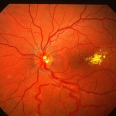
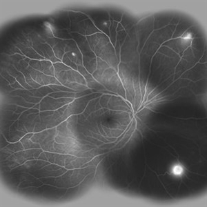
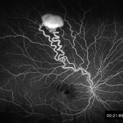 Retinal Hemangioblastoma Mid Phase FA
May 15 2013 by Robert T. Wendel, MD
20-year-old male. Genetic hx not yet defined.
Condition/keywords: Von Hippel-Lindau
 Loading…