Search results (402 results)
-
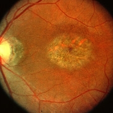
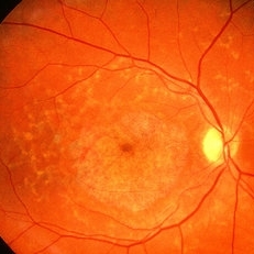 Fundus Flavimaculatus
May 2 2013 by Henry J. Kaplan, MD
Yellow pisciform flecks at the level of RPE accompanied by Bull's eye, Right eye; #1.
Condition/keywords: bull's eye maculopathy, fundus flavimaculatus, Stargardt disease
-
 Stargardt macular dystrophy slide 1
Oct 22 2012 by Ronald C. Gentile, MD
16-year-boy with difficulty in school seeing the black board. The macula area of the right eye had areas with a beaten bronze appearance and atrophy. Small pisci-form flecks can be seen surrounding the fovea.
Photographer: The New York Eye & Ear Infirmary Department of Medical Imaging
Condition/keywords: small pisci-form flecks, Stargardt disease
-
 Late Stage Stargardt's Disease
Mar 13 2013 by Hamid Ahmadieh, MD
Autofluorescence imaging of the left eye of a 46-year-old man with decreased VA due to advanced Stargardt's disease.
Photographer: Nayereh Hadipoor, Negah Eye Center, Tehran
Imaging device: Heidelberg Spectralis
Condition/keywords: autofluorescence imaging, Stargardt disease
-
 Stargardts Disease in FAF
Sep 14 2012 by Michael P. Kelly, FOPS
This is a scanning laser ophthalmoscopic FAF image of a patient with Stargardts Disease captured with a Heidelberg Spectralis imaging unit. Note, besides the obvious hyper-autofluorescent areas centrally, the much smaller, and in greater number, pinpoints of hyper-autofluorescence extending from the vascular arcades into the mid-periphery.
Photographer: Michael P. Kelly, FOPS, Director, Duke Eye Center Labs, Duke Universtiy Hospital
Imaging device: Heidelberg Spectralis
Condition/keywords: fundus autofluorescence (FAF), Stargardt disease
-
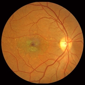
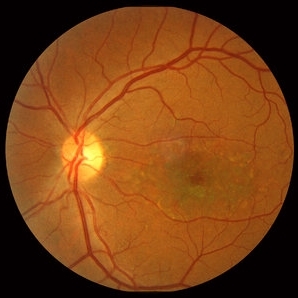
 Stargardt's Disease
May 2 2013 by Henry J. Kaplan, MD
Fundus photograph of the right eye in a patient with Stargardt's disease shows typical bull's eye maculopathy; #1.
Condition/keywords: bull's eye maculopathy, Stargardt disease
-
 Stargardt's Disease
Jun 6 2013 by Sharon Fekrat, MD FACS FASRS
Fundus photograph of left eye with Stargardt's Disease.
Photographer: Duke Eye Imaging, Duke University Eye Center, Durham, NC
Condition/keywords: Stargardt disease
-
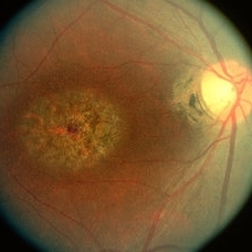
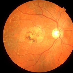
 Stargardt Disease
Feb 28 2013 by Theodore Leng, MD, MS, FASRS
Fundus photograph of a 63-year-old man with Stargardt disease. Multiple pisciform flecks are visible.
Imaging device: Zeiss FF450
Condition/keywords: flecks, Stargardt disease
-
 Stargardt macular dystrophy slide 1
Oct 22 2012 by Ronald C. Gentile, MD
45-year-old man with progressive central vision loss since the age of 10. Small flecks can be seen surrounding the central area of atrophy.
Photographer: The New York Eye & Ear Infirmary Department of Medical Imaging
Condition/keywords: Stargardt disease, vision loss
-
 Stargardt's disease
May 2 2013 by Henry J. Kaplan, MD
The same patient , left eye #2
Condition/keywords: bull's eye maculopathy, Stargardt disease
-
 Fundus Flavimaculatus
May 2 2013 by Henry J. Kaplan, MD
Widespread pisciform flecks in the fundus accompanied by macular beaten bronze lesion; Left eye; #2.
Condition/keywords: fundus flavimaculatus, Stargardt disease
-
 Stargardt's Disease
Oct 8 2012 by Susanna S. Park, MD, PhD
Fluorescein angiogram of a 12-year-old girl with progressive loss of vision in both eyes.
Photographer: Ellen Redenbo, University of California Davis Eye Center
Condition/keywords: bull's eye maculopathy, dark area on retina and choroid, Stargardt disease
-
 Stargardt macular dystrophy slide 2
Oct 22 2012 by Ronald C. Gentile, MD
The central areas of atrophy were symmetric in both eyes with increased visibility of the choroidal vessels within the atrophic macula.
Photographer: The New York Eye & Ear Infirmary Department of Medical Imaging
Condition/keywords: Stargardt disease
-
 Stargardt's Disease
Oct 18 2012 by Raj K. Maturi, MD
Photographer: Stephanie Morrow
Imaging device: HRA
Condition/keywords: red-free, Stargardt disease
-
 Stargardt's Disease
Oct 8 2012 by Susanna S. Park, MD, PhD
Fundus photograph of a 12-year-old girl with progressive loss of vision in both eyes.
Photographer: Ellen Redenbo, University of California Davis Eye Center
Condition/keywords: bull's eye maculopathy, dark area on retina and choroid, Stargardt disease
-
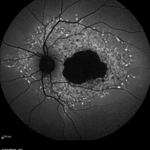
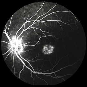
Stargardts Disease in Fundus Autofluorescence
Sep 12 2012 by Michael P. Kelly, FOPS
Fundus autofluorescence of a patient with Stargardts disease. Note the central area of hypo-autofluorescence indicating atrophy surrounded by smaller areas of hyper-autofluorescence. Note also the much smaller, and in greater number, pinpoints of hyper-autofluorescence extending from the vascular arcades into the mid-periphery.
Photographer: Michael P. Kelly, FOPS, Director, Duke Eye Labs, Duke University Hospital, Duke Eye Center
Imaging device: Heidelberg Spectralis
Condition/keywords: fundus autofluorescence (FAF), Stargardt disease
-
 Stargardt macular dystrophy slide 2
Oct 22 2012 by Ronald C. Gentile, MD
Fundus examination of the left eye had similar findings with centrally atrophic macula area with surrounding flecks.
Photographer: The New York Eye & Ear Infirmary Department of Medical Imaging
Condition/keywords: Stargardt disease
-
 Pattern Dystrophy slide 1
Oct 22 2012 by Ronald C. Gentile, MD
Asymptomatic middle-age man with normal vision and a multifocal pattern dystrophy. The pattern dystrophy simulates Stargardt disease/fundus flavimaculatus with irregular yellow-white flecks scattered throughout the posterior pole. Some lesions extend beyond the retinal vascular arcades.
Photographer: The New York Eye & Ear Infirmary Department of Medical Imaging
Condition/keywords: pattern macular dystrophy
-
 Bull's Eye Maculopathy Stargardt VS Cornea Dystrophy
Jul 31 2013 by From the Collections of Thomas M. Aaberg, MD and Thomas M. Aaberg Jr., MD
Bull's eye maculopathy stargardt vs cornea dystrophy.
Condition/keywords: bull's eye maculopathy, cornea dystrophy, corneal dystrophy, Stargardt disease
-
 Stargardt's Disease
Oct 18 2012 by Raj K. Maturi, MD
Photographer: Stephanie Morrow
Imaging device: HRA
Condition/keywords: Stargardt disease
-
 Late Stage Stargardt's Disease
Mar 13 2013 by Hamid Ahmadieh, MD
Color fundus photograph of the right eye of a 46-year-old man with decreased VA due to advanced Stargardt's disease.
Photographer: Nayereh Hadipoor, Negah Eye Center, Tehran
Imaging device: Heidelberg Spectralis
Condition/keywords: Stargardt disease
-
 Stargardt's Disease
Jun 6 2013 by Sharon Fekrat, MD FACS FASRS
Fluorescein angiogram of the left eye of a patient with Stargardt's Disease. Note the characteristic dark choroid.
Photographer: Duke Eye Imaging, Duke University Eye Center, Durham, NC
Condition/keywords: Stargardt disease
-
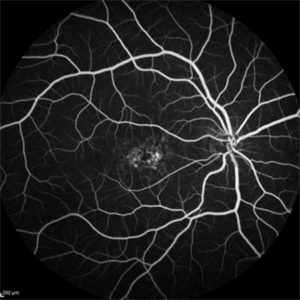
 Stargardt disease case 3 FA LE
Jan 11 2013 by Alex P. Hunyor, MD
Stargardt disease - left eye. Atrophic maculopathy without flecks. Fluorescein angiogram - note silent choroid.
Condition/keywords: Stargardt disease
-
 Stargardt's Disease - Dark Choroid
Dec 1 2016 by Courtney Crawford, MD, FACS
24-year-old male with progressive decreased vision to level of 20/200 OU.
Photographer: Kristen Dunn, North Texas Retina Consultants
Condition/keywords: choroid, Stargardt disease
-
 Stargardt's Disease
Oct 18 2012 by Raj K. Maturi, MD
Photographer: Stephanie Morrow
Imaging device: HRA
Condition/keywords: Stargardt disease
-
 Stargardt Disease
Feb 28 2013 by Theodore Leng, MD, MS, FASRS
Fundus photograph of a 63-year-old man with Stargardt disease. Multiple pisciform flecks are visible.
Imaging device: Zeiss FF450
Condition/keywords: flecks, Stargardt disease
 Loading…