Search results (550 results)
-
 Myelinated Nerve Fibers
Sep 17 2012 by Michael P. Kelly, FOPS
Retinal fundus photograph of a macular hole.
Photographer: Michael P. Kelly, FOPS Director, Duke Eye Labs, Duke University Hospital, Duke Eye Center
Imaging device: Topcon
Condition/keywords: macular hole, myelinated nerve fibers
-
 Traumatic Macular Hole with Retinal Detachment and PVR - montage
Sep 27 2012 by Pauline T Merrill, MD, FASRS
Fundus photo montage of a 13-year-old boy s/p soccer ball injury 1 month previously.
Photographer: Karen Parque, Illinois Retina Associates, Chicago, IL
Condition/keywords: proliferative vitreoretinopathy (PVR), traumatic macular hole
-
 Traumatic Macular Hole with Retinal Detachment and PVR
Sep 27 2012 by Pauline T Merrill, MD, FASRS
Fundus photo of a 13-year-old boy s/p soccer ball injury 1 month previously. In addition to full-thickness macular hole and total retinal detachment with grade C PVR, note pigment granules visible in vitreous over optic nerve.
Photographer: Karen Parque, Illinois Retina Associates, Chicago, IL
Condition/keywords: proliferative vitreoretinopathy (PVR), traumatic macular hole
-
 Traumatic Macular Hole
Aug 23 2012 by Gabriela Lopezcarasa Hernandez, MD
12-year-old boy with blunt trauma in right eye and central scotoma.
Photographer: Gabriela Lopezcarasa Hernandez, Hospital Angeles Lomas
Imaging device: ZEISS F4
Condition/keywords: blunt trauma, central scotoma, macular hole
-
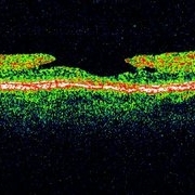
 Stage 1 Macular Hole
Jul 4 2012 by John T. Thompson, MD
Stage 1 macular hole with vitreomacular adhesion
Imaging device: Heidelberg Spectralis
Condition/keywords: macular hole, vitreomacular adhesion, vitreomacular traction (VMT)
-
 Traumatic Macular Hole
Aug 23 2012 by Gerardo Garcia-Aguirre, MD
Fundus photograph of a large macular hole with an area of pigment migration secondary to blunt trauma.
Photographer: Noemí Hernández, Asociación para Evitar la Ceguera en México
Imaging device: Zeiss FF4
Condition/keywords: deformity, macular hole
-
 Macular Hole, Autofluorescence
Sep 14 2012 by Michael P. Kelly, FOPS
Fundus autofluorescence (FAF) of a macular hole captured using a Heidelberg Spectralis.
Photographer: Michael P. Kelly, FOPS, Director, Duke Eye Cneter Labs, Duke Universty Hospital
Imaging device: Heidelberg Spectralis
Condition/keywords: fundus autofluorescence (FAF), macular hole
-
 Lamellar Macular Hole
Sep 18 2012 by Michael P. Kelly, FOPS
Photographer: Michael P. Kelly, FOPS Director, Duke Eye Center Labs, Duke University Hospital
Imaging device: Zeiss Cirrus
Condition/keywords: lamellar macular hole
-
 Chronic Macular Hole
Sep 2 2012 by Hyung-Woo Kwak, MD
A large hole with rolled everted edges, adjacent cystoid intraretinal spaces, a shallow rim of subretinal fluids.
Imaging device: Zeiss F450 plus
Condition/keywords: macular hole
-
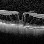 Myopic Macular Schisis with Lamellar Macular Hole
May 26 2014 by John T. Thompson, MD
Spectral domain OCT of patient with high myopia and myopic macular schisis resulting in lamellar macular hole.
Condition/keywords: lamellar macular hole, myopic macular schisis
-
 Traumatic macular hole
Dec 19 2012 by Eric A. Postel, MD
Color fundus photograph of a young male with a traumatic macular hole
Condition/keywords: blunt trauma, macular hole
-
 Stage 2 Macular Hole
-
 Stage 3 Macular Hole
-
 Macular Hole
Sep 27 2012 by Jeffrey G. Gross, MD, FASRS
Macular hole s/p 360 degree laser to fluid cuff.
Condition/keywords: macular hole, subretinal fluid
-
 Macular Hole
Sep 18 2012 by Michael P. Kelly, FOPS
Retinal fundus photograph of a macular hole.
Photographer: Michael P. Kelly, FOPS Director, Duke Eye Labs, Duke University Hospital, Duke Eye Center
Imaging device: Topcon
Condition/keywords: macular hole
-
 Temporal conus of optic nerve
Feb 19 2013 by From the Collections of Thomas M. Aaberg, MD and Thomas M. Aaberg Jr., MD
Associated with a macular hole and subretinal new vessel membrane.
Condition/keywords: conus of optic nerve, subretinal new vessel membrane
-
 Macular Hole
Sep 20 2012 by Jeffrey G. Gross, MD, FASRS
Macular hole, Stage 3, pre-op 20/400
Condition/keywords: macular hole, pre-op
-
 Macular Hole
Sep 27 2012 by Jeffrey G. Gross, MD, FASRS
Macular hole, 20/100.
Condition/keywords: 20/100, macular hole
-
 Full-thickness Macular Hole
Aug 28 2012 by Sharon Fekrat, MD FACS FASRS
65 year old woman with a recurrent full thickness macular hole following previous 20 g pars plana vitrectomy in the right eye as well as an iatrogenic retinal hole in the papillomacular bundle. Both retinal defects are captured here in this Zeiss Stratus OCT image.
Photographer: Michael P. Kelly, FOPS Director, Duke Eye Labs, Duke University Eye Center, Durham, NC
Imaging device: Zeiss Cirrus
Condition/keywords: retinal break
-
 Macular Hole
Sep 14 2012 by Michael P. Kelly, FOPS
Photographer: Michael P. Kelly, FOPS, Director, Duke Eye Center Labs, Duke University Hospital
Condition/keywords: macular hole
-
 Macular Hole Post-Op
Sep 27 2012 by Jeffrey G. Gross, MD, FASRS
Macular hole post-op, hole closed 20/40.
Condition/keywords: 20/40, hole closed, macular hole, post-op
-
 Macular Hole
Sep 27 2012 by Jeffrey G. Gross, MD, FASRS
Macular hole magnified with cuff of SRF.
Condition/keywords: cuff, macular hole, subretinal fluid
-
 Macular Hole Stage 3
Sep 27 2012 by Jeffrey G. Gross, MD, FASRS
Macular hole stage 3 post op with gas bubble 20/60.
Condition/keywords: 20/60, gas bubble, macular hole, post-op
-
 Lamellar Macular Hole
Jul 3 2012 by Antonio Capone, MD
OCT of lamellar macular hole
Condition/keywords: lamellar macular hole
-
 One Week Post Blunt Trauma Macular HD OCT
Dec 31 2012 by Humberto Ruiz-Garcia, MD
One week after blunt trauma, closure of macular hole is seen, persistent thinning and subretinal fluid are observed. Vision is CF.
Photographer: Humberto Ruiz-Garcia
Imaging device: Cirrus HD OCT
Condition/keywords: blunt trauma, macular hole, optical coherence tomography (OCT)
 Loading…