Search results (40 results)
-
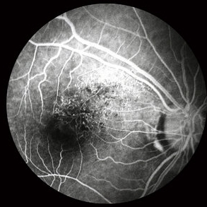
 Sickle SC Sea Fan FA Early Phase
Oct 8 2012 by Jeffrey G. Gross, MD, FASRS
Sickle SC sea fan, FA, early phase.
Condition/keywords: early phase, sea fan, sickle cell
-
 BRVO FA, Early Phase
Oct 1 2012 by Jeffrey G. Gross, MD, FASRS
BRVO-FA early phase.
Condition/keywords: branch retinal vein occlusion (BRVO), capillary nonperfusion, early phase, microaneurysms
-
 Macular Telangiectasia (FA Early Phase)
May 16 2014 by Avris Romario Diparaja Siahaan
FA (early phase) image of a 58-year-old-man with a macular telangiectasia condition on his left eye.
Photographer: Avris Romario Diparaja Siahaan, Klinik Mata Nusantara
Imaging device: Topcon TRC 50 DX Type IA
Condition/keywords: FA early phase, macular telangiectasia
-
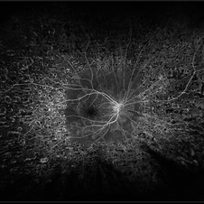
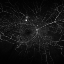
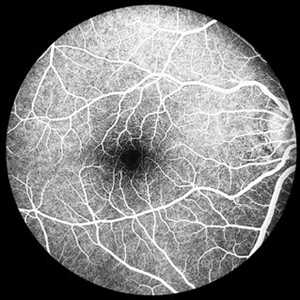 Normal FA Early Phase
-
 Coats' Disease FA
Apr 27 2018 by Brenda Fallas
3-year-old boy with unilateral Coats' Disease FA photo.
Photographer: Brenda Fallas, Bascom Palmer Eye Institute, Miami, FL
Imaging device: Retcam III 130 degree lens
Condition/keywords: Coats' disease, FA early phase, fluorescein angiogram (FA), retinal telangiectasia
-
 Central Serous Chorioretinopathy with Smokestack FA Early Phase
Oct 8 2012 by Jeffrey G. Gross, MD, FASRS
CSCR with smokestack, FA, early phase.
Condition/keywords: central serous chorioretinopathy (CSCR), FA early phase, smokestack
-
 Sickle SC Sea Fan
Oct 8 2012 by Jeffrey G. Gross, MD, FASRS
Sickle SC sea fan, partial regression, FA early phase.
Condition/keywords: FA early phase, partial regression, sickle cell
-
 Retinal Arterio-Venous Malformations
Apr 7 2017 by Deepak Bhojwani, MS
Multimodal imaging of a 16-year-old boy with retinal arterio-venous malformations(AVM). He also had cerebral AVM's on MRI-contrast studies suggesting Wyburn-Mason syndrome.
Photographer: DEEPAK BHOJWANI, RAGHUDEEP EYE HOSPITAL, AHMEDABAD.
Imaging device: Zeiss VISUCAM
Condition/keywords: color fundus photograph, FA early phase, optical coherence tomography (OCT), Wyburn-Mason
-
 FA Early Phase Optic Disc Edema
Oct 1 2012 by Jeffrey G. Gross, MD, FASRS
FA early phase optic disc edema with blockage of hemorrhages in patient with subdural hematoma.
Condition/keywords: subdural hematoma
-
 Circumscribed Choroidal Hemangioma
Oct 20 2012 by Hyung-Woo Kwak, MD
Fundus and OCT examination showed an oval mass at the posterior pole with indistinct margins that blend with surrounding choroid. FA early phase showed hyperfluorescence.
-
 BRVO, FA, Hemorrhage, Diabetic
Mar 13 2014 by James B. Soque, CRA, OCT-C, COA, FOPS
51-year-old white male, diabetes, and with BRVO left eye, early phase 36 seconds. Flame heme from ON, showing microaneurysims, and fine capillary detail of this FA.
Photographer: James B Soque, CRA COA
Imaging device: Topcon TRC 50DX with MERGE software
Condition/keywords: branch retinal vein occlusion (BRVO), diabetes, FA early phase, microaneurysms
-
 Circumscribed Choroidal Hemangioma
Oct 20 2012 by Hyung-Woo Kwak, MD
Fundus and OCT examination showed an oval mass at the posterior pole with indistinct margins that blend with surrounding choroid. FA early phase showed hyperfluorescence.
-
 Circumscribed Choroidal Hemangioma
Oct 20 2012 by Hyung-Woo Kwak, MD
Fundus and OCT examination showed an oval mass at the posterior pole with indistinct margins that blend with surrounding choroid. FA early phase showed hyperfluorescence.
-
 Circumscribed Choroidal Hemangioma
Oct 20 2012 by Hyung-Woo Kwak, MD
Fundus and OCT examination showed an oval mass at the posterior pole with indistinct margins that blend with surrounding choroid. FA early phase showed hyperfluorescence.
-
 BRVO and VMT Vitreo Macular Traction, FA Early Phase
Apr 18 2013 by James B. Soque, CRA, OCT-C, COA, FOPS
Early FA photo, 50 degrees, mag 2X of 79-year old white female, VA sc 20/40, with BRVO OS, and VMT OS, diagnosed on exam and SD OCT. See accompanying FC and RF photos reveal BRVO IT OS, and SD OCT reveal VMT OS.
Photographer: James B. Soque, CRA, COA, Island Retina. Shirley, NY
Imaging device: Topcon TRC-50DX with MERGE software
Condition/keywords: branch retinal vein occlusion (BRVO), vitreomacular traction (VMT)
-
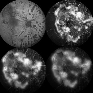 Proliferative Diabetic Retinopathy
May 28 2016 by Olivia Rainey
Fluorescein angiogram series of an 30-year-old male with proliferative diabetic retinopathy affecting his right eye. The patient presented with worsening neovascularization and scar tissue contracting in macula in the right eye. He experienced a decline in vision secondary to macula ischemia. Patient was seeing 20/400 and with PH 20/200 in the right eye and HM in the left eye.
Photographer: Olivia Rainey
Imaging device: Heidelberg Spectralis
Condition/keywords: diabetes, FA early phase, FA late phase, FA mid phase, fluorescein leakage, fundus autofluorescence (FAF), neovascularization (NV), proliferative diabetic retinopathy (PDR)
-
---thumb.jpg/image-square;max$300,300.ImageHandler) Pattern Dystrophy
Pattern Dystrophy
Aug 9 2013 by From the Collections of Thomas M. Aaberg, MD and Thomas M. Aaberg Jr., MD
Early FA of patient with pattern dystrophy.
Condition/keywords: FA early phase, pattern macular dystrophy
-
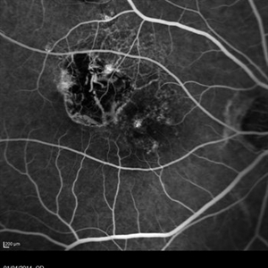 Polypoidal Choroidal Vasculopathy (FA Early Phase)
May 16 2014 by Avris Romario Diparaja Siahaan
FA image (early phase) a 66-year-old woman with a polypoidal choroidal vasculopathy (PCV) on her right eye. She had a injection (Avastin) for several times
Photographer: Avris Romario Diparaja Siahaan, Klinik Mata Nusantara
Imaging device: Heidelberg HRA + OCT Spectralis
Condition/keywords: FA early phase, polypoidal choroidal vasculopathy (PCV)
-
 Coats' disease early fluorescein angiogram of telangiectasia
Apr 3 2018 by Victor M Villegas, MD
6-year-old male with unilateral exudative retinopathy.
Photographer: Brenda Fallas
Imaging device: RetCam3
Condition/keywords: Coats' disease, FA early phase, fluorescein angiogram (FA), fluorescein leakage
-
 Choroidal Melanoma - Stable, Fluorescein Angiogram, Early Phase
Mar 13 2019 by James B. Soque, CRA, OCT-C, COA, FOPS
Early FA, right eye, with choroidal melanoma-stable, and a few tiny microaneurysms showing leakage in re-circulation phase.
Photographer: James Soque, CRA, OCT-C, FOPS
Imaging device: Topcon TRC-50DX with MERGE Eye Station software
Condition/keywords: FA early phase, fluorescein angiogram (FA), MERGE, microaneurysms
-
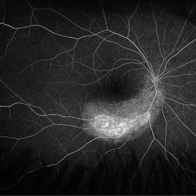
 Acute Multifocal Placoid Pigment Epitheliopathy
Sep 15 2014 by Thomas A. Ciulla, MD, MBA, FASRS
AMPPE in a 42-year-old woman. Early phase angiography show blockage of multiple focal lesions in the superior macula and peripapillary region of the right eye.
Photographer: Thomas Steele
Condition/keywords: acute multifocal placoid pigment epitheliopathy (AMPPE), FA early phase
-
 Von Hippel-Lindau (FA Early Phase)
May 17 2014 by Avris Romario Diparaja Siahaan
Fluorescein angiogram (early phase) Photograph of a 57-year-old woman with a Von Hippel-Lindau
Photographer: Avris Romario Diparaja Siahaan, Klinik Mata Nusantara
Imaging device: Topcon TRC 50 DX Type IA
Condition/keywords: FA early phase, Von Hippel-Lindau
-
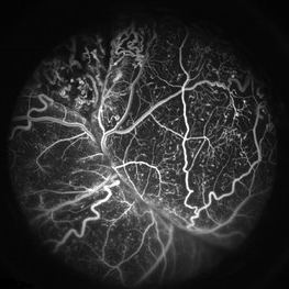
 Acute Zonal Occult Outer Retinopathy (AZOOR) FA, Fluorescein Angiography, Peripheral Vasculitis
Jan 19 2022 by James B. Soque, CRA, OCT-C, COA, FOPS
Acute Zonal Occult Outer Retinopathy (AZOOR). Peripheral Vasculitis OD. Fluorescein angiography showing vasculitis in the far right periphery 8-10 o'clock. 46-year-old white male, VA CC 20/16, 20/12.5, has had recurrent vasculitis for 11 years. No treatment.
Photographer: James Soque, CRA, OCT-C, COA, FOPS, Island Retina, Shirley, NY
Imaging device: Optos California
Condition/keywords: acute zonal occult outer retinopathy (AZOOR), FA early phase, fluorescein angiogram (FA), Peripheral Vasculitis, ultra-wide field imaging
-
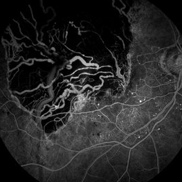
 Proliferative Diabetic Retinopathy
Nov 13 2019 by Olivia Rainey
Ultra-wide field fluorescein angiogram at 29 seconds of a 52-year-old male with proliferative diabetic retinopathy affecting his right eye. Patient is receiving Eylea intravitreal injections and has had panretinal photocoagulation in the past. Patient's vision tested 20/40 and with pinholes to 20/30.
Photographer: Olivia Rainey
Imaging device: Optos California
Condition/keywords: diabetes, diabetic macular edema, early phase, FA early phase, fluorescein angiogram (FA), intravitreal injection, ischemia, pan-retinal photocoagulation (PRP), proliferative diabetic retinopathy (PDR)
-
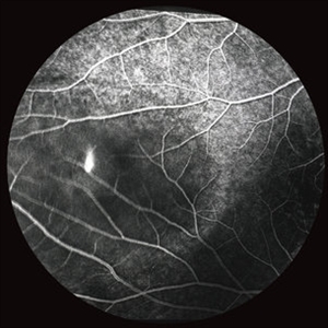
 PDR with Ischemia
Jul 7 2020 by Stephanie Burke
Early frame of a 45-year-old male with Type II diabetes.
Photographer: Stephanie Burke, CRA, OCT-C
Condition/keywords: FA early phase, ischemia, microaneurysms, neovascularization (NV), proliferative diabetic retinopathy (PDR), ultra-wide field imaging, venous beading
 Loading…