Search results (120 results)
-
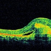
 Plaquenil Toxicity
Apr 30 2013 by Theodore Leng, MD, MS, FASRS
SD-OCT scan from a 44-year-old woman with bilateral plaquenil toxicity. There is damage visible in the outer retina in a perifoveal distribution.
Condition/keywords: hydroxychloroquine toxicity, plaquenil toxicity
-
 Plaquenil Toxicity
Apr 30 2013 by Theodore Leng, MD, MS, FASRS
SD-OCT scan from a 44-year-old woman with bilateral plaquenil toxicity. There is damage visible in the outer retina in a perifoveal distribution.
Condition/keywords: hydroxychloroquine toxicity, plaquenil toxicity
-
 Macular Pseudohole - OCT
Jan 11 2013 by Gerardo Garcia-Aguirre, MD
OCT scan showing a hyperreflective line that is partially separated from the retina in the fovea and temporal macula, corresponding to an epiretinal membrane. Note the discontinuity of the line just above the fovea, which clinically corresponds to the pseudohole.
Photographer: Gerardo Garcia-Aguirre, MD
Imaging device: Topcon 3DOCT 1000
Condition/keywords: epiretinal membrane (ERM), macular pseudohole
-
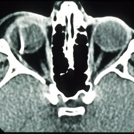
 Metallic intraocular foreign body - CT scan
Dec 25 2012 by Alex P. Hunyor, MD
Axial CT scan showing right metallic intraocular foreign body.
Imaging device: CT scan
Condition/keywords: intraocular foreign body
-
 Optic Disc Coloboma
Apr 25 2017 by Nimrod Dar
9 year-old patient, noticed a gradual deterioration in her visual acuity at her LE (6/15). On her examination, a double optic disc can be seen. OCT scan revealed an intra retinal fluid and macular schisis.
Photographer: Nimrod Dr, MD
Condition/keywords: coloboma of the optic nerve
-
 Solar Retinopathy
Mar 1 2013 by Theodore Leng, MD, MS, FASRS
OCT scan of a 15-year-old male with solar retinopathy after staring directly at a solar eclipse. Note the hyperreflectivity in the foveola.
Photographer: Erich Hagan
Imaging device: Heidelberg
Condition/keywords: solar retinopathy
-
 Severe vitreomacular traction
Dec 23 2012 by Alex P. Hunyor, MD
OCT scan of the right eye of an 82-year-old male with 20/40 vision despite severe vitreomacular traction (VMT).
Condition/keywords: vitreomacular traction (VMT)
-
 Hypertensive Choroidopathy - Right Eye
Dec 21 2016 by Maciej Czepita
Fundus photograph and SD-OCT scan as well as fundus autofluorescence image (FAF) of the right eye of a 70-year-old woman with hypertensive choroidopathy. In the fundus image numerous Elschnig's spots are visible. Note the Hollenhorst plaque in the superior temporal artery. In the SD-OCT scan (green line on the fundus image) the RPE layer is uneven. Numerous hypo and hyperautofluorescent patches can be seen in the fundus autofluorescence image.
Photographer: Maciej Czepita, M.D., Ph.D., Pomeranian Medical University, Szczecin, Poland
Imaging device: Heidelberg Spectralis HRA+OCT
Condition/keywords: hypertensive choroidopathy
-
 Intraocular Foreign Body, Metallic, CT Scan Orbits
Oct 1 2012 by Jeffrey G. Gross, MD, FASRS
IOFB, metallic, CT scan orbits.
Condition/keywords: CT scan, intraocular foreign body, orbits
-
 Choroidal Granuloma Secondary to Tuberculosis
Mar 14 2013 by Eduardo Torres-Porras, MD
OCT scan through the granuloma shows attachment of the retinal pigment epithelial-choriocapillaris layer and the neurosensory retina over the granuloma (“contact” sign), inflammatory retinal infiltrate in the deeper retinal layers and subretinal fluid.
Photographer: Eduardo Torres Porras, Laser y ultrasonido ocular de Puebla
Imaging device: Cirrus
Condition/keywords: optical coherence tomography (OCT), tubercular choroidal granuloma
-
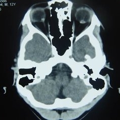 Orbital CT Scan in Optic Nerve Drusen
Mar 27 2013 by Henry J. Kaplan, MD
Axial CT scan of orbit demonstrates high density spot on optic nerve head on both sides #4.
Condition/keywords: drusen of optic disc, optic disc drusen, optic nerve drusen
-
---thumb.jpg/image-square;max$300,300.ImageHandler) Optic Disc Drusen
Optic Disc Drusen
Mar 27 2013 by Henry J. Kaplan, MD
An 11-year-old boy presented with transient blurry vision, VA:20/20 bilaterally. He has pseudo optic disc swelling only in the right eye ; margin is blurred but the pattern of vessels are normal and there are some yellowish deposits on the superior of ON #1. AF, B-scan, CT scan, and VF are uploaded in the following slides.
Condition/keywords: drusen of optic disc, optic disc drusen, optic nerve drusen
-
 Choroidal Granuloma Secondary to Tuberculosis
Mar 14 2013 by Eduardo Torres-Porras, MD
OCT scan through the granuloma shows attachment of the retinal pigment epithelial-choriocapillaris layer and the neurosensory retina over the granuloma (“contact” sign), inflammatory retinal infiltrate in the deeper retinal layers and subretinal fluid.
Photographer: Eduardo Torres Porras
Imaging device: Cirrus
Condition/keywords: optical coherence tomography (OCT), tubercular choroidal granuloma
-
 CME-OCT
Apr 28 2015 by Neha Goel, MS DNB FRCS (Glasg)
Horizontal spectral-domain OCT scan through the right macula.
Photographer: Neha Goel
Imaging device: RTVue
Condition/keywords: cystoid macular edema (CME)
-
 Metallic intraocular foreign body - colour composite image
Dec 25 2012 by Alex P. Hunyor, MD
4-up colour images: 1. anterior segment photo, 2. normal appearing posterior pole image, 3. temporal view showing streak of vitreous haemorrhage, and 4. superotemporal view of impact site where metallic IOFB disrupted superotemporal branch retinal vein leading to vitreous haemorrhage. The IOFB is located in the inferior vitreous cavity, obscured by haemorrhage (see associated CT scan).
Condition/keywords: intraocular foreign body
-
---thumb.jpg/image-square;max$300,300.ImageHandler) Floaters
Floaters
Oct 9 2013 by Maurice F. Rabb
KR is a 25 year old white female who presented with a one month history of floaters OD. Past ocular and systemic history were unremarkable. On clinical examination, the visual acuity was 20/20 OU, and the anterior segments were normal. There was a very mild degree of vitreous cell OD, though no cystoid macular edema nor vasculitis. A lobulated white mass was noted overlying the vitreous base inferotemporally OD (thickness 3.3mm). There was no calcification, though prominent cysts were noted on the surface of the lesion. A fluorescein angiogram, echogram, and CT scan were obtained, along with a thorough systemic evaluation.
Condition/keywords: floaters
-
 Mild VMT plus drusen
Dec 23 2012 by Alex P. Hunyor, MD
Fundus photograph of the right eye of a 62-year-old female referred with drusen in the right macula. The accompanying OCT scans document the evolution of posterior vitreous separation, with mild vitreomacular traction (VMT) which then spontaneously releases.
Condition/keywords: vitreomacular traction (VMT)
-
---thumb.jpg/image-square;max$300,300.ImageHandler) Floaters
Floaters
Oct 9 2013 by Maurice F. Rabb
KR is a 25 year old white female who presented with a one month history of floaters OD. Past ocular and systemic history were unremarkable. On clinical examination, the visual acuity was 20/20 OU, and the anterior segments were normal. There was a very mild degree of vitreous cell OD, though no cystoid macular edema nor vasculitis. A lobulated white mass was noted overlying the vitreous base inferotemporally OD (thickness 3.3mm). There was no calcification, though prominent cysts were noted on the surface of the lesion. A fluorescein angiogram, echogram, and CT scan were obtained, along with a thorough systemic evaluation.
Condition/keywords: floaters
-
 Evolution of VMT - OCT 2 at 6 months
Dec 23 2012 by Alex P. Hunyor, MD
OCT scan 6 months later, showing further separation of posterior cortical vitreous with early VMT.
Condition/keywords: vitreomacular traction (VMT)
-
 Macular Disciform Scar
Jun 8 2015 by ARIEL WANG
Fundus photograph and OCT scan of an 86-year-old man with long-standing type I diabetic proliferative retinopathy.
Photographer: Suber Huang, Retina Center of Ohio
Imaging device: Heidelberg Spectralis
Condition/keywords: central disciform scar
-
 Vitreopapillary traction - OCT
Dec 25 2012 by Alex P. Hunyor, MD
High definition raster OCT scan through optic nerve showing vitreopapillary traction with peripapillary oedema.
Imaging device: Zeiss Cirrus OCT
Condition/keywords: vitreopapillary traction
-
 Angioid Streaks With CNVM OCT RE
Jun 17 2014 by Neha Goel, MS DNB FRCS (Glasg)
Horizontal OCT scan through the right macula.
Photographer: Neha Goel
Imaging device: RTVue
Condition/keywords: angioid streaks, choroidal neovascularization (CNV)
-
 Solar Retinopathy
Mar 1 2013 by Theodore Leng, MD, MS, FASRS
OCT scan of a 15-year-old male with solar retinopathy after staring directly at a solar eclipse. Note the hyperreflectivity in the foveola.
Photographer: Erich Hagan
Imaging device: Heidelberg
Condition/keywords: solar retinopathy
-
 IOFB-Endophthalmitis Slide 2
Oct 22 2012 by Ronald C. Gentile, MD
Anterior segment examination revealed a self sealing peripheral corneal wound at the 7:30 position with iris defect. There was a small hypopyon layering in the inferior angle. CT scan revealed a small intra-ocular foreign body.
Photographer: The New York Eye & Ear Infirmary Department of Medical Imaging
Condition/keywords: intraocular foreign body
-
---thumb.jpg/image-square;max$300,300.ImageHandler) Floaters
Floaters
Oct 9 2013 by Maurice F. Rabb
KR is a 25 year old white female who presented with a one month history of floaters OD. Past ocular and systemic history were unremarkable. On clinical examination, the visual acuity was 20/20 OU, and the anterior segments were normal. There was a very mild degree of vitreous cell OD, though no cystoid macular edema nor vasculitis. A lobulated white mass was noted overlying the vitreous base inferotemporally OD (thickness 3.3mm). There was no calcification, though prominent cysts were noted on the surface of the lesion. A fluorescein angiogram, echogram, and CT scan were obtained, along with a thorough systemic evaluation.
Condition/keywords: floaters
 Loading…