Search results (92 results)
-
 Congenital Hypertrophy of the Retinal Pigment Epithelium (CHRPE)
Mar 1 2014 by Homayoun Tabandeh, MD, FASRS
Congenital hypertrophy of the retinal pigment epithelium (CHRPE).
Condition/keywords: congenital hypertrophy of the retinal pigment epithelium (CHRPE)
-
 Bear Tracks
Dec 31 2012 by Raj K. Maturi, MD
Photographer: Tom Steele, CRA Midwest Eye Institute Indianapolis, Indiana
Imaging device: Topcon 50ex 50 degree field
Condition/keywords: bear tracks, benign pigmented lesions, congenital hypertrophy of the retinal pigment epithelium (CHRPE), OD
-
 Operculated Hole and CHRPE
Jan 16 2018 by Carolyn Daley
58-year-old woman with an operculated hole and CHRPE in the right eye. Patient is asymptomatic so no treatment was recommended at this time.
Photographer: Carolyn Daley
Imaging device: Optos ultra wide field image
Condition/keywords: congenital hypertrophy of the retinal pigment epithelium (CHRPE), operculated retinal hole, Optos, ultra-wide field imaging
-
 Congenital Hypertrophy of the Retinal Pigment Epithelium (CHRPE)
Mar 1 2014 by Homayoun Tabandeh, MD, FASRS
Congenital hypertrophy of the retinal pigment epithelium (CHRPE).
Condition/keywords: congenital hypertrophy of the retinal pigment epithelium (CHRPE)
-
 CHRPE - grouped pigmentation
Jan 11 2013 by Alex P. Hunyor, MD
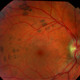
Congenital grouped pigmentation of the RPE ("bear tracks").
Condition/keywords: bear tracks, congenital hypertrophy of the retinal pigment epithelium (CHRPE)
-
---thumb.jpg/image-square;max$300,300.ImageHandler) Macular CHRPE
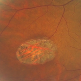
Macular CHRPE
Aug 11 2013 by Eric M. Shrier, DO
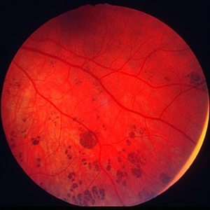
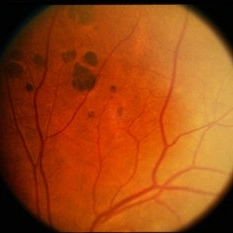
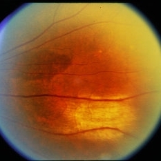
This a color fundus photograph of a 74-year-old black male with longstanding poor vision os, 20/200. He exhibits mild NPDR additionally.
Photographer: Christopher Bunce
Condition/keywords: congenital hypertrophy of the retinal pigment epithelium (CHRPE)
-
 Bear Tracks / CHRPE / Myelinated NFL
Jul 12 2014 by David Callanan, MD
58-year-old female, bear tracks / CHRPE / myelinated NFL.
Condition/keywords: bear tracks, congenital hypertrophy of the retinal pigment epithelium (CHRPE), myelinated nerve fibers
-
---thumb.jpg/image-square;max$300,300.ImageHandler) Congenital RPE Hypertrophy
Congenital RPE Hypertrophy
Aug 8 2013 by From the Collections of Thomas M. Aaberg, MD and Thomas M. Aaberg Jr., MD
Well - demarcated CHRPE inferonasal to the optic disc of right eye.
Condition/keywords: retinal pigment epithelium (RPE) hypertrophy
-
 Congenital Hypertrophy of the Retinal Pigment Epithelium (CHRPE)
Jul 14 2013 by Jason S. Calhoun
Fundus photo shows congenital hypertrophy of the retinal pigment epithelium (CHRPE), superior, nasally in the left eye.
Photographer: Jason S. Calhoun, Department of Ophthalmology, Mayo Clinic Jacksonville, Florida
Imaging device: TOPCON TRC 50-EX
Condition/keywords: congenital hypertrophy of the retinal pigment epithelium (CHRPE)
-
 CHRPE
CHRPE
Dec 13 2018 by Jason Griffith
50-year-old male being followed for typical CHRPE presentation.
Photographer: Jason Griffith, Tennessee Retina, Nashville, TN
Imaging device: Clarus 500
Condition/keywords: congenital hypertrophy of the retinal pigment epithelium (CHRPE)
-
 Bear Tracks / CHRPE / Myelinated NFL
Jul 12 2014 by David Callanan, MD
58-year-old female, bear tracks / CHRPE / myelinated NFL.
Condition/keywords: bear tracks, congenital hypertrophy of the retinal pigment epithelium (CHRPE), myelinated nerve fibers
-
 Bear Tracks / CHRPE / Myelinated NFL
Jul 12 2014 by David Callanan, MD
58-year-old female, bear tracks / CHRPE / myelinated NFL.
Condition/keywords: bear tracks, congenital hypertrophy of the retinal pigment epithelium (CHRPE), myelinated nerve fibers
-
 Congenital Hypertrophy of the Retinal Pigment Epithelium (CHRPE)
Aug 24 2012 by Andrew N. Antoszyk, MD FASRS
CHRPE lesion (black pigmented lesion) located along superior temporal arcade of left eye
Photographer: Lorainne Clark, Charlotte Eye Ear Nose and Throat Associates
-
 Adenocarcinoma Arising from CHRPE
Sep 17 2015 by Marc C. Peden, MD
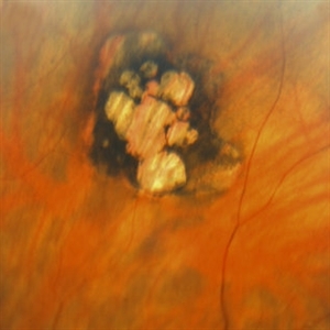
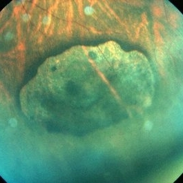
49-year-old female referred for presumed ocular melanoma. On examination was noted to have darkly pigmented lesion in the temporal retina of left eye. Lesion had characteristic scalloped edges with central lacunae, however, on ultrasonography was noted to have 1.8mm of elevation with high internal reflectivity. IVFA shows absence of dual circulation with areas of window defect. Findings were consistent with those described by Shields et al., in their April 2001 article in Archives of Ophthalmology.
Photographer: Janet Traynom
Imaging device: Optos P200MA
Condition/keywords: adenocarcinoma arising from CHRPE
-
 Gardner Syndrome
Dec 12 2018 by John S. King, MD
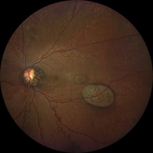
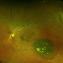
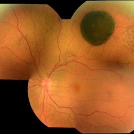
66-year-old white male with Gardner Syndrome (colon resection in 1991), who has two children with Gardner Syndrome, presented to Dr. Zocchi with an RD in the fellow eye that was successfully repaired with a pneumatic retinopexy. Currently 20/20 OU with IOP of 7 OD and 14 OS; no RAPD; PCIOL OU. Dr. Zocchi got oral permission by the patient to have these put into the Retina Image Bank. Although the CHRPE like lesions (2 OD) are not bilateral, we both think these lesions represent "retinal pigment epithelial hamartomas associated with familial adenomatous polyposis (RPEH-FAP)" as Shields described in their Intraocular Tumors book. One lesion is located superiorly and is pigmented with depigmented margins; the temporal lesion is atrophic with minimal remaining pigment hypertrophy.
Photographer: Karin Aletter
Imaging device: Optos CA
Condition/keywords: Gardner Syndrome, RPEH-FAP
-
 Congenital Hypertrophy of the Retinal Pigment Epithelium Wide Field Optomap
Sep 24 2019 by Sophia El Hamichi, MD
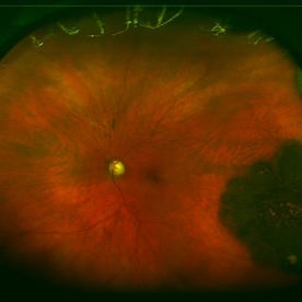
A 52-year-old female followed for 2 temporal lesions of CHRPE OD and white without pressure.
Photographer: Sophia El Hamichi,MD, Murray Ocular Oncology and Retina, Miami
Condition/keywords: congenital hypertrophy of the retinal pigment epithelium (CHRPE), Optomap, ultra-wide field imaging, white without pressure
-
 CHRPE
CHRPE
Oct 8 2019 by DIEGO TOLENTINO
CHRPE plus laser barricade around retinal break
Photographer: Diego Tolentino
Condition/keywords: congenital hypertrophy of the retinal pigment epithelium (CHRPE)
-
 Congenital Hypertrophy of the Retinal Pigment (CHRPE)
Nov 21 2013 by Gavin Thorsrud
Congenital hypertrophy of the retinal pigment.
Photographer: Gavin Thorsrud, COMT, CRA
Imaging device: scanned slide - original image Topcon TRC 50 VT
Condition/keywords: congenital hypertrophy of the retinal pigment epithelium (CHRPE)
-
 Congenital Retinal Pigment Epithelial Hypertrophy (CHRPE) Associated with Gardner's Syndrome
Mar 13 2018 by Olivia Rainey
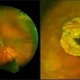
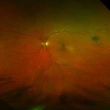
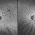
Ultra-wide field fundus autofluorescence images of a 14-year-old patient with congenital retinal pigment epithelial hypertrophy affecting both eyes as a manifestation of Gardner's Syndrome.
Photographer: Olivia Rainey
Imaging device: Optos
Condition/keywords: bilateral, familial adenomatous polyposis, fundus autofluorescence (FAF), Gardner Syndrome, hypofluorescent lesions, Optos, ultra-wide field imaging
-
 Gardner Syndrome
Dec 12 2018 by John S. King, MD
66-year-old white male with Gardner Syndrome (colon resection in 1991), who has two children with Gardner Syndrome, presented to Dr. Zocchi with an RD in the fellow eye that was successfully repaired with a pneumatic retinopexy. Currently 20/20 OU with IOP of 7 OD and 14 OS; no RAPD; PCIOL OU. Dr. Zocchi got oral permission by the patient to have these put into the Retina Image Bank. Although the CHRPE like lesions (2 OD) are not bilateral, we both think these lesions represent "retinal pigment epithelial hamartomas associated with familial adenomatous polyposis (RPEH-FAP)" as Shields described in their Intraocular Tumors book. One lesion is located superiorly and is pigmented with depigmented margins; the temporal lesion is atrophic with minimal remaining pigment hypertrophy.
Photographer: Karin Aletter
Imaging device: Optos CA
Condition/keywords: Gardner Syndrome, RPEH-FAP
-
 BRVO / CHRPE
Apr 1 2014 by David Callanan, MD
73-year-old white male, BRVO / CHRPE.
Condition/keywords: branch retinal vein occlusion (BRVO), congenital hypertrophy of the retinal pigment epithelium (CHRPE)
-
 BRVO / CHRPE
Apr 1 2014 by David Callanan, MD
73-year-old white male, BRVO / CHRPE.
Condition/keywords: branch retinal vein occlusion (BRVO), congenital hypertrophy of the retinal pigment epithelium (CHRPE)
-
 CHRPE
CHRPE
May 9 2014 by S. Natarajan, MD, FASRS, FRCS (GLASGOW) , FICO, D.Sc, FELA
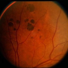
23-year-old male in for routine eye checkup with BCVA 6/6 (OU). Showed CHRPE lesion in INQ (OD )on fundus.
Photographer: ADITYA JYOT EYE HOSPITAL,MUMBAI,INDIA
Condition/keywords: congenital hypertrophy of the retinal pigment epithelium (CHRPE), myopic eye
-
 Bear Tracks / CHRPE / Myelinated NFL
Jul 12 2014 by David Callanan, MD
58-year-old female, bear tracks / CHRPE / myelinated NFL.
Condition/keywords: bear tracks, congenital hypertrophy of the retinal pigment epithelium (CHRPE), myelinated nerve fibers
-
 Multiple Uveal Choroidal Nevi
Jul 29 2013 by Jason S. Calhoun
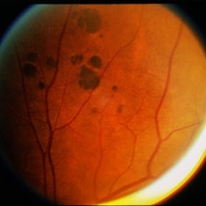
Patient referred for CHRPE which in this case has unusual appearance. VA is 20/20 in the both eyes. Distinguishing if there was any relation to polyposis. This is not CHRPE.
Photographer: Jason S. Calhoun, Department of Ophthalmology, Mayo Clinic Jacksonville, Florida
Imaging device: TOPCON TRC 50-EX
Condition/keywords: choroidal nevus, pigmented nevus
 Loading…