Search results (38 results)
-
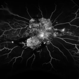
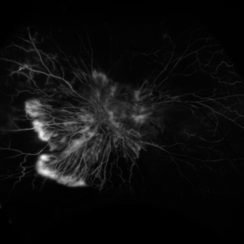
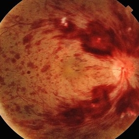
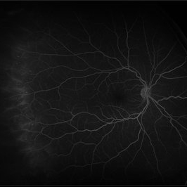
 Central Retinal Artery Occlusion
Apr 20 2018 by Kim Barrett
64-year-old female woke with no vision in her right eye. This image was taken at 6:11 minutes and the vessels have not filled. Patient has been treated with PRP laser and anti-VEGF therapy. Current vision is CF @ 2 ft.
Photographer: Kim Barrett C.O.A.
Imaging device: Heidelberg
Condition/keywords: central retinal artery occlusion (CRAO), diabetes, hypertension, smoker, uncontrolled
-
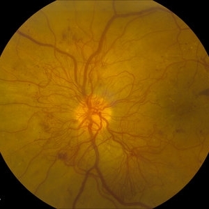
 Central Retinal Artery Occlusion
Jan 22 2021 by Renata Garcia Franco, Md
65-year-old male, history of uncontrolled systemic arterial hypertension. Fluorescein angiography (FA) shows a delay in filling of the retinal arteries.
Photographer: Fatima Hernandez, Instituto de la Retina del Bajio SC
Imaging device: Zeiss
Condition/keywords: central retinal artery occlusion (CRAO)
-
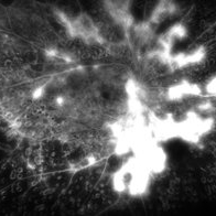
 Central Retinal Artery Occlusion
Jan 22 2021 by Renata Garcia Franco, Md
65-year-old male, history of uncontrolled systemic arterial hypertension. Segmentation of blood in retinal arterioles, retinal whitening and cherry red spot.
Photographer: Fatima Hernandez, Instituto de la Retina del Bajio SC
Imaging device: Zeiss
Condition/keywords: central retinal artery occlusion (CRAO)
-
 Proliferative Diabetic Retinopathy
Aug 16 2022 by Donnie Willis
51 yo female. Uncontrolled Diabetes. Active PDR.
Photographer: Donnie Willis, Tennessee Retina
Imaging device: Optos
Condition/keywords: capillary dropouts, Diabetes, FA, fluorescein angiogram (FA), Optos, proliferative diabetic retinopathy (PDR), vitreomacular traction (VMT)
-
 Severe NVD
Mar 26 2018 by Kristen Wagner
Fundus photograph of a young woman with uncontrolled Diabetes Type II with severe neovascularization of the disc (NVD) and PDR.
Photographer: Kristen Wagner, COT, OSC
Condition/keywords: diabetes, neovascularization (NV), neovascularization of the disc (NVD), optic disc, optic nerve, proliferative diabetic retinopathy (PDR)
-
 Tractional Retinal Detachment
Jan 23 2018 by Nilesh K Kanjani, MD
Fundus Photograph of 42-year-old female patient with uncontrolled diabetes show tractional retinal detachment involving macula.
Photographer: Nilesh K Kanjani, Dr Agarwal Eye Hospital, Ahmedabad, India
Condition/keywords: tractional retinal detachment
-
 Subhyaloid Hemorrhage With Flat Neovascular Vessels
Sep 26 2017 by Purva Patwari
60-year-old patient with uncontrolled diabetes.
Photographer: Dr Purva Patwari, Patwari Retina Clinic,Ahmedabad, India
Imaging device: Zeiss
Condition/keywords: flat neovascularization
-
 Active neovascularization in Proliferative Diabetic Retinopathy
Jan 10 2018 by Peter H. Tang, MD, PhD
Fluorescein angiography image from a 46-year-old woman with uncontrolled proliferative diabetic retinopathy shows extensive dye leakage from active neovascularization.
Imaging device: Optos California
Condition/keywords: diabetes, diabetic retinopathy, fluorescein leakage, neovascularization elsewhere (NVE), neovascularization of the disc (NVD), pan-retinal photocoagulation (PRP), proliferative diabetic retinopathy (PDR)
-
 Active Proliferative Diabetic Retinopathy
Aug 16 2022 by Donnie Willis
51 yo female. Uncontrolled Diabetes. Active Proliferative Diabetic Retinopathy.
Photographer: Donnie Willis, Tennessee Retina
Imaging device: Optos
Condition/keywords: capillary dropouts, Diabetes, fluorescein angiogram (FA), OPTOS, proliferative diabetic retinopathy (PDR), tractional retinal detachment
-
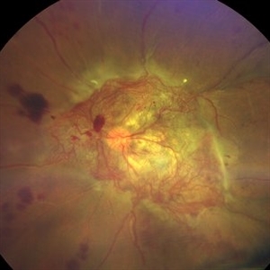 An Intricate Web of Vasculature
Jan 5 2022 by SHISHIR VERGHESE, MS, FVRS, FAICO (Retina)
Fundus photograph of a 55-year-old gentleman with decreased vision in the left eye for 6 months. History of uncontrolled diabetes and hypertension for 15 years. Best corrected visual acuity in the left eye was 5/60.
Photographer: SHISHIR VERGHESE
Imaging device: ZEISS CLARUS
Condition/keywords: diabetes, proliferative diabetic retinopathy (PDR)
-
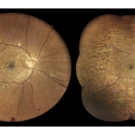
 Bilateral Macroaneurysm
Aug 4 2017 by Eitae Kim, MD
UWF fundus photograph of 81-year-old woman with diabetic retinopathy. Recently the blood pressure was abnormally high and uncontrolled. The above is 6-month- ago fundus photograph and below is recent photograph.
Photographer: Eitae Kim, BOIM retina center, Pureun eye hospital
Condition/keywords: ultra-wide field imaging
-
 Branch Retinal Vein Occlusion
Apr 15 2023 by Yousef A Fouad, MBBCh, MSc
BRVO in a young female with uncontrolled hypertension
Photographer: Yousef Fouad, Ain Shams University, Egypt
Condition/keywords: branch retinal vein occlusion (BRVO), fundus photograph, Hypertension, venous occlusion
-
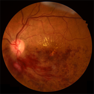
 Central Retinal Artery Occlusion
Oct 25 2017 by satar Baghrizabehi
Fundus photograph of an 67-year-old man with uncontrolled arterial hypertension.
Photographer: Satar Baghrizabehi MD Educ. Hospital Rakican
Condition/keywords: central retinal artery occlusion (CRAO)
-
 Central Retinal Vein Occlusion
Jul 21 2024 by César Adrián Gómez Valdivia, MD
Central Retinal Vein Occlusion found in a 72 year old patient with history of uncontrolled Hypertension. Non-Ischemic Variant.
Photographer: Erika Paulina Ornelas Cazares
Imaging device: TOPCON TRC-50DX
Condition/keywords: central retinal vein occlusion (CRVO)
-
 Extensive Neovascularization
Jul 21 2024 by César Adrián Gómez Valdivia, MD
Macular Traction and Extensive Neovascularization found in a 66 year-old patient with history of uncontrolled Type 2 Diabetes Mellitus.
Photographer: Erika Paulina Ornelas Cazares
Imaging device: TOPCON TRC-50DX
Condition/keywords: diabetic retinopathy, neovascularization (NV)
-
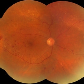
 Hypertensive Retinopathy
May 1 2024 by Marco Antonio Sauza
36 year old male with uncontrolled systemic hypertension.
Photographer: MARCO SAUZA CASTILLEJOS
Imaging device: VISUCAM ZEISS
Condition/keywords: choroidal infarction, hypertensive retinopathy, macular star
-
 Hypertensive Retinopathy Grade IV
May 1 2024 by Marco Antonio Sauza
Fundus photograph of an 36-year-old male with uncontrolled systemic hypertension, >200/100mmhg, presenting decreased vision in the left eye.
Photographer: MARCO SAUZA CASTILLEJOS
Imaging device: VISUCAM ZEISS
Condition/keywords: choroidal infarction, hypertensive retinopathy, macular star
-
 Macular OCT Image of a Patient With Central Retinal Artery Occlusion
Jul 7 2024 by Thiago Mazzeo
This is a macular OCT image of a patient that presented sudden visual loss in the right eye (Light perception) after leaving the hospital due to uncontrolled systemic arterial hypertension.
Photographer: Thiago Mazzeo
Imaging device: Zeiss Cirrus 5000
Condition/keywords: Central Retinal Artery Occlusion, macular changes, OCT
-
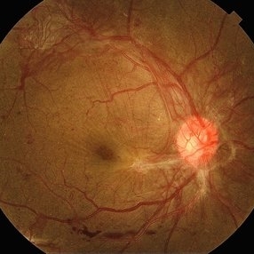
 Neovascularisation of Disc
Oct 3 2017 by Purva Patwari
52-year-old with uncontrolled DM, unilateral NVD, other eye moderate NPDR.
Photographer: Dr Purva Patwari, Patwari Retina Clinic, Ahmedabad INDIA
Condition/keywords: neovascularization of the disc (NVD)
-
 Neovascularisation of Disc
Oct 3 2017 by Purva Patwari
52-year-old with uncontrolled DM, unilateral NVD, other eye moderate NPDR.
Photographer: Dr Purva Patwari, Patwari Retina Clinic, Ahmedabad INDIA
Condition/keywords: neovascularization (NV), neovascularization of the disc (NVD)
-
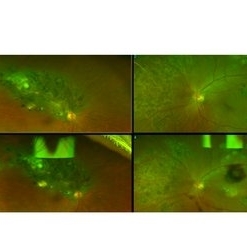
 Peripheral Retinal Vasculitis
May 27 2020 by Olivia Rainey
Ultra-widefield fluorescein angiogram of a 58-year-old female with possible peripheral vasculitis. There was no venous access for this patient, so the fluorescein was administered orally. The image was taken at 7:33 after oral administration. The physician stated that the peripheral nonperfusion could be a sign of previous vasculitis, although could also be a result of uncontrolled diabetes. She was asked to obtain additional bloodwork in order to rule out sarcoidosis, as well as sickle cell. It does not appear the nonperfusion has progressed since her last evaluation. Her vision was 20/40 in the right eye at the time the image was taken.
Photographer: Olivia Rainey, OCT-C, COA
Imaging device: Optos California
Condition/keywords: diabetes, fluorescein angiogram (FA), hypertensive retinopathy, non-perfusion, Optos, oral fluorescein, peripheral retinal vasculitis, ultra-wide field imaging
-
 Post Treatment
Sep 26 2017 by Purva Patwari
60-year-old patient with uncontrolled diabetes.
Photographer: Dr Purva Patwari, Patwari Retina Clinic,Ahmedabad , India
Condition/keywords: flat neovascularization
-
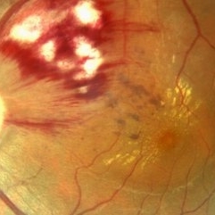
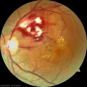
 Proliferative Diabetic Retinopathy S/P Pan Retinal Photocoagulation
Mar 4 2025 by Prithvi Chandrakanth
A 52-year-old female patient presented with complaints of diminishing vision, compounded by uncontrolled diabetes mellitus. Her Fundus examination revealed proliferative diabetic retinopathy, characterized by neovascularization of the disc and elsewhere, and sclerosed vessels. To address this, Pan Retinal Photocoagulation was performed, and the condition stabilized, halting the progression of the disease.
Photographer: DR PRITHVI CHANDRAKANTH, DR CHANDRAKANTH NETHRALAYA, KOZHIKODE, KERALA, INDIA
Imaging device: EIDON
Condition/keywords: Diabetic Retinopathy, Neovascularisation at the Disc (NVD), neovascularization of the disc (NVD), NVD, pan-retinal photocoagulation (PRP), PDR, PDR with NVE (periphery), PRP
-
 Red free Proliferative DR
Sep 25 2023 by firdaus sukhi, MD
Red Free photograph in a sick patient detected by optometrist for prompt retinal intervention Renal failure uncontrolled systemic parameters FFA avoided
Photographer: Naseem Akhtar Optometrist
Condition/keywords: FLUORESCEIN ANGIOGRAPHY, proliferative diabetic retinopathy (PDR), Red free
-
 Red free reval of proliferative diabetic retinopathy
Sep 25 2023 by firdaus sukhi, MD
56 year old diabetic not on any systemic treatment reported in optometry clinic with blurred vision. A simple red free pictures by optometrist could detect the florid NVD and NVE in optometry clinic Poor renal profile uncontrolled systemic parameters limits fluorescein angiography and such simple tool could help referring the urgent cases to the retina clinic for prompt treatment.
Photographer: Naseem Akhtar -optometrist SKMC Ajman UAE
Condition/keywords: proliferative diabetic retinopathy (PDR)
 Loading…