Search results (38 results)
-
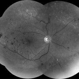
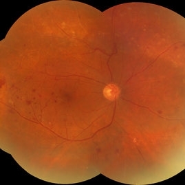
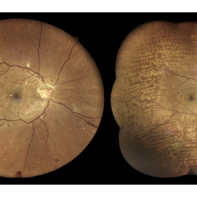 Proliferative Diabetic Retinopathy S/P Pan Retinal Photocoagulation
Mar 4 2025 by Prithvi Chandrakanth
A 52-year-old female patient presented with complaints of diminishing vision, compounded by uncontrolled diabetes mellitus. Her Fundus examination revealed proliferative diabetic retinopathy, characterized by neovascularization of the disc and elsewhere, and sclerosed vessels. To address this, Pan Retinal Photocoagulation was performed, and the condition stabilized, halting the progression of the disease.
Photographer: DR PRITHVI CHANDRAKANTH, DR CHANDRAKANTH NETHRALAYA, KOZHIKODE, KERALA, INDIA
Imaging device: EIDON
Condition/keywords: Diabetic Retinopathy, Neovascularisation at the Disc (NVD), neovascularization of the disc (NVD), NVD, pan-retinal photocoagulation (PRP), PDR, PDR with NVE (periphery), PRP
-
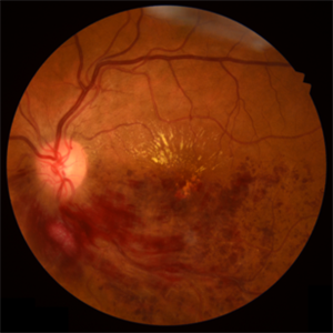
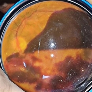 Subhyaloid Hemorrhage
Jan 22 2025 by DR Rohit Gupta
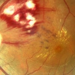
48 year old female presented with right eye diminution of vision, on fundus examination a large hemorrhage was seen in subhyaloid space with multiple retinal hemorrhages. Patients was known case of diabetes with uncontrolled blood sugar level.
Photographer: Dr Rohit gupta
Imaging device: Samsung S21
Condition/keywords: SUB ILM hemorrhage, subhyaloid blood, subhyaloid hemorrhage
-
 Severe Exudative Diabetic Retinopathy - Left Eye
Aug 19 2024 by Nishikant J Borse, MS, FMRF, FASRS
52-year-old diabetic lady presented with diminution of vision for 7 months. She had uncontrolled Diabetes mellitus with an HbA1C of 11.5. On examination she showed Severe Non-Proliferative Diabetic Retinopathy with exudates filling the macular area up to the arcades.
Photographer: Dr Nishikant Borse , Insight eye Clinic , Mumbai
Imaging device: Topcon Triton
Condition/keywords: Diabetic Retinopathy, foveal hard exudates
-
 Severe Exudative Diabetic Retinopathy - Right Eye
Aug 19 2024 by Nishikant J Borse, MS, FMRF, FASRS
52-year-old diabetic lady presented with diminution of vision for 7 months. She had uncontrolled Diabetes mellitus with an HbA1C of 11.5. On examination she showed Severe Non-Proliferative Diabetic Retinopathy with exudates filling the macular area up to the arcades.
Photographer: Dr Nishikant Borse , Insight eye Clinic , Mumbai
Imaging device: Topcon Triton
Condition/keywords: Diabetic Retinopathy, foveal hard exudates
-
 Subhyaloid Hemorrhage
Jul 31 2024 by Arthi Mohankumar , MS,MRCS ED, FICO,FAICO
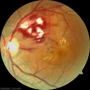
A 35 year old male presented with complaints of seeing a black spot in left eye for past one day after working out in the gym the previous day. He has history of uncontrolled diabetes and hypertension. Fundus exam of the left eye revealed a sub hyaloid hemorrhage nasal to the disc with minimal background Diabetic and hypertensive changes. His baseline CBG was 200 mg/dl and BP was 170/100 He was suggested observation initially considering the nasal location. But patient found the scotoma very disturbing and eventually underwent yag hyaloidotomy
Photographer: Arthi Mohankumar
Condition/keywords: Sub hyaloid haemorrhage, valsalva retinopathy
-
 Extensive Neovascularization
Jul 21 2024 by César Adrián Gómez Valdivia, MD
Macular Traction and Extensive Neovascularization found in a 66 year-old patient with history of uncontrolled Type 2 Diabetes Mellitus.
Photographer: Erika Paulina Ornelas Cazares
Imaging device: TOPCON TRC-50DX
Condition/keywords: diabetic retinopathy, neovascularization (NV)
-
 Central Retinal Vein Occlusion
Jul 21 2024 by César Adrián Gómez Valdivia, MD
Central Retinal Vein Occlusion found in a 72 year old patient with history of uncontrolled Hypertension. Non-Ischemic Variant.
Photographer: Erika Paulina Ornelas Cazares
Imaging device: TOPCON TRC-50DX
Condition/keywords: central retinal vein occlusion (CRVO)
-
 Macular OCT Image of a Patient With Central Retinal Artery Occlusion
Jul 7 2024 by Thiago Mazzeo
This is a macular OCT image of a patient that presented sudden visual loss in the right eye (Light perception) after leaving the hospital due to uncontrolled systemic arterial hypertension.
Photographer: Thiago Mazzeo
Imaging device: Zeiss Cirrus 5000
Condition/keywords: Central Retinal Artery Occlusion, macular changes, OCT
-
 Hypertensive Retinopathy
May 1 2024 by Marco Antonio Sauza
36 year old male with uncontrolled systemic hypertension.
Photographer: MARCO SAUZA CASTILLEJOS
Imaging device: VISUCAM ZEISS
Condition/keywords: choroidal infarction, hypertensive retinopathy, macular star
-
 Hypertensive Retinopathy Grade IV
May 1 2024 by Marco Antonio Sauza
Fundus photograph of an 36-year-old male with uncontrolled systemic hypertension, >200/100mmhg, presenting decreased vision in the left eye.
Photographer: MARCO SAUZA CASTILLEJOS
Imaging device: VISUCAM ZEISS
Condition/keywords: choroidal infarction, hypertensive retinopathy, macular star
-
 Red free Proliferative DR
Sep 25 2023 by firdaus sukhi, MD
Red Free photograph in a sick patient detected by optometrist for prompt retinal intervention Renal failure uncontrolled systemic parameters FFA avoided
Photographer: Naseem Akhtar Optometrist
Condition/keywords: FLUORESCEIN ANGIOGRAPHY, proliferative diabetic retinopathy (PDR), Red free
-
 Red free reveal proliferative diabetic retinopathy
Sep 25 2023 by firdaus sukhi, MD
56 year old diabetic not on any systemic treatment reported in optometry clinic with blurred vision. A simple red free pictures by optometrist could detect the florid NVD and NVE in optometry clinic . Poor renal profile uncontrolled systemic parameters limits fluorescein angiography and such simple tool could help referring the urgent cases to the retina clinic for prompt treatment.
Photographer: Naseem Akhtar -optometrist SKMC Ajman UAE
Condition/keywords: proliferative diabetic retinopathy (PDR)
-
 Red free reveal of proliferative diabetic retinopathy
Sep 25 2023 by firdaus sukhi, MD
56 year old diabetic not on any systemic treatment reported in optometry clinic with blurred vision. A simple red free pictures by optometrist could detect the florid NVD and NVE in optometry clinic Poor renal profile uncontrolled systemic parameters limits fluorescein angiography and such simple tool could help referring the urgent cases to the retina clinic for prompt treatment.
Photographer: Naseem Akhtar -optometrist SKMC Ajman UAE
Condition/keywords: proliferative diabetic retinopathy (PDR)
-
 Red free reval of proliferative diabetic retinopathy
Sep 25 2023 by firdaus sukhi, MD
56 year old diabetic not on any systemic treatment reported in optometry clinic with blurred vision. A simple red free pictures by optometrist could detect the florid NVD and NVE in optometry clinic Poor renal profile uncontrolled systemic parameters limits fluorescein angiography and such simple tool could help referring the urgent cases to the retina clinic for prompt treatment.
Photographer: Naseem Akhtar -optometrist SKMC Ajman UAE
Condition/keywords: proliferative diabetic retinopathy (PDR)
-
 RIGHT EYE- DIABETIC PAPILLOPATHY
Jun 20 2023 by Deepak Bhojwani, MS
FUNDUS IMAGE OF A 62 YEAR OLD LADY WITH UNCONTROLLED DIABETES SHOWING EXTENSIVE TELAGIECTATIC VESSELS WITH HAEMORRHAGES PREDOMINANTLY OVER OPTIC DISC SURROUNDED BY BACKGROUND DIABETIC RETINOPATHY.
Photographer: DEEPAK BHOJWANI
Condition/keywords: Diabetes, papillary involvement
-
 Branch Retinal Vein Occlusion
Apr 15 2023 by Yousef A Fouad, MBBCh, MSc
BRVO in a young female with uncontrolled hypertension
Photographer: Yousef Fouad, Ain Shams University, Egypt
Condition/keywords: branch retinal vein occlusion (BRVO), fundus photograph, Hypertension, venous occlusion
-
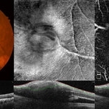 Retinal Arterial Macroaneurysm
Apr 8 2023 by Yousef A Fouad, MBBCh, MSc
Multimodal imaging of a retinal arterial macroaneurysm in the right eye of a 73-year-old male patient with uncontrolled hypertension. Fundus photography shows hemorrhage surrounding an arterial branch of the upper temporal arcade. Optical coherence tomography (OCT) through the lesion shows inner retinal hyperreflectivity with back shadowing, and adjacent cystoid macular edema in the outer retina. En face OCT centered on the lesion delineates the fusiform dilatation of the affected vessel, and OCT angiography confirms the presence of blood flow within the aneurysmal dilatation.
Photographer: Yousef Fouad, Ain Shams University, Egypt
Condition/keywords: arteriolar macroaneurysm, enface imaging, macroaneurysm, macroarterial aneurysm, OCT Angiography, OCTA
-
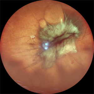 Table Top Tractional Retinal Detachment
Apr 4 2023 by SHISHIR VERGHESE, MS, FVRS, FAICO (Retina)
Fundus photograph of a 65 year old male with history of uncontrolled diabetes mellitus showing table top tractional retinal detachment
Photographer: Dr Shishir Verghese
Imaging device: Nidek Mirante
Condition/keywords: proliferative diabetic retinopathy (PDR), tractional retinal detachment
-
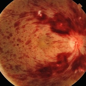
 Active Proliferative Diabetic Retinopathy
Aug 16 2022 by Donnie Willis
51 yo female. Uncontrolled Diabetes. Active Proliferative Diabetic Retinopathy.
Photographer: Donnie Willis, Tennessee Retina
Imaging device: Optos
Condition/keywords: capillary dropouts, Diabetes, fluorescein angiogram (FA), OPTOS, proliferative diabetic retinopathy (PDR), tractional retinal detachment
-
 Proliferative Diabetic Retinopathy
Aug 16 2022 by Donnie Willis
51 yo female. Uncontrolled Diabetes. Active PDR.
Photographer: Donnie Willis, Tennessee Retina
Imaging device: Optos
Condition/keywords: capillary dropouts, Diabetes, FA, fluorescein angiogram (FA), Optos, proliferative diabetic retinopathy (PDR), vitreomacular traction (VMT)
-
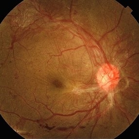
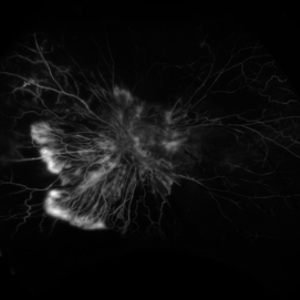
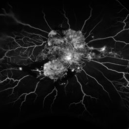
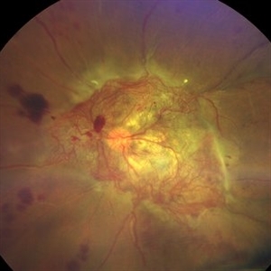 An Intricate Web of Vasculature
Jan 5 2022 by SHISHIR VERGHESE, MS, FVRS, FAICO (Retina)
Fundus photograph of a 55-year-old gentleman with decreased vision in the left eye for 6 months. History of uncontrolled diabetes and hypertension for 15 years. Best corrected visual acuity in the left eye was 5/60.
Photographer: SHISHIR VERGHESE
Imaging device: ZEISS CLARUS
Condition/keywords: diabetes, proliferative diabetic retinopathy (PDR)
-
 Right Central Retinal Vein Occlusion
Jun 25 2021 by Ahmed Almuhaylib, MD
Fundus photograph of a 65-year-old man with uncontrolled hypertension.
Photographer: Ahmed Almuhaylib, MD, Qassim University, Kingdom of Saudi Arabia
Condition/keywords: central retinal vein occlusion (CRVO), central vein occlusion, non-ischemic central retinal vein occlusion (CRVO)
-
 Central Retinal Artery Occlusion
Jan 22 2021 by Renata Garcia Franco, Md
65-year-old male, history of uncontrolled systemic arterial hypertension. Segmentation of blood in retinal arterioles, retinal whitening and cherry red spot.
Photographer: Fatima Hernandez, Instituto de la Retina del Bajio SC
Imaging device: Zeiss
Condition/keywords: central retinal artery occlusion (CRAO)
-
 Central Retinal Artery Occlusion
Jan 22 2021 by Renata Garcia Franco, Md
65-year-old male, history of uncontrolled systemic arterial hypertension. Fluorescein angiography (FA) shows a delay in filling of the retinal arteries.
Photographer: Fatima Hernandez, Instituto de la Retina del Bajio SC
Imaging device: Zeiss
Condition/keywords: central retinal artery occlusion (CRAO)
 Loading…