Search results (118 results)
-
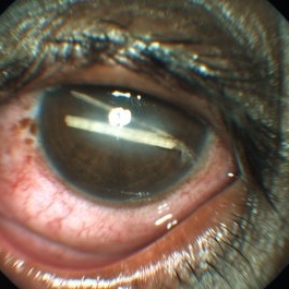
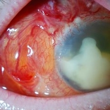
 Rubeosis
Jul 14 2013 by Jason S. Calhoun
Slit lamp photo shows active rubeosis in the left eye.
Photographer: Jason S. Calhoun, Department of Ophthalmology, Mayo Clinic Jacksonville, Florida
Imaging device: TOPCON D-90 SL NIKON CAMERA
Condition/keywords: rubeosis
-
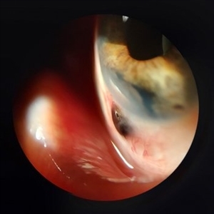
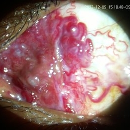 Conjunctival AV Malformation
Dec 18 2023 by siddharth sheth
33 year old male presented with a complaint of redness since 15 years in left eye.
Photographer: Gaurav Kamble, Isha Netralaya
Imaging device: Dyanmic slit lamp imaging
Condition/keywords: conjunctival AV malformation, slit lamp photo, slit lamp photography, unilateral
-
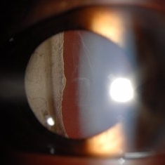
 Dislocated IOL
Jun 4 2024 by Marlee Curnutt
Slit lamp photo of a 64 year old woman presenting with worsening vision and depth perception after trauma induced by a dog, which dislocated her IOL. The patient's IOL haptic was seen in the AC, and almost abutting cornea. Patient's vision upon presentation was DCC CF@1 feet. Patient was counseled and underwent an IOL exchange.
Photographer: Marlee Curnutt, COA
Imaging device: Galaxy A42
Condition/keywords: dislocated intraocular lens (IOL), haptic, IOL, right eye, slit lamp photo, slit lamp photography, trauma
-
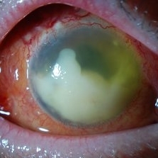
 Endophthalmitis
Apr 9 2014 by Aleksandra V. Rachitskaya, MD, FASRS
Slit lamp photo of a patient with endophthalmitis after cataract surgery. An infectious infiltrate is noted next to the clear corneal incision.
Photographer: Bascom Palmer Eye Institute
Condition/keywords: cataract surgery, endophthalmitis
-
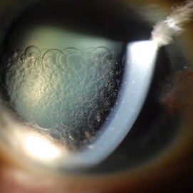
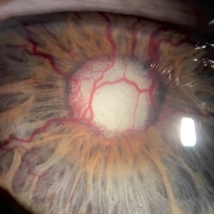
 Epicapsular Stars
Jan 28 2025 by Korey Starkey
Epicapsular stars and cataract noted in natural lens of 68-year-old patient.
Photographer: Korey Starkey
Imaging device: Slit lamp camera
Condition/keywords: cataract, chicken tracks, epicapsular stars, slit lamp photography
-
 Iris Nevus
Jan 28 2025 by Korey Starkey
Slit-lamp image of an 89-year-old patient with an iris nevus. Nevus appeared stable on exam, will continue to monitor.
Photographer: Korey Starkey
Imaging device: Slit lamp camera
Condition/keywords: ectropion uveae, iris nevus, slit lamp photo
-
 Metastatic Breast Carcinoma
Jan 21 2021 by Jamin S. Brown, MD
This anterior segment photograph was taken with a smartphone camera attached to a regular Haag Streit slit lamp ocular demonstrates unusual clustering of white cells on the posterior surface of the intraocular lens. The clinical diagnosis is metastatic breast carcinoma to the vitreous, which is very rare.
Photographer: Stefanie Palmer CRA, Retina Vitreous Surgeons of CNY
Imaging device: Cell phone camera
Condition/keywords: anterior segment, breast cancer, cell phone camera, slit lamp photo
-
 Multiple Intra Ocular Foreeign Body
Apr 23 2015 by Mehul A Shah
A slit lamp photograph showing multiple foreign bodies in anterior chamber.
Photographer: Mehul Shah
Condition/keywords: penetrating trauma
-
 Retisert Implant Migration Into the Anterior Chamber
Feb 11 2025 by Niloofar Piri, MD
Slit Lamp photograph demonstrating spontaneous dislocation and migration of old Retisert implant into the anterior chamber inferiorly with secondary corneal decompensation. Please notice that patient is aphakic. Implant was removed surgically.
Photographer: Hossein Asghari MD, Saint Louis University
Condition/keywords: Corticosteroid implant, implant migration, Retisert
-
 Scleral Ectasia Post Radiation for Iris Melanoma
Jul 5 2024 by Zach Seim
Slit-Lamp Photograph of a 52 year old male with Scleral Ectasia post radiation for Iris Melanoma.
Photographer: Zach Seim
Imaging device: Slit Lamp Photography on Samsung Galaxy 7
Condition/keywords: Iris, iris melanoma, scleral ectasia, slit lamp photo, slit lamp photography
-
---thumb.JPG/image-square;max$300,300.ImageHandler) Scleritis
Scleritis
Jul 14 2013 by Jason S. Calhoun
Patient complains of severe eye pain with drainage. Eye is very red. Slit lamp photo shows active scleritis.
Photographer: Jason S. Calhoun, Department of Ophthalmology, Mayo Clinic Jacksonville, Florida
Imaging device: TOPCON D-90 SL NIKON CAMERA
Condition/keywords: scleritis
-
 Stickler Syndrome
Sep 28 2016 by Philip J. Polkinghorne, MD
Slit lamp photograph
Photographer: Alex Fraser
Condition/keywords: giant retinal tear, membranous vitreous, Stickler Syndrome
-
 Retention of Perfluorocarbon in Anterior Chamber
Mar 1 2017 by Philip J. Polkinghorne, MD
Slit lamp photograph takien one week after retinal detachment surgery where perfluorocarbon liquid was used to re-attach the retina.
Photographer: Alex Fraser
Condition/keywords: perfluorocarbon fluid, retained perfluorocarbon, retina surgery complications
-
 Vitreous Amyloidosis Slit Lamp Photo
Oct 23 2019 by Alexander D Port, MD
Slit lamp photograph preoperatively demonstrating dense symptomatic vitreous opacity in the setting of amyloidosis. The patient elected to undergo pars plana vitrectomy.
Condition/keywords: slit lamp photo, vitreous amyloidosis
-
 Endophthalmitis
Apr 10 2014 by Jason S. Calhoun
Slit lamp photos shows endophthalmitis.
Photographer: Jason S. Calhoun, Mayo Clinic Jacksonville, Department of Ophthalmology
Imaging device: TOPCON D-90 SL/NIKON
Condition/keywords: endophthalmitis
-
 Fluocinolone Implant
Sep 12 2012 by Pauline T Merrill, MD, FASRS
Slit lamp photograph of a Retisert fluocinolone implant in a 52-year-old male with birdshot chorioretinopathy.
Photographer: Pauline Merrill, MD
Imaging device: iPhone photo through slit lamp
Condition/keywords: birdshot, chronic uveitis, fluocinolone implant
-
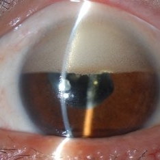 Inverse Hypopyon
Mar 4 2018 by Yoshihiro Yonekawa, MD, FASRS
Slit lamp photograph of a 40-year-old man with previous retinal detachment surgery with silicone oil tamponade, presenting with an inverse hypopyon from emulsified silicone oil.
Photographer: Steven A Bennett, COA, CRA
Imaging device: Nikon D200 / Topcon Slit lamp
Condition/keywords: hypopyon, silicone oil
-
 NVI
NVI
Nov 10 2012 by Pauline T Merrill, MD, FASRS
Slit lamp photo of a 58-year-old woman with severe proliferative diabetic retinopathy and florid neovascularization of the iris.
Photographer: Pauline Merrill, Illinois Retina Associates
Imaging device: iPhone through slit lamp
Condition/keywords: neovascularization of iris (NVI)
-
 Shafer's Sign
Jan 3 2020 by Manuel Ángel Alcántara Delgado, MD
Slit lamp photograph of a 58-year-old man with rhegmatogenous retinal detachment and tobacco dust presence.
Photographer: Manuel Ángel Alcántara Delgado, CMN SXXI, Mexico City
Condition/keywords: acute retinal detachment, retina surgery, vitrectomy
-
 Neovascular Glaucoma
Jan 4 2020 by Manuel Ángel Alcántara Delgado, MD
Slit lamp photograph of a 69-year-old man with diabetic retinopathy and poor metabolic control.
Photographer: Manuel Ángel Alcántara Delgado, CMN SXXI, Mexico City.
Condition/keywords: diabetes, diabetic retinopathy vitrectomy study (DRVS), neovascular glaucoma
-
 Endophthalmitis
Apr 10 2014 by Jason S. Calhoun
Slit lamp photos shows endophthalmitis.
Photographer: Jason S. Calhoun, Mayo Clinic Jacksonville, Department of Ophthalmology
Imaging device: TOPCON D-90 SL/NIKON
Condition/keywords: endophthalmitis
-
 Acute Anterior Uveitis
Apr 14 2022 by Divya Jain
Anterior Segment Slit Lamp photograph of a 33 year old woman with first episode of acute granulomatous anterior uveitis showing circumcorneal congestion, mutton fat KP'S, 3+ cells, 2+ flare and Koeppe's nodules at pupillary margin.
Photographer: Divya Jain
Condition/keywords: acute anterior uveitis
-
 Anterior Basement Membrane Dystrophy
Dec 22 2014 by H. Michael Lambert, MD
Slit lamp photo of dots.
Condition/keywords: anterior basement membrane dystrophy
-
 Anterior Basement Membrane Dystrophy
Dec 22 2014 by H. Michael Lambert, MD
Slit lamp photo of lines.
Condition/keywords: anterior basement membrane dystrophy
-
 Anterior Basement Membrane Dystrophy
Dec 22 2014 by H. Michael Lambert, MD
Slit lamp photo of lines.
Condition/keywords: anterior basement membrane dystrophy
 Loading…