Search results (109 results)
-
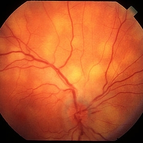
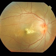
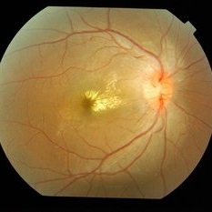
 Bilateral Optic Nerve Involvement in Sarcoidosis
Feb 25 2013 by Henry J. Kaplan, MD
Optic nerve head granuloma of sarcoidosis with severe infiltration and exudation in the left eye of the same patient #2.
Condition/keywords: bilateral involvement, sarcoid granuloma
-
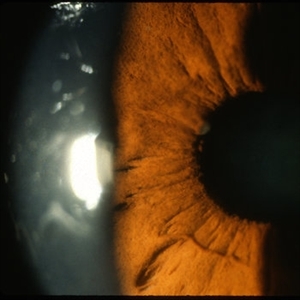
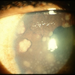 Sarcoid Bussaca Iris Nodules
Oct 11 2012 by Jeffrey G. Gross, MD, FASRS
Sarcoid bussaca iris nodules.
Condition/keywords: autoimmunity, sarcoid bussaca iris nodules, sarcoidosis
-
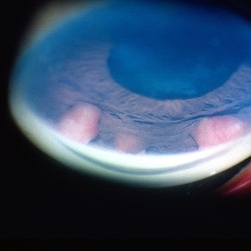 Berlin's Nodules
May 2 2013 by Henry J. Kaplan, MD
Granulomatous berlin's nodules in the angle secondary to sarcoidosis.
Condition/keywords: Berlin's nodules, sarcoidosis
-
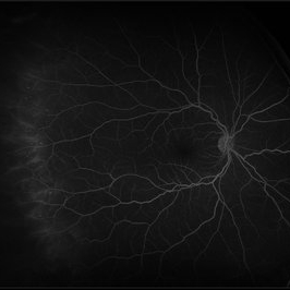
 Sarcoidosis Choroiditis
Feb 25 2013 by Henry J. Kaplan, MD
Sarcoidosis multifocal choroiditis in a case with a known diagnosis of sarcoidosis.
Condition/keywords: sarcoidosis choroiditis
-
 Sarcoid Vasculitis
Oct 11 2012 by Jeffrey G. Gross, MD, FASRS
Sarcoid vasculitis.
Condition/keywords: autoimmunity, sarcoid vasculitis, sarcoidosis
-
 Sarcoid Vasculitis
Oct 23 2012 by Larry Halperin, MD
Sarcoid vasculitis
Condition/keywords: sarcoid vasculitis, sarcoidosis
-
 Sarcoidosis Panuveitis Slide 2
Oct 22 2012 by Ronald C. Gentile, MD
Anterior segment photo of the iris and pupillary margin shows a Koeppe nodule at the 9:30 position. Koeppe nodules consist of inflammatory cell precipitates.
Photographer: The New York Eye & Ear Infirmary Department of Medical Imaging
Condition/keywords: sarcoidosis panuveitis
-
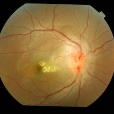
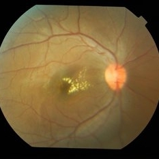
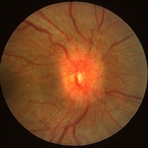 Sarcoidosis
Feb 25 2013 by Henry J. Kaplan, MD
Optic nerve head infiltration of sarcoidosis presenting as optic nerve swelling.
Condition/keywords: sarcoidosis
-
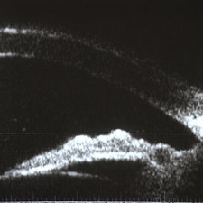 Sarcoidosis Panuveitis Slide 4
Oct 22 2012 by Ronald C. Gentile, MD
High frequency ultrasound biomicroscopy of the anterior chamber and angle images a granuloma involving the iris root and Bussaca nodules on the iris surface consistent with granulomatous uveitis.
Photographer: The New York Eye & Ear Infirmary Department of Medical Imaging
Condition/keywords: sarcoid granuloma, sarcoidosis panuveitis
-
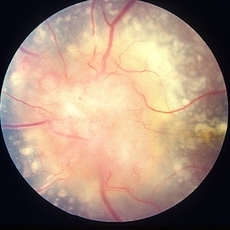
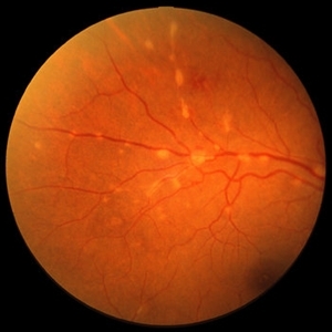
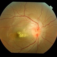
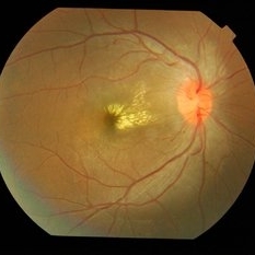
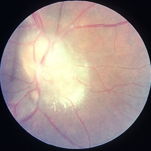 Bilateral Optic Nerve Involvement in Sarcoidosis
Feb 25 2013 by Henry J. Kaplan, MD
Optic nerve granuloma of sarcoidosis in the right eye of a patient with bilateral involvement #1. Left eye is in the following slide.
Condition/keywords: bilateral involvement, sarcoid granuloma
-
 Collateral Vessels in Resolved HRVO
Feb 12 2018 by John S. King, MD
Initial presentation: 25-year-old AAM; Hx DM, Sarcoidosis, Renal Disease, HTN with recent hypertensive crisis. Collateral vessels likely due to old HRVO with one front of NVE inferiorly; veins attenuated proximal to the collateral vessels.
Condition/keywords: collaterals, hemicentral retinal vein occlusion, retinal neovascularization
-
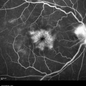 Cystoid Macular Edema Secondary to Panuveitis
Jan 15 2019 by Olivia Rainey
Fluorescein angiogram of a 55-year-old female with cystoid macular edema secondary to uveitis affecting her right eye. Patient was diagnosed with sarcoidosis.
Photographer: Olivia Rainey
Imaging device: Heidelberg Spectralis
Condition/keywords: 30 degrees, cystoid macular edema (CME), fluorescein angiogram (FA), fluorescein leakage, Heidelburg Spectralis, sarcoidosis, uveitis
-
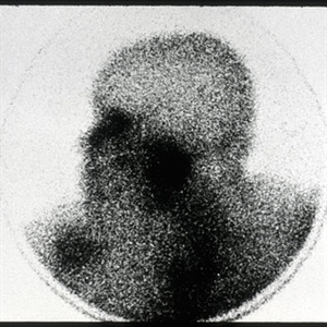 Gadolinium Scan in Sarcoidosis
Oct 11 2012 by Jeffrey G. Gross, MD, FASRS
Gadolinium scan in patient with sarcoidosis showing glandular uptake.
Condition/keywords: autoimmunity, gadolinium scan, glandular uptake, sarcoidosis
-
 Multifocal choroiditis secondary to sarcoidosis- Quiescent
May 6 2023 by Niloofar Piri, MD
Montage fundus photograph of the left eye in a patient with sarcoidosis demonstrating peripheral inactive multifocal chorioretinal scars after systemic immunomodulatory therapy.
Photographer: Sean Kelso, Saint Lousi University
Condition/keywords: multifocal chorioretinitis (MCP), multifocal choroiditis, sarcoid uveitis, sarcoidosis
-
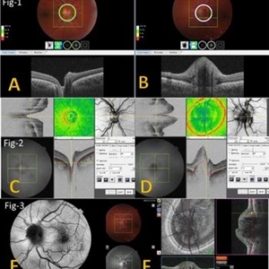 Multimodal Imaging for Differentiating Unilateral Pseudo Optic Disc Swelling(Buried Drusen) From True Optic Disc Swelling
Feb 7 2024 by Fawwaz F Al Mamoori, MD, Medical Retina Consultant
A 27-year-old male patient, medically free, presented with unilateral left optic disc swelling. BCVA=1.0(OU), color vision, and contrast sensitivity were normal (OU) with no RAPD in the left eye. SS-OCT: showed left optic disc elevation with hyporeflective mass lesion (Fig-1 B). Enface OCT: showed left peripapillary hyperreflective ovoid mass lesions(Fig-2 D, Fig-3 F), FAF: showed left superonasal hyperautofluorescent drusenoid lesions. Orbital MRI with contrast was requested to exclude any optic nerve compressive lesions like (tumors: like mengioma or inflammatory lesions like granuloma (sarcoidosis). the result of orbital MRI was normal.
Photographer: Hana.S.Owais
Imaging device: TRITON(TOPCON,Swept Source OCT)
Condition/keywords: fundus autofluorescence (FAF), multimodal imaging, OCT EN FACE, optic disc drusen, optic disc edema
-
 Neuroretinitis
Apr 19 2014 by Mallika Goyal, MD
Right eye fundus of a 25-year-old female patient with idiopathic neuroretinitis shows disc edema and congestion with macular exudates and edema at presentation. All relevant investigations (to rule out sarcoidosis, TB, SLE, leptospirosis, syphilis, borrelia bergdorferi, HIV returned negative). She was treated with pulsed intravenous steroids with resolution.
Photographer: Mallika Goyal, MD, Apollo Health City, Hyderabad, India
Condition/keywords: neuroretinitis
-
 Neuroretinitis
Apr 19 2014 by Mallika Goyal, MD
Right eye fundus of a 25-year-old female patient shows resolving macular exudates and disc edema 2 days after initiating pulsed intravenous steroids for idiopathic neuroretinitis. All relevant investigations (to rule out sarcoidosis, TB, SLE, leptospirosis, syphilis, borrelia bergdorferi, HIV returned negative).
Photographer: Mallika Goyal, MD, Apollo Health City, Hyderabad, India
Condition/keywords: neuroretinitis
-
 Neuroretinitis
Apr 19 2014 by Mallika Goyal, MD
Right eye fundus of a 25-year-old female patient shows resolving macular exudates and disc edema 5 days after initiating pulsed intravenous steroids for idiopathic neuroretinitis. All relevant investigations (to rule out sarcoidosis, TB, SLE, leptospirosis, syphilis, borrelia bergdorferi, HIV returned negative).
Photographer: Mallika Goyal, MD, Apollo Health City, Hyderabad, India
Condition/keywords: neuroretinitis
-
 Neuroretinitis
May 15 2014 by Mallika Goyal, MD
Right eye fundus of a 25-year-old female patient shows resolving macular exudates and completely resolved disc edema 4 weeks after initiating pulsed intravenous steroids for idiopathic neuroretinitis. All relevant investigations (to rule out sarcoidosis, TB, SLE, leptospirosis, syphilis, borrelia bergdorferi, HIV returned negative).
Photographer: Mallika Goyal, MD, Apollo Health City, Jubilee Hills, Hyderabad, India
Condition/keywords: neuroretinitis
-
 Peripheral Retinal Vasculitis
May 27 2020 by Olivia Rainey
Ultra-widefield fluorescein angiogram of a 58-year-old female with possible peripheral vasculitis. There was no venous access for this patient, so the fluorescein was administered orally. The image was taken at 7:33 after oral administration. The physician stated that the peripheral nonperfusion could be a sign of previous vasculitis, although could also be a result of uncontrolled diabetes. She was asked to obtain additional bloodwork in order to rule out sarcoidosis, as well as sickle cell. It does not appear the nonperfusion has progressed since her last evaluation. Her vision was 20/40 in the right eye at the time the image was taken.
Photographer: Olivia Rainey, OCT-C, COA
Imaging device: Optos California
Condition/keywords: diabetes, fluorescein angiogram (FA), hypertensive retinopathy, non-perfusion, Optos, oral fluorescein, peripheral retinal vasculitis, ultra-wide field imaging
-
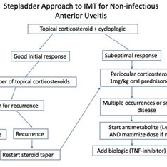 Posterior Manifestations of Sarcoidosis – Management of noninfectious anterior uveitis
Mar 29 2023 by Joshua Friedman
Management of noninfectious anterior uveitis.
Condition/keywords: uveitis
-
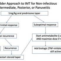 Posterior Manifestations of Sarcoidosis – Management of noninfectious intermediate, posterior, or panuveitis
Mar 29 2023 by Joshua Friedman
Management of noninfectious intermediate, posterior, or panuveitis.
Condition/keywords: panuveitis, sarcoidosis
-
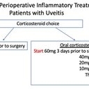 Posterior Manifestations of Sarcoidosis – Perioperative inflammatory supplementation
Mar 29 2023 by Joshua Friedman
Perioperative inflammatory supplementation.
Condition/keywords: sarcoidosis
 Loading…