Search results (100 results)
-
 Rod Cone dystrophy
Nov 29 2022 by Niloofar Piri, MD
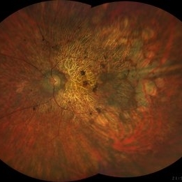
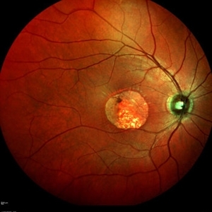
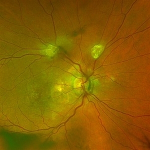
Fundus photograph of the left eye in a 58 yo male with rod cone dystrophy. He presented with night blindness and peripheral vision loss since youth and recent decrease in central vision for the past 10 years. Notice waxy pallor of the nerve, severe arterial narrowing and chorioretinal atrophy mainly around the arcades as well as posterior pole along with RPE hyperplastic changes and atrophy. RPE atrophy in midperiphery has coin shaped appearance. FAF has characteristic appearance (uploaded separately) He has one pathogenic variants of both CEP290 and PRPH2 genes.
Photographer: Sean Kelso, Saint Louis University
Condition/keywords: hereditary retinal deg, hereditary retinal dystrophy, Rod cone dystrophy
-
 Rod Cone dystrophy
Nov 29 2022 by Niloofar Piri, MD
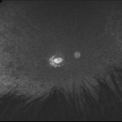
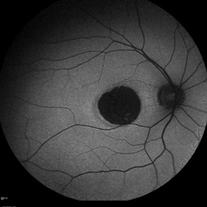
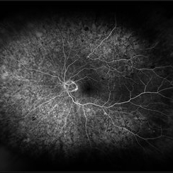
Fundus autofluorescence of the left eye in a 58 yo male with rod cone dystrophy. He presented with night blindness and peripheral vision loss since youth and recent decrease in central vision for the past 10 years. Notice multiple coin shaped hypoautofluorescent pacthes within central 20 degrees which are coalescing centrally. (fundus photo uploaded separately) He has one pathogenic variants of both CEP290 and PRPH2 genes.
Photographer: Sean Kelso, Saint Louis University
Condition/keywords: hereditary retinal degeneration, hereditary retinal dystrophy, rod cone dystrophy
-
 Pericentral Retinitis Pigmentosa
Sep 6 2024 by Mauricio Bayram-Suverza, MD
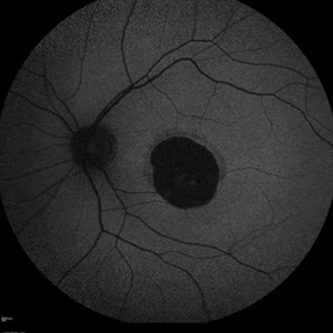
A 65-year-old male patient reports experiencing bilateral blind spots that have gradually intensified over time. Genetic testing was unrevealing. The fundus autofluorescence image shows a hypoautofluorescent ring in the posterior pole, especially nasal to the nerve and along arcades.
Photographer: Mauricio Bayram-Suverza, Casey Eye Institute, OHSU.
Imaging device: Optos California
Condition/keywords: fundus autofluorescence (FAF), inherited retinal disease, nyctalopia, retinal dystrophy, retinitis pigmentosa
-
 Choroideremia
Sep 21 2022 by Zach Seim
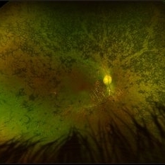
Ultra-widefield fundus photo of a 74 year old male presenting with severe vision loss beginning at age 55. Patient sought a second opinion with our office and was diagnosed with Choroideremia. Patient denies hearing loss, heart problems, balance issues, polydactyly, kidney problems, and dental problems. Patient reports that nobody in the family had blindness. Choroideremia is an X-linked chorioretinal dystrophy characterized by the diffuse, progressive degeneration of the retinal pigment epithelium (RPE), photoreceptors and choriocapillaris. It is caused by a mutation in the CHM gene.
Photographer: Zach Seim
Imaging device: Optos California
Condition/keywords: choroideremia, hereditary choroidal atrophy, hereditary retinal dystrophy, Optos, pseudocolor, ultra-wide field imaging
-
 Choroideremia
Sep 21 2022 by Zach Seim
Ultra-widefield fundus photo of a 74 year old male presenting with severe vision loss beginning at age 55. Patient sought a second opinion with our office and was diagnosed with Choroideremia. Patient denies hearing loss, heart problems, balance issues, polydactyly, kidney problems, and dental problems. Patient reports that nobody in the family had blindness. Choroideremia is an X-linked chorioretinal dystrophy characterized by the diffuse, progressive degeneration of the retinal pigment epithelium (RPE), photoreceptors and choriocapillaris. It is caused by a mutation in the CHM gene.
Photographer: Zach Seim
Imaging device: Optos California
Condition/keywords: choroideremia, hereditary choroidal atrophy, hereditary retinal dystrophy, left eye, light perception, low vision, Optos, pseudocolor, ultra-wide field imaging
-
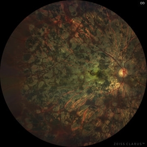 Cone-Rod Dystrophy
Jul 20 2023 by Harsh Vardhan Singh, MS
52-year-old male with a advanced stage of cone-rod dystrophy
Photographer: Harsh Vardhan Singh, AIIMS, Guwahati
Imaging device: Zeiss Clarus 700
Condition/keywords: cone dystrophy, Cone-Rod Dystrophy, pigmentary retinal dystrophy, retinal dystrophy
-
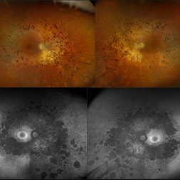 Retinitis Pigmentosa
Nov 7 2023 by Jolee Rodriguez
Bilateral fundus photography and fundus autofluorescence imaging of a 62-year-old male with Retinitis Pigmentosa. Patient reported visual field defects and dark adapting issues. Patient's vision at the time images were taken were sc20/20 of the right eye and sc20/25 of the left eye. Dr. Sutherland determined that based on the patient's lack of family history, the most likely route of inheritance is autosomal recessive.
Photographer: Jolee Rodriguez
Imaging device: Optos California RGB
Condition/keywords: autofluorescence imaging, fundus photography, hereditary retinal dystrophy, Optos, OPTOS CALIFORNIA RGB, retinitis pigmentosa, ultra-wide field imaging, Ultra-wide field retinal imaging, ultra-widefield image
-
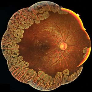 Gyrate Atrophy
Jan 6 2019 by Hashim Ali Khan, OD, FAAO
Montage of Multiple Fundus Photographs from the right eye of a 25-year-old woman with gyrate atrophy.
Photographer: Ahmed Abbass
Imaging device: Topcon TRC-NW8F
Condition/keywords: gyrate atrophy, hereditary retinal dystrophy, retinal dystrophy
-
 Reticular Drusen, Doyne's Honeycomb Retinal Dystrophy, Malattia Leventinese, Familial Dominant Drusen
Feb 22 2018 by Nichole Lewis
Reticular Drusen, Doyne's Honeycomb Retinal Dystrophy, Malattia Leventinese, Familial Dominant Drusen
Photographer: Nichole Lewis
Condition/keywords: Doyne's Honeycomb, Familial Dominant Drusen, Malattia Leventinese, reticular drusen
-
 Central Areolar Choroidal Dystrophy
Apr 14 2018 by Hamza Ahmed Shawky
Right fundus color photograph of a 35-year-old man with central areolar choroidal dystrophy, BCVA is 6/60
Photographer: Hamza Shawky, Alferdaws eye hospital, Retina unit
Imaging device: Heidelberg Spectralis
Condition/keywords: central areolar choroidal dystrophy (CACD), hereditary retinal dystrophy, macular dystrophy, retinal dystrophy
-
 Pigmentary Retinal Dystrophy
Mar 29 2019 by Jessica Norkus
Optos ultra wide field image of 41-year-old male patient with pigmentary retinal dystrophy. Atypical findings due to unilateral presentation. Patient has been experiencing symptoms for 15 years, notes significant nyctalopia.
Photographer: Jessica Norkus
Imaging device: Optos Ultra Wide Field Camera
Condition/keywords: abnormal fundus, bone spicule, color fundus photograph, color photo, fundus autofluorescence (FAF), fundus photograph, Optos, peripheral bone spicules, pigment changes, ultra-wide field imaging, unilateral blindness
-
 Pigmentary Retinal Dystrophy
Mar 29 2019 by Jessica Norkus
Optos ultra wide field image of 41-year-old male patient with pigmentary retinal dystrophy. Atypical findings due to unilateral presentation. Patient has been experiencing symptoms for 15 years, notes significant nyctalopia.
Photographer: Jessica Norkus
Imaging device: Optos Ultra Wide Field Camera
Condition/keywords: abnormal fundus, bone spicule, color fundus photograph, color photo, fundus photograph, Optos, peripheral bone spicules, pigment changes, ultra-wide field imaging, unilateral blindness
-
 Rod cone dystrophy autofluorescence
Sep 19 2022 by Kenneth Fong
34 year old male with colour blindness and loss of visual field
Condition/keywords: retinal dystrophy
-
 Central Areolar Choroidal Dystrophy
Apr 14 2018 by Hamza Ahmed Shawky
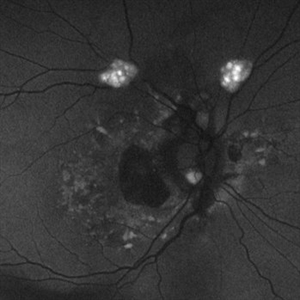
Left fundus autofluorescence photograph of a 35-year-old man with central areolar choroidal dystrophy, BCVA is 6/60
Photographer: Hamza Shawky, Alferdaws eye hospital, Retina unit
Imaging device: Heidelberg Spectralis
Condition/keywords: central areolar choroidal dystrophy (CACD), hereditary retinal dystrophy, macular dystrophy, retinal dystrophy
-
 Central Areolar Choroidal Dystrophy
Apr 14 2018 by Hamza Ahmed Shawky
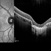
Right fundus OCT of a 35-year-old man with central areolar choroidal dystrophy, BCVA is 6/60
Photographer: Hamza Shawky, Alferdaws eye hospital, Retina unit
Imaging device: Heidelberg Spectralis
Condition/keywords: central areolar choroidal dystrophy (CACD), hereditary retinal dystrophy, macular dystrophy, retinal dystrophy
-
 Central areolar choroidal dystrophy
Apr 14 2018 by Hamza Ahmed Shawky
Right fundus autofluorescence photograph of a 35-year-old man with central areolar choroidal dystrophy, BCVA is 6/60
Photographer: Hamza Shawky, Alferdaws eye hospital, Retina unit
Imaging device: Heidelberg Spectralis
Condition/keywords: central areolar choroidal dystrophy (CACD), hereditary retinal dystrophy, macular dystrophy, retinal dystrophy
-
 Asteroid Hyalosis in Retinitis Pigmentosa
Dec 9 2024 by Mauricio Bayram-Suverza, MD
A 54 year-old male patient presented with asteroid hyalosis. Retinal examination revealed the presence of bone spicules, primarily located in the mid-periphery. Genetic testing identified a pathogenic variant in the RHO gene.
Photographer: Mauricio Bayram-Suverza, Casey Eye Institute, OHSU.
Imaging device: Optos California
Condition/keywords: Asteroid hyalosis, retinal dystrophy, Retinitis Pigmentosa, vitreous
-
 Astrocytic Hamartoma
Feb 27 2025 by Daniel Davis, OCT-C
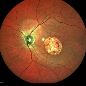
Color fundus photo of 55-year-old female with Astrocytic Hamartoma in association with tuberous sclerosis. No treatment options available, benign. Other findings include; Posterior Vitreous Detachment, Vitreous Hemorrhage, Hereditary Retinal Dystrophy, Vitreous Opacities, Hypertensive Retinopathy.
Photographer: Daniel Davis, OCT-C
Imaging device: Optos California
Condition/keywords: color fundus photograph
-
 Astrocytic Hamartoma
Feb 27 2025 by Daniel Davis, OCT-C
Fundus autofluorescence photo of 55-year-old female with astrocytic hamartoma in association with tuberous sclerosis. No treatment options available, benign. Other findings include; Posterior Vitreous Detachment, Vitreous Hemorrhage, Hereditary Retinal Dystrophy, Vitreous Opacities, Hypertensive Retinopathy.
Photographer: Daniel Davis, OCT-C
Imaging device: Optos California
Condition/keywords: astrocytic hamartoma, fundus autofluorescence (FAF)
-
 Branch Retinal Vein Occlusion With Peripheral Pigmentary Change
Jan 15 2019 by Olivia Rainey
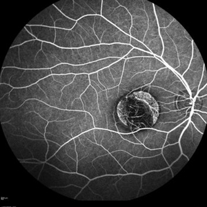
Ultra-wide field fluorescein angiogram of an 85-year-old female with a branch retinal vein occlusion with peripheral pigmentary changes. Patient developed a BRVO after a PPV for an epiretinal membrane.
Photographer: Olivia Rainey
Imaging device: Optos
Condition/keywords: branch retinal vein occlusion (BRVO), epiretinal membrane (ERM), fluorescein angiogram (FA), left eye, Optos, pigmentary retinal dystrophy
-
 Central Areolar Choroidal Dystrophy
Apr 14 2018 by Hamza Ahmed Shawky
Right fundus late FFA photograph of a 35-year-old man with central areolar choroidal dystrophy, BCVA is 6/60
Photographer: Hamza Shawky, Alferdaws eye hospital, Retina unit
Imaging device: Heidelberg Spectralis
Condition/keywords: central areolar choroidal dystrophy (CACD), hereditary retinal dystrophy, macular dystrophy, retinal dystrophy
-
 Central Areolar Choroidal Dystrophy
Apr 14 2018 by Hamza Ahmed Shawky
Left fundus smartphone photograph of a 35-year-old man with central areolar choroidal dystrophy, BCVA is 6/60
Photographer: Hamza Shawky, Alferdaws eye hospital, Retina unit
Imaging device: smartphone fundus photography
Condition/keywords: central areolar choroidal dystrophy (CACD), hereditary retinal dystrophy, macular dystrophy, retinal dystrophy
-
 Central Areolar Choroidal Dystrophy
Apr 14 2018 by Hamza Ahmed Shawky
Right fundus smartphone photograph of a 35-year-old man with central areolar choroidal dystrophy, BCVA is 6/60
Photographer: Hamza Shawky, Alferdaws eye hospital, Retina unit
Imaging device: smartphone fundus photography
Condition/keywords: central areolar choroidal dystrophy (CACD), hereditary retinal dystrophy, macular dystrophy, retinal dystrophy
-
 Central Areolar Choroidal Dystrophy
Apr 14 2018 by Hamza Ahmed Shawky
Left fundus OCT of a 35-year-old man with central areolar choroidal dystrophy, BCVA is 6/60
Photographer: Hamza Shawky, Alferdaws eye hospital, Retina unit
Imaging device: Heidelberg Spectralis
Condition/keywords: central areolar choroidal dystrophy (CACD), hereditary retinal dystrophy, macular dystrophy, retinal dystrophy
-
 Central Areolar Choroidal Dystrophy
Apr 14 2018 by Hamza Ahmed Shawky
Left fundus color photograph of a 35-year-old man with central areolar choroidal dystrophy, BCVA is 6/60.
Photographer: Hamza Shawky, Alferdaws eye hospital, Retina unit
Imaging device: Heidelberg Spectralis
Condition/keywords: central areolar choroidal dystrophy (CACD), hereditary retinal dystrophy, macular dystrophy, retinal dystrophy
 Loading…