Search results (41 results)
-
 Proliferative Retinopathy
Mar 28 2023 by Harold Rodriguez
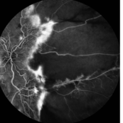
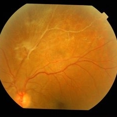
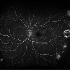
Fluorescein Angiogram on a 43 Year Old female with Proliferative Retinopathy.
Photographer: Harold Rodriguez
Condition/keywords: proliferative retinopathy
-
 Proliferative Sickle Cell Retinopathy
Feb 1 2023 by Olivia Rainey
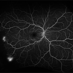
Ultra-widefield fluorescein angiography of a 25-year old male with Proliferative Sickle Cell Retinopathy affecting his right eye. Patient stated that he was born with Sickle disease (SC), and has yearly eye exams. He noted no vision concerns over the last year but has typically experienced sickle attacks about 1-2 per year. The physician noted that the fluorescein obtained showed peripheral nonperfusion affecting the patient's nasal and temporal retina as well as neovascularization affecting his left eye more than his right. He recommended pan retinal photocoagulation in his left eye for his temporal and nasal retina, as as well as his right eye following.
Photographer: Olivia Rainey, OCT-C, COA
Imaging device: Optos California
Condition/keywords: early phase, fluorescein angiogram (FA), fluorescein leakage, neovascularization (NV), non-perfusion, proliferative retinopathy, right eye, sickle cell retinopathy, ultra-wide field imaging, ultra-widefield image
-
 Diabetic Spider Web
Nov 5 2021 by Joana Roque
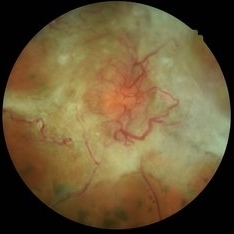
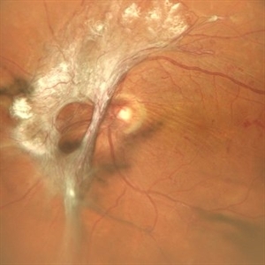
59 year-old poorly controlled diabetic patient with proliferative retinopathy and tractional retinal detachment.
Photographer: Joana Roque
Condition/keywords: proliferative diabetic retinopathy (PDR)
-
---thumb.jpg/image-square;max$300,300.ImageHandler) Extensive neovascularization of the disc
Extensive neovascularization of the disc
Feb 15 2013 by From the Collections of Thomas M. Aaberg, MD and Thomas M. Aaberg Jr., MD
Color fundus photograph showing extensive neovascularization of the disc.
Condition/keywords: proliferative retinopathy, retinal neovascularization
-
 Vasoproliferative Tumor (VPT)
Apr 25 2017 by Christopher G Fuller, MD
Fundus photograph of a presumptive vasoproliferative tumor (with resultant total exudative retinal detachment) in a 54-year-old white truck driver. Image is taken on post-operative day 4, after 25/27 gauge vitrectomy with drainage retinotomy, air-fluid exchange, endoscopic laser blanching of VPT, oil, and Ozurdex.
Photographer: Ray Garner, Texas Retina Associates [Lubbock, TX]
Condition/keywords: vasoproliferative retinopathy
-
 Active Vasculitis with Proliferative Retinopathy
Jan 30 2021 by Raja Rami P Reddy, MD FRCS FASRS
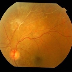
25-year-old boy with unilateral recent onset visual loss. Fundus shows areas of active vasculitis nasally and large neovascular complexes temporally and on the disc and early fibrous membrane formation. Fellow eye fundus is normal. Further investigations suggested tubercular etiology
Photographer: Raja Rami Reddy P
Imaging device: fundus camera
Condition/keywords: proliferative retinopathy, tuberculosis, vasculitis
-
---thumb.jpg/image-square;max$300,300.ImageHandler) Binder3 P12 Slide82
Binder3 P12 Slide82
Feb 15 2013 by From the Collections of Thomas M. Aaberg, MD and Thomas M. Aaberg Jr., MD
Color fundus photograph showing peripheral retinal nonperfusion, retinal neovascularization elsewhere (NVE), venous beading and dilatation, retinal microaneurysms, and intraretinal hemorrhage.
Condition/keywords: peripheral retinal nonperfusion, proliferative retinopathy, retinal neovascularization
-
 Branch Retinal Vein Occlusion With Proliferative Retinopathy
Jul 29 2014 by Mallika Goyal, MD
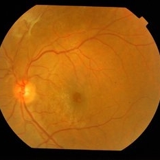
Left eye of a 54-year-old visually asymptomatic male shows superonasal BRVO with proliferation and gliosis on routine fundus exam.
Photographer: Mallika Goyal, MD, Apollo Health City, Jubilee Hills, Hyderabad-500033
Condition/keywords: branch retinal vein occlusion (BRVO)
-
 Branch Retinal Vein Occlusion With Proliferative Retinopathy
Jul 29 2014 by Mallika Goyal, MD
Left eye of a 54-year-old visually asymptomatic male shows superonasal BRVO with proliferation and gliosis on routine fundus exam.
Photographer: Mallika Goyal, MD, Apollo Health City, Jubilee Hills, Hyderabad-500033
Condition/keywords: branch retinal vein occlusion (BRVO)
-
 BRVO With Proliferative Retinopathy
Sep 22 2014 by Mallika Goyal, MD
Left fundus of 54-year-old male shows ST BRVO with preretinal gliosis suggestive of proliferative retinopathy.
Photographer: Mallika Goyal, MD, Apollo Health City, Jubilee Hills, Hyderabad-500033
Condition/keywords: branch retinal vein occlusion (BRVO)
-
 BRVO With Proliferative Retinopathy
Sep 22 2014 by Mallika Goyal, MD
Left fundus of 54-year-old male shows ST BRVO with cystoid macular edema.
Photographer: Mallika Goyal, MD, Apollo Health City, Jubilee Hills, Hyderabad-500033
Condition/keywords: branch retinal vein occlusion (BRVO)
-
 Diabetic Proliferative Retinopathy
Dec 1 2019 by Lucas Zago Ribeiro, MD
Fundus photograph of 75-year-old man with diabetic proliferative retinopathy with fibrovascular proliferation over the optic disc.
Photographer: Lucas Zago Ribeiro, Federal University of São Paulo
Imaging device: Zeiss Visucam 524
Condition/keywords: diabetic retinopathy, fibrovascular proliferation, neovascularization (NV)
-
---thumb.jpg/image-square;max$300,300.ImageHandler) Leakage from peripheral retinal neovascularization and peripheral nonperfusion
Leakage from peripheral retinal neovascularization and peripheral nonperfusion
Feb 15 2013 by From the Collections of Thomas M. Aaberg, MD and Thomas M. Aaberg Jr., MD
Late-phase fluorescein angiograph showing leakage from peripheral retinal neovascularization and peripheral nonperfusion
Condition/keywords: peripheral retinal nonperfusion, proliferative retinopathy, retinal neovascularization
-
 Macular Disciform Scar
Jun 8 2015 by ARIEL WANG
Fundus photograph and OCT scan of an 86-year-old man with long-standing type I diabetic proliferative retinopathy.
Photographer: Suber Huang, Retina Center of Ohio
Imaging device: Heidelberg Spectralis
Condition/keywords: central disciform scar
-
---thumb.jpg/image-square;max$300,300.ImageHandler) peripheral retinal nonperfusion, capillary abnormalities, leaking retinal microaneurysms, and blocked fluorescence
peripheral retinal nonperfusion, capillary abnormalities, leaking retinal microaneurysms, and blocked fluorescence
Feb 15 2013 by From the Collections of Thomas M. Aaberg, MD and Thomas M. Aaberg Jr., MD
Mid-phase fluorescein angiograph showing peripheral retinal nonperfusion, capillary abnormalities, leaking retinal microaneurysms, and blocked fluorescence from intraretinal hemorrhage.
Condition/keywords: peripheral retinal nonperfusion, proliferative retinopathy
-
---thumb.jpg/image-square;max$300,300.ImageHandler) Peripheral retinal nonperfusion, capillary abnormalities, retinal microaneurysms, and intraretinal hemorrhage
Peripheral retinal nonperfusion, capillary abnormalities, retinal microaneurysms, and intraretinal hemorrhage
Feb 15 2013 by From the Collections of Thomas M. Aaberg, MD and Thomas M. Aaberg Jr., MD
Color fundus photograph showing peripheral retinal nonperfusion, capillary abnormalities, retinal microaneurysms, and intraretinal hemorrhage.
Condition/keywords: peripheral retinal nonperfusion, proliferative retinopathy
-
---thumb.jpg/image-square;max$300,300.ImageHandler) Peripheral retinal nonperfusion, venous beading and dilatation, retinal microaneurysms, and intraretinal hemorrhage
Peripheral retinal nonperfusion, venous beading and dilatation, retinal microaneurysms, and intraretinal hemorrhage
Feb 15 2013 by From the Collections of Thomas M. Aaberg, MD and Thomas M. Aaberg Jr., MD
Color fundus photograph corresponding to slide titled "staining of retinal vessels, leakage from peripheral retinal neovascularization and peripheral nonperfusion." Shows peripheral retinal nonperfusion, venous beading and dilatation, retinal microaneurysms, and intraretinal hemorrhage.
Condition/keywords: peripheral retinal nonperfusion, proliferative retinopathy, retinal neovascularization
-
---thumb.jpg/image-square;max$300,300.ImageHandler) Primary Hyperoxaluria and Oxalosis
Primary Hyperoxaluria and Oxalosis
Jul 24 2013 by Hamid Ahmadieh, MD
Color fundus photograph of the left eye of a 55-year-old man with primary hyperoxaluria and oxalosis. Vitreous hemorrhage originating from NVD due to vasoproliferative retinopathy is seen.
Photographer: Hanieh Payab, Ophthalmic Research Center, Tehran
Imaging device: Topcon Fundus Camera
Condition/keywords: neovascularization of the disc (NVD), oxalosis, primary hyperoxaluria, vasoproliferative retinopathy
-
---thumb.jpg/image-square;max$300,300.ImageHandler) Primary Hyperoxaluria and Oxalosis
Primary Hyperoxaluria and Oxalosis
Jul 24 2013 by Hamid Ahmadieh, MD
Late phase FA image of the left eye of a 55-year-old man with primary hyperoxaluria and oxalosis. Profound leakage from disc due to NVD is visible. Vasoproliferative retinopathy has occurred secondary to retinal ischemia due to intravascular deposition of calcium oxalate crystals.
Photographer: Hanieh Payab, Ophthalmic Research Center, Tehran
Imaging device: Topcon Fundus Camera
Condition/keywords: oxalosis, primary hyperoxaluria, vasoproliferative retinopathy
-
 Proliferative Retinopathy
Nov 4 2024 by Tejaswita Verma
Fundus photograph of a middle aged male with diabetes showing large FVP following NVD.
Photographer: DR. TEJASWITA VERMA
Imaging device: MIRANTE
Condition/keywords: FVPs, neovascularization of the disc (NVD), proliferative diabetic retinopathy (PDR)
-
 Proliferative Sickle Cell Retinopathy
Feb 1 2023 by Olivia Rainey
Ultra-widefield fluorescein angiography of a 25-year old male with Proliferative Sickle Cell Retinopathy affecting his left eye. Patient stated that he was born with Sickle disease (SC), and has yearly eye exams. He noted no vision concerns over the last year but has typically experienced sickle attacks about 1-2 per year. The physician noted that the fluorescein obtained showed peripheral nonperfusion affecting the patient's nasal and temporal retina as well as neovascularization affecting his left eye more than his right. He recommended pan retinal photocoagulation in his left eye for his temporal and nasal retina, as as well as his right eye following.
Photographer: Olivia Rainey, OCT-C, COA
Imaging device: Optos California
Condition/keywords: early phase, fluorescein angiogram (FA), fluorescein leakage, left eye, neovascularization (NV), proliferative retinopathy, sickle cell retinopathy, ultra-wide field imaging, ultra-widefield image
-
 Proliferative Sickle Cell Retinopathy, Color OD
May 23 2018 by Hosam Attia, MD
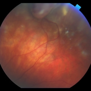
45-year-old African American, male with sickle cell anemia (SC disease) with arteriolar attenuation, mild venous tortuosity, Sunburst (S) and large, partially auto-infarcted sea fan with fresh heme, OD.
Imaging device: Optos California Ultra-Wide Field Fundus Camera
Condition/keywords: neovascularization elsewhere (NVE), proliferative retinopathy, sea fan, sickle cell, sickle cell retinopathy
-
 Proliferative Sickle Cell Retinopathy, Color OD
May 23 2018 by Hosam Attia, MD
45-year-old African American, male with sickle cell anemia (SC disease) with arteriolar attenuation, mild venous tortuosity, Sunburst (S) and large, partially auto-infarcted Seafan with fresh heme, OD.
Imaging device: Optos California Ultra-Wide Field Fundus Camera
Condition/keywords: neovascularization elsewhere (NVE), proliferative retinopathy, sea fan, sickle cell, sickle cell retinopathy
 Loading…