Search results (19 results)
-
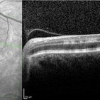
 PAMM-OCTA
Nov 29 2023 by Daniel Davis, OCT-C
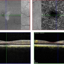
OCT-A of a 30 yo female with PAMM OD.
Photographer: Daniel Davis, OCT-C
Imaging device: Heidelberg Spectralis
Condition/keywords: OCTA, paracentral acute middle maculopathy
-
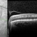
Paracentral Acute Middle Maculopathy
Nov 29 2023 by Daniel Davis, OCT-C
30 yo female OCT with Paracentral Acute Middle Maculopathy (PAMM) OD VA OD: sc20/60+1
Condition/keywords: OCT, PAMM
-
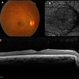
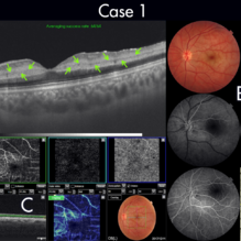 Figure-1 Paracentral Acute Middle Maculopathy (PAMM)
Dec 21 2018 by Fawwaz F Al Mamoori, MD, Medical Retina Consultant
25-year-old male patient medically free, had sudden deterioration in his left eye vision. Visual acuity on presentation was counting fingers at 3 meter distance. Marked Relative Pupillary Afferent Defect (RAPD) was detected and fundoscopic exam showed abnormal foveal reflex. SS OCT B scan: showed a hypereflectivity of the inner plexiform layer (IPL), inner nuclear layer (INL) and OPL layer (fig 1, A).FA images were normal (fig 1, B). Angiography shows remarkable perifoveal capillary drop out within middle retinal layer correlating with perfusion density map which reveals significant decrease in capillary density at the same level (Fig 1, C). Enface ads more proof to PAMM by delineating ischemic distribution in a fern like pattern of hyper reflective areas within DCP (fig1, D).
Photographer: Dr.Fawwaz Al Mamoori (Al Mamoori Eye Clinic)
Imaging device: Triton Swept Source OCT (TOPCON)
Condition/keywords: optical coherence tomography (OCT), paracentral acute middle maculopathy
-
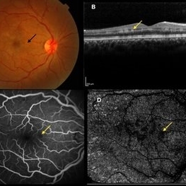 Paracentral Acute Middle Maculopathy
Oct 25 2019 by Gayathri Mohan
Multimodal images of a case of a 29-year-old female with paracentral acute middle maculopathy. A-color fundus photograph showing multiple confluent white retinal patches. B- On OCT the acute lesions of PAMM characteristically appear as placoid, hyperreflective bands at the level of the INL C-Fundus fluorescein angiography showing a capillary nonperfusion area D-flow void areas in deep capillary plexus
Photographer: Akshar Soni
Imaging device: Heidelberg, Nidek
Condition/keywords: fundus albipunctatus, optical coherence tomography (OCT), paracentral acute middle maculopathy
-
 Figure-2 Paracentral Acute Middle Maculopathy (PAMM)
Dec 21 2018 by Fawwaz F Al Mamoori, MD, Medical Retina Consultant
70-year-old female patient known to have hypertension, presented with acute deterioration of left eye vision, best corrected visual acuity was 6/60. Fundoscopic exam showed abnormal foveal reflex whiting .SS-OCT B scan showed also a hypereflectivity of the inner plexiform layer (IPL), inner nuclear layer (INL) and OPL layer(figure-2, A). FA images were also normal(figure-2 B). Segmented angiographic images elucidate ischemia and capillary drop out predominantly at the level of DCP but less severe than Case 1 (fig 2, C). Correspondingly, Enface highlights hyper reflective areas in a fern like distribution in the middle retina at similar depth of ischemic lesions demonstrated on B scans and OCTA (fig 2, D)
Photographer: Dr.Fawwaz Al Mamoori (Al Mamoori Eye Clinic)
Imaging device: Triton Swept Source OCT (TOPCON)
Condition/keywords: optical coherence tomography (OCT), paracentral acute middle maculopathy
-
 Fundus Fluorescein Angiography of Paracentral Acute Middle Maculopathy
Oct 22 2019 by Rengin Aslihan Sonmez, MD, FEBO
36-year-old female patient who had started on oral contraceptives 3 weeks ago presents with right scotoma.
Condition/keywords: fluorescein angiogram (FA), fundus photograph, paracentral acute middle maculopathy
-
 PAMM
PAMM
May 14 2019 by Paola Brito, MD
OCT image of a 52-year-old male, no systemic disease. Left eye with central scotoma and VA 20/25. Hyperreflective lesion in the inner nuclear layer. OCT-A decreased capilar density in superficial and deep capilar plexus. Irregular FAZ.
Photographer: Paola Brito Sandoval, Fundación Hospital Nuestra Señora de la Luz
Imaging device: Angioplex
Condition/keywords: paracentral acute middle maculopathy
-
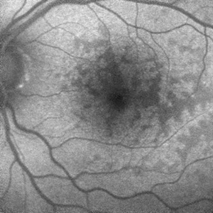
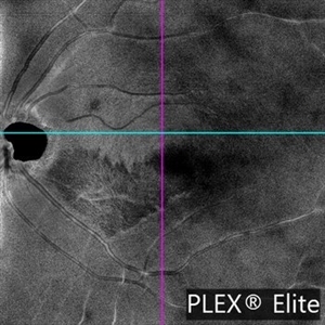 PAMM
PAMM
May 24 2024 by Gustavo Del Castillo-Marquez, MD
EN FACE OCT ANGIOPLEX ELITE image of an 44-year-old man with Paracentral Acute Middle Maculopathy of early onset.
Photographer: Gustavo Del Castillo-Márquez, Asociación Para Evitar la Ceguera en México, CDMX
Imaging device: Zeis Ciruss Angioplex 5000
Condition/keywords: enface imaging, PAMM
-
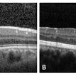 Paracentral Acute Middle Maculopathy
Nov 27 2019 by Alexander D Port, MD
(a) acute PAMM (2) chronic PAMM after 3 months of follow up
Condition/keywords: PAMM, paracentral acute middle maculopathy
-
 Paracentral Acute Middle Maculopathy
Oct 22 2019 by Jeffrey G. Gross, MD, FASRS
FA of 75-year-old white male with 6 day history of acute vision loss. 20/40
Photographer: Tammy Mclaughlin
Imaging device: Zeiss Visucam
Condition/keywords: FA late phase, paracentral acute middle maculopathy, retinal ischemia
-
 Paracentral Acute Middle Maculopathy
Mar 24 2018 by Rania G Estawro, FRCS
Enface OCT & Line scan at different segmentation levels, in male patient with sudden diminution of vision , showing para-central acute middle maculopathy.
Photographer: Rania Estawro, Al Watany Eye Hospital, Cairo, Egypt.
Imaging device: RTVue XR; Optovue, Fremont, CA, USA
Condition/keywords: paracentral acute middle maculopathy
-
 Paracentral Acute Middle Maculopathy
Oct 25 2019 by Gayathri Mohan
Multimodal images of follow up visit of a case of Paracentral acute middle maculopathy 2 months post oral steroid therapy. Color fundus photograph(A) showing resolution of whitish retinal lesions . OCT and OCTA (B,C) also show resolution of lesions post treatment.
Photographer: Akshar Soni
Imaging device: Heidelberg, Nidek
Condition/keywords: fundus albipunctatus, optical coherence tomography (OCT), paracentral acute middle maculopathy
-
 Paracentral Acute Middle Maculopathy
Oct 22 2019 by Jeffrey G. Gross, MD, FASRS
Fundus autofluorescence photograph of 75-year-old white male with 6 day history of acute vision loss. 20/40
Photographer: Tammy McLaughlin
Imaging device: Heidelberg Spectralis
Condition/keywords: paracentral acute middle maculopathy, retinal ischemia
-
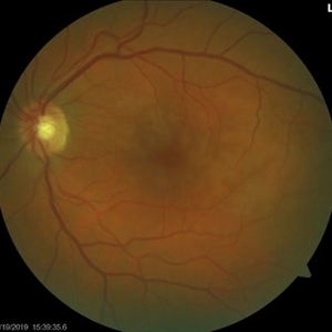 Paracentral Acute Middle Maculopathy
Oct 22 2019 by Jeffrey G. Gross, MD, FASRS
Color photograph of 75-year-old white male with 6 day history of acute vision loss. 20/40
Photographer: Tammy McLaughlin
Imaging device: Zeiss Visucam
Condition/keywords: paracentral acute middle maculopathy, retinal ischemia
-
 Paracentral Acute Middle Maculopathy
Oct 22 2019 by Jeffrey G. Gross, MD, FASRS
OCT of 75-year-old white male with 6 day history of acute vision loss. 20/40
Photographer: Tammy McLaughlin
Imaging device: Heidelberg Spectralis
Condition/keywords: paracentral acute middle maculopathy, retinal ischemia
-
 Paracentral Acute Middle Maculopathy (PAMM)
Mar 21 2019 by Jonathan C. Tsui, MD
26-year-old female with hypertension presenting with chief complaint of "darkening" in her nasal visual field in the right eye. No flashes, floaters, or vision loss. Va 20/60 and nasal VF defect OD. SD-OCT demonstrated hyperreflectivity in the INL consistent with paracentral acute middle maculopathy. She was referred to her PCP for blood pressure optimization and a cardiovascular work-up. She returned for follow-up two months later with 20/80 OD, 20/20 OS. Repeat SD-OCT demonstrated inner retinal atrophy.
Photographer: Zellers, Diane
Condition/keywords: paracentral acute middle maculopathy
-
 Paracentral Acute Middle Maculopathy Fundus Fluorescein Angiography Early Phase
Oct 22 2019 by Rengin Aslihan Sonmez, MD, FEBO
36-year-old female patient who had started on oral contraceptives 3 weeks ago presents with right scotoma.
Condition/keywords: paracentral acute middle maculopathy
-
 Paracentral Acute Middle Maculopathy OCT
Oct 22 2019 by Rengin Aslihan Sonmez, MD, FEBO
OCT of the same patient.
Condition/keywords: optical coherence tomography (OCT), paracentral acute middle maculopathy
 Loading…