Search results (308 results)
-
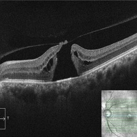
 Whole Eye OCT
Jan 4 2019 by Netan Choudhry, MD, FRCS(C) FASRS
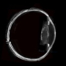
Swept-Source OCT montage of a 45-year-old male with Alports disease and posterior subcapsular cataract.
Photographer: John Golding BA, Vitreous Retina Macula Specialists of Toronto
Imaging device: Topcon DRI Triton
Condition/keywords: Alports disease, optical coherence tomography (OCT), swept source
-
 Retinal Arterio-Venous Malformations
Apr 7 2017 by Deepak Bhojwani, MS
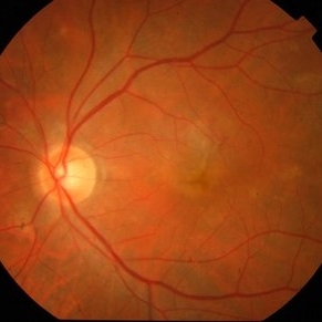
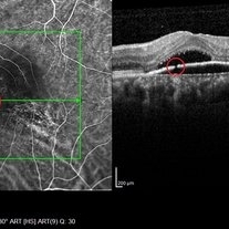
Multimodal imaging of a 16-year-old boy with retinal arterio-venous malformations(AVM). He also had cerebral AVM's on MRI-contrast studies suggesting Wyburn-Mason syndrome.
Photographer: DEEPAK BHOJWANI, RAGHUDEEP EYE HOSPITAL, AHMEDABAD.
Imaging device: Zeiss VISUCAM
Condition/keywords: color fundus photograph, FA early phase, optical coherence tomography (OCT), Wyburn-Mason
-
 Active CNVM
Jul 11 2016 by Manish Nagpal, MD, FRCS (UK), FASRS
Colour photo showing an active CNVM.
Photographer: pooja barot
Condition/keywords: choroidal neovascular membrane (CNVM), optical coherence tomography (OCT)
-
 Active CNVM on Angio OCT
Jul 11 2016 by Manish Nagpal, MD, FRCS (UK), FASRS
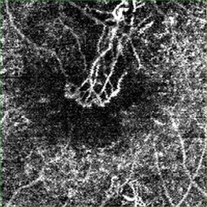
Angio OCT picture showing neovascularization corresponding to the area of CNVM.
Photographer: pooja barot
Condition/keywords: choroidal neovascular membrane (CNVM), optical coherence tomography (OCT)
-
 OCT Image of Epiretinal Membrane
Aug 29 2017 by Carolyn Daley
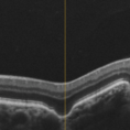
OCT photograph of a 64-year-old women with an epiretinal membrane in the right eye. Patient has not noticed any decline in vision so surgery was not recommended at this time.
Photographer: Carolyn Daley
Imaging device: Heidelberg Spectralis
Condition/keywords: epiretinal membrane (ERM), optical coherence tomography (OCT)
-
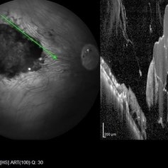
 3D OCT of juxtapapillary melanoma
May 15 2020 by Sophia El Hamichi, MD
A 63-year-old male with juxtapapillary melanoma of the right eye. Visual acuity at presentation was 20/25 OD. Patient treated with brachytherapy Iodine125 plaque
Photographer: Belinda Rodriguez
Condition/keywords: optical coherence tomography (OCT)
-
 Choroidal Osteoma Plus CNV
Sep 2 2012 by Hamid Ahmadieh, MD
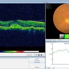
Color fundus photograph and OCT imaging of a 47-year-old man with a juxtafoveal CNV superimposed on a choroidal osteoma.
Photographer: Hamid Ahmadieh, Ophthalmic Research Center, Labbafinejad Medical Center
Imaging device: Topcon
Condition/keywords: choroidal neovascularization (CNV), choroidal osteoma, optical coherence tomography (OCT)
-
 Discontinuity RPE
Oct 17 2014 by Avris Romario Diparaja Siahaan
A simultan ICG angiography + OCT of 56-year-old man that shows a image of discontinuity retinal pigment ephitelial.
Photographer: Harni Christine Damanik, Klinik Mata Nusantara
Imaging device: Heidelberg Spectralis
Condition/keywords: indocyanine green (ICG) angiography, optical coherence tomography (OCT), retinal pigment epithelium
-
 Flat Fovea in Oculocutaneous Albinism
Oct 24 2020 by Guilherme Daher
Optical coherence tomography of a patient with oculocutaneous albinism showing a flat fovea.
Photographer: Jefferson Rocha, Instituto Suel Abujamra, Sao Paulo Brazil
Imaging device: Zeiss Cirrus HD-OCT 5000
Condition/keywords: albinism, fovea, foveal hypoplasia, nystagmus, oculocutaneous albinism, optical coherence tomography (OCT)
-
 Focal Choroidal Excavation
Jan 6 2019 by Aristofanes Canamary jr
A 51-year-old female who reported low visual acuity on AO, worse in the OE. Fundoscopy of OE is observed color and brightness alteration in macular region. Focal concave-shaped chorioretinal anomaly in the foveal region and other two anomaly peripapilary and temporal to the fovea with a hyporreflective subretinal space distinguishing from each other.
Photographer: Aristófanes Canamary Jr, UPO ophthalmology, Sao Paulo
Condition/keywords: excavation, optical coherence tomography (OCT), pachychoroid
-
 Geographic Atrophy
Mar 27 2013 by Michael P. Kelly, FOPS
This is a combined FAF/SD-OCT in EDI mode of a patient with geographic atrophy and foveal sparing.
Photographer: Michael P. Kelly, FOPS. Director, Duke Eye Labs, Duke University Eye Center
Imaging device: Heidelberg Spectralis
Condition/keywords: enhanced depth imaging, foveal sparing, fundus autofluorescence (FAF), geographic atrophy, optical coherence tomography (OCT)
-
 Hemangioma of Retina
Sep 11 2018 by Carolyn Daley
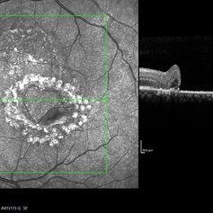
50 degree OCT imaging of a 20-year-old with multiple bilateral hemangiomas. Patient was diagnosed with Von Hippel-Lindau Syndrome.
Photographer: Carolyn Daley, Retina Specialists of Michigan
Imaging device: Heidelberg Spectralis
Condition/keywords: 50 degrees, edema, hemangioma, optical coherence tomography (OCT), Von Hippel-Lindau
-
 Idiopathic Choroidal Neovascularization
Mar 2 2023 by Corey Grant
Optical coherence tomography and ultra-wide field fundus photograph of a 51 year old male with idiopathic choroidal neovascularization affecting his right eye. The patient had no symptoms at the time of the appointment and his vision was Dcc20/20-2 OU. The physcian stated that there wasn't active exudation on the exam or ocular imaging and based on the clinical findings, he has recommended we defer any treatments.
Photographer: Corey Grant
Imaging device: Heidelberg Spectralis, OPTOS California
Condition/keywords: choroidal neovascularization (CNV), CNVM, fundus photograph, OCT, optical coherence tomography (OCT), Optos, Right Eye, ultra-wide field imaging
-
 Macula Off Retinal Detachment
Jan 2 2018 by Carolyn Daley
55-year-old with macula off retinal detachment post cataract surgery.
Photographer: Carolyn Daley, Retina Specialists of Michigan
Imaging device: Heidelberg Spectralis
Condition/keywords: Heidelburg Spectralis, optical coherence tomography (OCT)
-
 Macular Tear
May 14 2014 by Avris Romario Diparaja Siahaan
OCT a 40-year-old man with macular tear (had a photocoagulation laser).
Photographer: Avris Romario Diparaja Siahaan
Imaging device: Heidelberg HRA + OCT Spectralis
Condition/keywords: macular hole, optical coherence tomography (OCT)
-
 Myelinated Nerve Fibers
Apr 18 2025 by DR Rohit Gupta
The **myelinated nerve fibers of the optic disc** (also known as **medullated nerve fibers**) are retinal nerve fibers that retain their myelin sheath as they pass through the optic nerve head. Normally, retinal nerve fibers are unmyelinated to allow for light transparency, but in some cases, myelination extends anteriorly into the retina, appearing as a striking white, feathery patch on the optic disc or peripapillary retina. ### **Key Features:** 1. **Appearance:** - Dense, white, striated patches with feathery edges. - Typically located at the superior or inferior pole of the optic disc. - May obscure retinal vessels underneath. 2. **Clinical Significance:** - Usually **benign** and asymptomatic. - **Congenital** (present at birth or early childhood). - Rarely associated with **visual field defects** (e.g., scotomas corresponding to the area of myelination). - Occasionally linked with **high myopia** or **amblyopia** if extensive. 3. **Pathophysiology:** - Failure of oligodendrocytes or Schwann cells to stop myelination at the lamina cribrosa. - Normally, myelination stops at the optic nerve head, but in this condition, it extends into the retina. 4. **Diagnosis:** - **Fundoscopy:** Classic white, feathery appearance. - **Optical Coherence Tomography (OCT):** Shows thickened retinal nerve fiber layer (RNFL). - **Visual Field Testing:** May detect defects if large. 5. **Differential Diagnosis:** - Optic disc edema - Cotton wool spots - Retinoblastoma (rarely, but must be ruled out in children) 6. **Management:** - No treatment required if asymptomatic. - Monitor for amblyopia in children. - Rare cases with significant visual impairment may need further evaluation. ### **Fun Fact:** Myelinated nerve fibers are seen in **~0.5-1%** of the population and are usually an incidental finding.
Photographer: Dr Rohit gupta
Imaging device: Samsung S21
Condition/keywords: Medulated Nerve fibre, Medullated Nerve fibres, myelinated nerve fibers, Myelinated Nerve Fibres, optic disc drusen
-
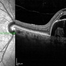
 OCT Evidence of VMT Resulting in Full Thickness Macular Hole
Dec 24 2020 by Deepak Bhojwani, MS
OCT image of a patient (with past history of focal VMT ) progressing to full thickness macular hole. Note the posterior hyaloid attachment over the torn edges of fovea.
Photographer: DEEPAK BHOJWANI
Condition/keywords: full thickness macular hole, optical coherence tomography (OCT), vitreomacular traction (VMT)
-
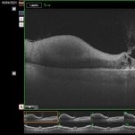
 Pigment Epithelium Detachment, Secondary to AMD
Mar 17 2023 by Ceara Donovan
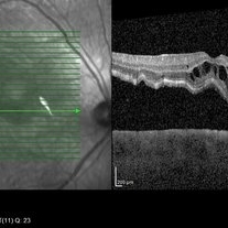
Optical coherence tomography of a 76 year old woman with a Pigment Epithelium Detachment, Secondary to AMD affecting her right eye. Patient had no significant response to Avastin, Eylea, Lucentis 0.5, or Vabysmo and was switched to Beovu. Following Beovu intravitreal injection her edema improved on OCT. Patient's vision was sc20/200+1 at the time the image was taken.
Photographer: Ceara Donovan
Imaging device: Heidelberg Spectralis
Condition/keywords: exudative age-related macular degeneration, heidelberg spectralis, macular degeneration, optical coherence tomography (OCT), pigment epithelial detachment (PED), Sub-retinal fluid
-
 Proliferative Diabetic Retinopathy
May 11 2020 by Gayathri Mohan
Color fundus photograph of a patient with PDR, showing neovascularisation infero-temporal to macula.
Photographer: Gayathri Mohan, Retina Foundation
Imaging device: Mirante, Nidek
Condition/keywords: neovascularization (NV), optical coherence tomography (OCT), proliferative diabetic retinopathy (PDR)
-
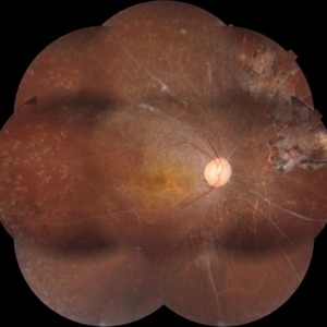 Proliferative Diabetic Retinopathy
Mar 1 2021 by Avris Romario Diparaja Siahaan
Fundus photograph (montage photography) of a 57-year-old woman with proliferative diabetic retinopathy in her both eyes.
Photographer: Nanda Lessi Hafni Eka Putri, MD (Ophthalmologist) & Ryan Mishbahuddin (Nurse), Ciawi General Hospital (Rumah Sakit Umum Daerah Ciawi)
Imaging device: DRI OCT Triton Plus
Condition/keywords: fundus photograph, montage, optical coherence tomography (OCT), swept source, wide angle imaging
-
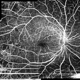 Proliferative Diabetic Retinopathy
Mar 1 2021 by Avris Romario Diparaja Siahaan
Swept source OCT angiography (montage photography) of a 62-year-old woman with proliferative diabetic retinopathy in her both eyes.
Photographer: Nanda Lessi Hafni Eka Putri, MD (Ophthalmologist) & Ryan Mishbahuddin (Nurse), Ciawi General Hospital (Rumah Sakit Umum Daerah Ciawi)
Imaging device: DRI OCT Triton Plus
Condition/keywords: fundus photograph, montage, optical coherence tomography (OCT), swept source, wide angle imaging
-
 Proliferative Diabetic Retinopathy OCT -A
Apr 2 2018 by Eduardo Torres-Porras, MD
OCT-A 57-year-old male with proliferative diabetic retinopathy affecting his left eye, 6x6 and 9x9 Angioplex maps
Photographer: EDUARDO TORRES PORRAS, PROVISSIA, PUEBLA, PUE. MEXICO
Imaging device: Angioplex cirrus 5000
Condition/keywords: CIRRUS 5000 ANGIOPLEX, optical coherence tomography (OCT), proliferative diabetic retinopathy (PDR)
-
 Retinal Cavernous Hemangioma
Oct 22 2020 by Olivia Rainey
Widefield OCT of a 31-year-old male presenting with a retinal cavernous hemangioma affecting his left eye. Patient was 18-years-old when he was diagnosed with a retinal cavernous hemangioma. He has had a few episodes of vitreous hemorrhages since then. His vision was 20/20-1 in both eyes.
Photographer: Becca Harris
Imaging device: Heidelberg Spectralis
Condition/keywords: 50 degrees, cavernous hemangioma of the retina, Heidelburg Spectralis, left eye, optical coherence tomography (OCT), wide angle imaging
-
 Retinal Detachment Sparing Fovea By Microns
Sep 24 2018 by samarth mishra
A 29-year-old young female presented with complaint of blurring of vision in the right eye since one year. Best corrected visual acuity was 20/40. On routine examination inferior retinal detachment was noted. Optical coherence tomography (OCT) showed the retinal detachment sparing the fovea by few microns.
Photographer: Aditya Birla Sankara Nethralaya, Kolkata , West Bengal , India
Condition/keywords: color fundus photograph, multicolor, optical coherence tomography (OCT)
-
 Retinal Hemorrhage
Sep 2 2021 by Avris Romario Diparaja Siahaan
Swept source OCT angiography of a 58-year-old man with hemorrhage in his left eye.
Photographer: Nanda Lessi Hafni Eka Putri, MD (Ophthalmologist) & Ryan Mishbahuddin (Nurse), Ciawi General Hospital (Rumah Sakit Umum Daerah Ciawi)
Imaging device: DRI OCT Triton Plus (Topcon)
Condition/keywords: fundus photograph, optical coherence tomography (OCT)
 Loading…