Search results (152 results)
-
 Post Traumatic Optic Nerve Head Avulsion
Nov 18 2017 by Vishal Agrawal, MD, FRCS,FACS,FASRS
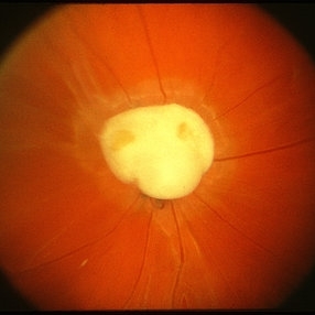
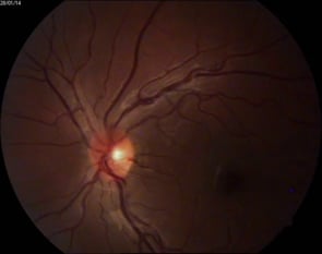
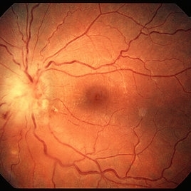
Right eye fundus picture of a 24-year-old male patient who suffered blunt trauma 7 days back with a wooden stick . He presented with NLP vision with a non reacting dilated pupil. Fundus montage picture shows ONH avulsion,CRAO,peripapillary resolving hemorrhages and cicatricial tissue at the edge.
Photographer: Vishal Agrawal, MD, SMS Medical College, Jaipur, India
Imaging device: Zeiss 524
Condition/keywords: avulsion, central retinal artery occlusion (CRAO)
-
 Toxocara Granuloma
Feb 25 2013 by Henry J. Kaplan, MD
Toxocara granuloma of the optic nerve head.
Condition/keywords: ocular toxoplasmosis, toxocara granuloma, toxocariasis
-
 Optic Nerve Head Drusen With Idiopathic CNV
Feb 17 2017 by Kristen Wagner
22-year-old female fundus photograph of a right eye with Optic Nerve Drusen with Idiopathic CNV.
Photographer: Kristen Wagner, COT, OSC Ophthalmic Photographer, Tennessee Retina, Nashville TN
Condition/keywords: choroidal neovascularization (CNV), drusen of optic disc, optic disc drusen
-
 Ophthalmic Artery Occlusion in a 39-Year-Old with Rheumatoid Arthritis
Oct 6 2020 by Michael Izzo, MD
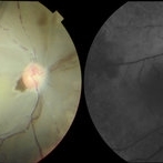
Left image: fundus photograph of a 39-year-old male with rheumatoid arthritis found to have ophthalmic artery occlusion depicting boxcar segmentation of blood in retinal vasculature and macular ischemia demonstrated by retinal whitening without cherry red fovea. Right image: early phase fluorescein angiography demonstrating patchy choroidal filling, arterial non-perfusion and optic nerve head leakage.
Photographer: Karen Rivera, COA; Washington National Eye Center
Condition/keywords: fluorescein angiogram (FA), ophthalmic artery occlusion, rheumatoid arthritis
-
 Optic Nerve Head Avulsion
Sep 24 2024 by Gustavo Uriel Fonseca Aguirre
A 14-year-old male with a history of blunt ocular trauma in the right eye presented partial avulsion of the optic nerve head and submacular hemorrhage that was managed with neumatic displacement.
Photographer: Gustavo U. Fonseca Aguirre, Fundación Hospital Nuestra Señora de la Luz, Ciudad de México
Condition/keywords: optic nerve head avulsion
-
 Optic Nerve Melanocytoma
Apr 3 2023 by Gustavo Aguirre Suarez
Fundus photograph of a 36-year-old female with a lesion dependent on the optic nerve head with subretinal extension, elevated, about 1.5 disc diameters, dark brown to black in color, involving more than three quarters of the neuroretinal ring towards the inferonasal area.
Photographer: Dr. Gustavo Aguirre-Suarez
Imaging device: Zeiss Visucam 500
Condition/keywords: melanocytic lesion, Melanocytoma
-
 Bilateral Optic Nerve Involvement in Sarcoidosis
Feb 25 2013 by Henry J. Kaplan, MD
Optic nerve head granuloma of sarcoidosis with severe infiltration and exudation in the left eye of the same patient #2.
Condition/keywords: bilateral involvement, sarcoid granuloma
-
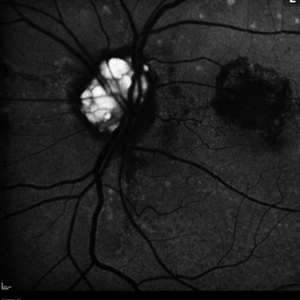
 Blue autofluroscence of Right eye optic nerve head showing auto fluorescence of the drusen
Aug 5 2022 by Kavitha Duraipandi, MD DNB FICO FRCS
A 20 year old patient referred to the clinic with blurred disc margins to rule out papilledema.
Photographer: Natalie Fox- Bussell
Condition/keywords: Blue autofluroscence, Heidelburg Spectralis
-
 Choroidal Melanoma (USG)
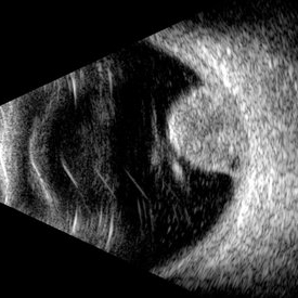
Jul 5 2025 by Gustavo Uriel Fonseca Aguirre
This B-mode transverse ultrasound scan reveals a mushroom-shaped choroidal tumor in the inferior nasal quadrant adjacent to the optic nerve head. The lesion appears solid with homogeneous internal reflectivity and is associated with minimal surrounding subretinal fluid and scleral excavation. It measures 6.54 mm in height × 7.52 mm in base diameter (transverse view) and extends 9.52 mm longitudinally. The vitreous contains abundant punctate opacities consistent with pigment dispersion. The retina and choroid remain attached elsewhere.
Photographer: Gustavo U. Fonseca Aguirre, Hospital Conde de Valenciana, Ciudad de México
Condition/keywords: choroidal melanoma
-
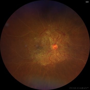 Fibrotic Vascular Tissue Proliferation
Feb 20 2024 by BENITO VERGARA, MD
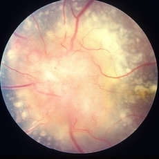
Fundus image of a 63-year-old woman with fibrotic vascular tissue proliferation that starts at the optic nerve head through the inferior temporal arcade, with the formation of new vessels in both nasal arcades and obliteration of peripheral vessels.
Photographer: Benito Vergara Flores, Asociación Para Evitar la Ceguera en México, I.A.P.
Imaging device: Zeiss Clarus 700
Condition/keywords: fibrotic neovascularization
-
 Melanocytoma
May 2 2013 by Henry J. Kaplan, MD
Melanocytoma of the optic nerve head.
Condition/keywords: melanocytoma
-
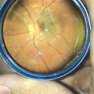 Melanocytoma of the Optic Nerve Head
Jan 13 2020 by Marlon García Roa, MD
Female 49-years-old, VA 20/100 OD and 20/20 OI. The patient does not associate low visual in right eye. Does not have heredofamiliar cancer background, and with a single pathographic unique personal background. Peripapilary injury of 3000 microns of diameter, elevated, with little defined edges, and orange pigment on the edge. in right eye.
Photographer: Marlon García Roa , INSTITUTO MEXICANO DE OFTALMOLOGIA, QUERETARO, MEXICO
Imaging device: SMARTPHONE
Condition/keywords: melanocytoma
-
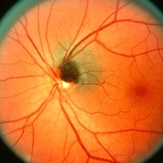
 Myelinated Nerve Fibers
Apr 18 2025 by DR Rohit Gupta
The **myelinated nerve fibers of the optic disc** (also known as **medullated nerve fibers**) are retinal nerve fibers that retain their myelin sheath as they pass through the optic nerve head. Normally, retinal nerve fibers are unmyelinated to allow for light transparency, but in some cases, myelination extends anteriorly into the retina, appearing as a striking white, feathery patch on the optic disc or peripapillary retina. ### **Key Features:** 1. **Appearance:** - Dense, white, striated patches with feathery edges. - Typically located at the superior or inferior pole of the optic disc. - May obscure retinal vessels underneath. 2. **Clinical Significance:** - Usually **benign** and asymptomatic. - **Congenital** (present at birth or early childhood). - Rarely associated with **visual field defects** (e.g., scotomas corresponding to the area of myelination). - Occasionally linked with **high myopia** or **amblyopia** if extensive. 3. **Pathophysiology:** - Failure of oligodendrocytes or Schwann cells to stop myelination at the lamina cribrosa. - Normally, myelination stops at the optic nerve head, but in this condition, it extends into the retina. 4. **Diagnosis:** - **Fundoscopy:** Classic white, feathery appearance. - **Optical Coherence Tomography (OCT):** Shows thickened retinal nerve fiber layer (RNFL). - **Visual Field Testing:** May detect defects if large. 5. **Differential Diagnosis:** - Optic disc edema - Cotton wool spots - Retinoblastoma (rarely, but must be ruled out in children) 6. **Management:** - No treatment required if asymptomatic. - Monitor for amblyopia in children. - Rare cases with significant visual impairment may need further evaluation. ### **Fun Fact:** Myelinated nerve fibers are seen in **~0.5-1%** of the population and are usually an incidental finding.
Photographer: Dr Rohit gupta
Imaging device: Samsung S21
Condition/keywords: Medulated Nerve fibre, Medullated Nerve fibres, myelinated nerve fibers, Myelinated Nerve Fibres, optic disc drusen
-
 Optic Disc Pit
Nov 8 2021 by Michael Grinton
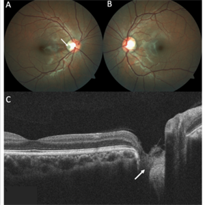
Optic disc pits are rare congenital abnormalities of the optic nerve head. Colour fundus image of an asymptomatic 18-year old male shows an optic disc pit in the right eye (A, white arrow); a small, grey, oval shaped excavation in the temporal segment of the optic disc. These pits are usually unilateral (B shows normal colour fundus of left eye) and asymptomatic. Imaging with optical coherence tomography (C) shows the optic disc pit in cross section (white arrow) and normal macular structure. In some patients with the condition, fluid can accumulate underneath the macular (serous macular detachment).
Condition/keywords: Optic disc pit, Optic nerve pit, Optic pit
-
 Optic Nerve Head Cannonball
Dec 15 2019 by Veer Singh, MS, FVRS, FMRF, FICO (Retina)
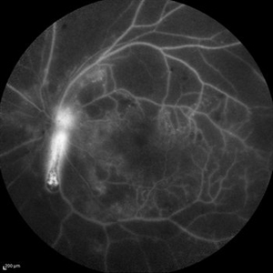
This is the fundus fluorescein angiography (FFA) of the left eye of a 62-year-old diabetic patient with proliferative diabetic retinopathy and neovascularization of disc who bled from the disc while he was undergoing an FFA procedure. The bleed from the disc gives the appearance of a cannonball fired from a cannon hence the caption "Optic Nerve Head Cannonball".
Photographer: Dr. Veer Singh
Imaging device: Heidelberg Spectralis HRA
Condition/keywords: fluorescein angiogram (FA), neovascularization of the disc (NVD), optic nerve head, proliferative diabetic retinopathy (PDR), vitreous hemorrhage
-
 Optic Nerve Head Drusen
Feb 12 2015 by Timothy S Fuller, MD
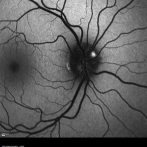
Fundus autofluorescence image of a 34-year-old woman with striking, asymptomatic optic nerve head drusen.
Photographer: Nice Hesse, Texas Retina Associates
Imaging device: Heidelberg Spectralis
Condition/keywords: drusen of optic disc
-
 Partial Optic Disc Avulsion with Optic Disc Pit
Jul 1 2018 by John S. King, MD
16-year-old with acute loss of vision after blunt finger injury to eye while playing football. This photo is three weeks post-injury. Vision HM. Retinal striae with subhyaloid heme. Decreased retinal whitening. Peripapillary heme clearing, and temporal optic disc avulsion with optic disc pit can be seen.
Photographer: Maisee Yang
Imaging device: Topcon
Condition/keywords: epiretinal membrane (ERM), optic nerve head avulsion, optic nerve pit, traumatic optic neuropathy
-
 Remnant of Hyaloidal Artery
Feb 5 2014 by Gerardo Garcia-Aguirre, MD
Video of the fundus of the left eye of a 14-year-old asymptomatic female, where a prepapillary vitreous opacity is observed. The opacity is attached to the origin of the retinal vessels in the optic nerve head, and is considered to be a remnant of the hyaloidal artery.
Photographer: Gerardo Garcia-Aguirre, MD
Condition/keywords: persistence of the hyaloid artery
-
 Retina
May 31 2014 by ruth pav
A 32-year-old woman with a history of drug abuse was admitted due to acute manifestation of multiple infarcts, including acute stroke, splenic and renal infarcts, and multiple cutaneous hematomas. Due to decreased vision in her left eye the patient was referred for ophthalmic evaluation. On exam, visual acuity was 6/10 in the right eye and no light perception in her left eye. Ophthalmoscopic examination was normal in the right eye but showed pallor of the optic nerve head with attenuated retinal vessels in the left eye. Fluorescein angiography showed an oval area of hyperfluorescence from from non-perfusion involving the macular center with staining of overlying retinal capillaries.
Photographer: Ruth Pav, Rambam medical center,Hifa Israel.
Imaging device: Zeiss FF4
Condition/keywords: retina
-
 Acute Traumatic Optic Nerve Avulsion
Feb 19 2016 by Mahdi Mwas
Fundus photograph of a 24-year-old gentleman, involved in a road traffic accident resulting in left no perception of light.
Photographer: Mahdi Mwas, FRCS, DRCOphth, Jordan
Condition/keywords: optic nerve head avulsion
-
 Drusen of Optic Disc
Mar 6 2018 by JEFFERSON R SOUSA, Tecg.º (Biomedical Systems Technology)
Female patient, 37 years old, Caucasian, with complaint of low lateral stroke in abos the eyes. In the retinal mapping examination and retinography, important alterations in the optic nerve head suggestive of DRUSAS DE PAPILA were observed. After being confirmed in the Autofluorescence examination, we observed Autohyperfluorescence compatible with deposits of calcified hyaline material, as well as another complementary exam such as USG and OCT.
Photographer: JEFFERSON R SOUSA - Study Center and Ophthalmological Research Dr. Andre M V Gomes, Institute Dr. Suel Abujamra, Clinic Marco Antonio Albhy Ophthalmology / São Paulo-Brazil
Imaging device: Heidelberg - HRA Angiograph, Autofluorescence com 30 degrees.
Condition/keywords: calcified drusen, drusen of optic disc
-
 Gyrate Atrophy
Apr 12 2023 by Ahmed Abbas Hashmi, OD
Left eye fundus of a 53-year-old male patient with advanced gyrate atrophy of the choroid and retina with macular sparing. Optic nerve head is healthy.
Photographer: Ahmed Abbas Hashmi
Imaging device: Topcon TRC-NW8F
Condition/keywords: chorioretinal atrophy
-
 Toxoplasma Neuroretinitis (Jensen`s Disease)
Feb 25 2013 by Henry J. Kaplan, MD
Toxoplasma neuroretinitis in the left eye of a patient with macular star formation, retinitis adjacent to the optic nerve head with disc swelling.
Condition/keywords: Jensen disease, ocular toxoplasmosis, toxoplasmosis
-
 Optic Nerve Head Drusen
Feb 9 2018 by Olivia Rainey
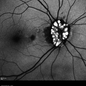
Fundus autofluorescence of a 49-year-old female with optic nerve head drusen affecting her left eye. The patient has pseudoxanthoma elasticum with choroidal neovascularization and has been receiving anti-VEGF treatment for many years.
Photographer: Olivia Rainey
Imaging device: Heidelberg Spectralis
Condition/keywords: 30 degrees, anti-VEGF, choroidal neovascularization (CNV), fundus autofluorescence (FAF), Heidelburg Spectralis, left eye, optic disc, optic nerve drusen, pseudoxanthoma elasticum (PXE)
-
---thumb.jpg/image-square;max$300,300.ImageHandler) Optic Disc Drusen
Optic Disc Drusen
Mar 27 2013 by Henry J. Kaplan, MD
Autofluorescence imaging shows heper AF on the optic nerve head specially superiorly due to drusen in the same patient #2.
Imaging device: Heidelberg spectralis
Condition/keywords: drusen of optic disc, optic disc drusen, optic nerve drusen
 Loading…