Search results (883 results)
-
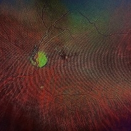
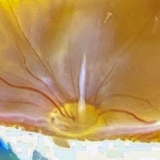
 Post Traumatic Optic Nerve Head Avulsion
Nov 18 2017 by Vishal Agrawal, MD, FRCS,FACS,FASRS
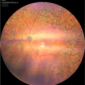
Right eye fundus picture of a 24-year-old male patient who suffered blunt trauma 7 days back with a wooden stick . He presented with NLP vision with a non reacting dilated pupil. Fundus montage picture shows ONH avulsion,CRAO,peripapillary resolving hemorrhages and cicatricial tissue at the edge.
Photographer: Vishal Agrawal, MD, SMS Medical College, Jaipur, India
Imaging device: Zeiss 524
Condition/keywords: avulsion, central retinal artery occlusion (CRAO)
-
 Venous Loop
Feb 20 2024 by Soobien Lee
A 77-year-old male with a history of bilateral optic neuropathy from bilateral optic nerve sheath meningiomas S/P radiation/proton-beam therapies. Presented with radiation retinopathy OS and a known venous loop OS.
Photographer: Gavin Bragdon, Elman Retina Group
Imaging device: Optos Ultra-Widefield Imaging
Condition/keywords: Optos, OPTOS CALIFORNIA, radiation retinopathy, retinal vascular disease, venous loop
-
 Venous Loop
Feb 20 2024 by Soobien Lee
A 77-year-old male with a history of bilateral optic neuropathy from bilateral optic nerve sheath meningiomas S/P radiation/proton-beam therapies. Presented with radiation retinopathy OS and a known venous loop OS.
Photographer: Gavin Bragdon, Elman Retina Group
Imaging device: Optos Ultra-Widefield Fluorescein Angiography
Condition/keywords: fluorescein angiogram (FA), Optos, radiation retinopathy, retinal vascular disease, venous loop
-
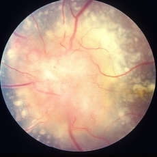
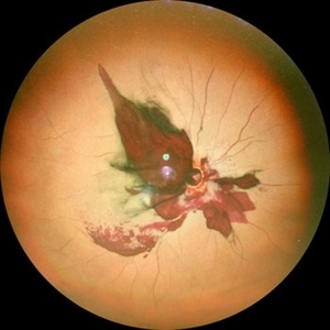 Optic Nerve Avulsion with Vitreous Hemorrhage and Pale Retina
Jan 25 2021 by Sham Talati, DOMS
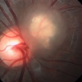
A 30-year-old male presented with history of trauma to RE with NO Perception of light in the affected eye.
Photographer: Dr. Sham Talati,Retina Foundation,Ahmedabad
Imaging device: Nidek Mirante
Condition/keywords: optic nerve, pale retina
-
 Toxocara Granuloma
Feb 25 2013 by Henry J. Kaplan, MD
Toxocara granuloma of the optic nerve head.
Condition/keywords: ocular toxoplasmosis, toxocara granuloma, toxocariasis
-
 Optic Nerve Head Drusen With Idiopathic CNV
Feb 17 2017 by Kristen Wagner
22-year-old female fundus photograph of a right eye with Optic Nerve Drusen with Idiopathic CNV.
Photographer: Kristen Wagner, COT, OSC Ophthalmic Photographer, Tennessee Retina, Nashville TN
Condition/keywords: choroidal neovascularization (CNV), drusen of optic disc, optic disc drusen
-
 Cat Eye Syndrome
Feb 11 2020 by Sophia El Hamichi, MD
A 3-year-old female with cat eye syndrome including iris, chorioretinal and optic nerve colobomas. Note the CNV temporally to the optic nerve coloboma (blue arrows)
Photographer: Giselle De Oliveira, Bascom Palmer Eye Institute, Miami
Imaging device: RetCam
Condition/keywords: cat eye syndrome, chorioretinal coloboma, choroidal neovascularization (CNV), coloboma, coloboma of optic disc, optic nerve coloboma
-
 HHPlaqueON
Aug 13 2021 by Jeffrey Barker
Hollenhorst Plaque
Photographer: Jeffrey P. Barker, B.S. Retina Vitreous Surgeons of C.N.Y.
Condition/keywords: hollenhorst plaque, optic nerve
-
 Melanocytoma of the Optic Nerve
Apr 6 2024 by Hector Gabriel Moreno Solano, MD, MHA
Fundus photograph of a 57-year-old male presented for an ophthalmological evaluation with a chief complaint of progressive visual loss. Indirect ophthalmoscopy revealed proliferative diabetic retinopathy, without macular edema, and a hyperpigmented lesion at the optic disc which corresponds to a melanocytoma.
Photographer: Héctor Gabriel Moreno-Solano
Imaging device: Clarus 700
Condition/keywords: diabetic retinopathy, intraocular tumor, melanocytoma, optic nerve
-
 Melanocytoma of the Optic Nerve
Apr 6 2024 by Hector Gabriel Moreno Solano, MD, MHA
Optic Nerve laser scan image reconstruction of a 57-year-old male presented for an ophthalmological evaluation with a chief complaint of progressive visual loss. Indirect ophthalmoscopy revealed proliferative diabetic retinopathy, without macular edema, and a hyperpigmented lesion at the optic disc which corresponds to a melanocytoma.
Photographer: Héctor Gabriel Moreno-Solano, MD, MHA
Imaging device: Mirante
Condition/keywords: intraocular tumor, macular edema, melanocytoma, optic nerve
-
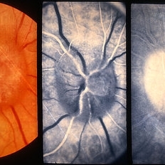
 Ophthalmic Artery Occlusion in a 39-Year-Old with Rheumatoid Arthritis
Oct 6 2020 by Michael Izzo, MD
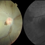
Left image: fundus photograph of a 39-year-old male with rheumatoid arthritis found to have ophthalmic artery occlusion depicting boxcar segmentation of blood in retinal vasculature and macular ischemia demonstrated by retinal whitening without cherry red fovea. Right image: early phase fluorescein angiography demonstrating patchy choroidal filling, arterial non-perfusion and optic nerve head leakage.
Photographer: Karen Rivera, COA; Washington National Eye Center
Condition/keywords: fluorescein angiogram (FA), ophthalmic artery occlusion, rheumatoid arthritis
-
 Optic Nerve Head Avulsion
Sep 24 2024 by Gustavo Uriel Fonseca Aguirre
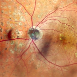
A 14-year-old male with a history of blunt ocular trauma in the right eye presented partial avulsion of the optic nerve head and submacular hemorrhage that was managed with neumatic displacement.
Photographer: Gustavo U. Fonseca Aguirre, Fundación Hospital Nuestra Señora de la Luz, Ciudad de México
Condition/keywords: optic nerve head avulsion
-
 Optic Nerve Melanocytoma
Apr 3 2023 by Gustavo Aguirre Suarez
Fundus photograph of a 36-year-old female with a lesion dependent on the optic nerve head with subretinal extension, elevated, about 1.5 disc diameters, dark brown to black in color, involving more than three quarters of the neuroretinal ring towards the inferonasal area.
Photographer: Dr. Gustavo Aguirre-Suarez
Imaging device: Zeiss Visucam 500
Condition/keywords: melanocytic lesion, Melanocytoma
-
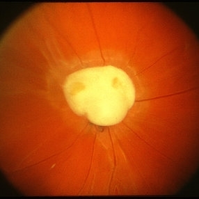
 Optic Nerve Pit
Aug 30 2012 by Raj K. Maturi, MD
congenital optic nerve pit with chronic pigment changes in macula due to detachment
Photographer: Tom Steele, CRA, Midwest Eye Institute
Imaging device: Topcon Ex
Condition/keywords: optic nerve pit
-
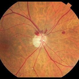
 Optic Nerve Pit
Aug 30 2012 by Raj K. Maturi, MD
Photographer: Tom Steele, CRA, Midwest Eye Institute
Imaging device: Topcon Ex
Condition/keywords: optic nerve pit
-
 Amelanotic Mushroom-Shaped Choroidal Melanoma
May 18 2020 by McGill University Health Centre
This enucleation specimen demonstrates an amelanotic, mushroom-shaped, slightly hemorrhagic tumor near the optic nerve (arrow). True retinal detachment is present, and the retina is folded (arrowhead). The subretinal fluid is hazy (*).
Condition/keywords: amelanotic melanoma, enucleation, mushroom-shaped
-
 Astrocytic Hamartoma
Oct 10 2012 by Anat Loewenstein, MD
Five year-old girl came for regular eye examination with no complaints. Medical and ophthalmic history was unremarkable. On examination, visual acuity was 20/25 in both eyes. A Retinal Astrocytic Hamartoma was seen adjacent to the right optic nerve (picture). The rest of the eye examination was normal and no Lisch nodules were seen. The patient was referred for a pediatric neurologist examination and MRI scan of the brain.
Photographer: Galit Yair-Pur
Imaging device: ZEISS FF450 PLUS IR
-
 Bergmeister Papilla
Feb 20 2020 by Nisarg Joshi, MD
Gross pathology photo of a Bergmeister Papilla. It is a remnant of incompletely resorbed hyaloid vasculature from ocular development. This glial tissue is seen emminating from the optic nerve, which also shows glaucomatous cupping. The eye was enucleated due to a choroidal melanoma.
Photographer: Nisarg Joshi, MD, Geisinger Medical Center
Imaging device: Digital camera
Condition/keywords: Bergmeister's Papillae, hyaloid artery, persistent fetal vasculature (PFV)
-
 Bilateral Optic Nerve Involvement in Sarcoidosis
Feb 25 2013 by Henry J. Kaplan, MD
Optic nerve head granuloma of sarcoidosis with severe infiltration and exudation in the left eye of the same patient #2.
Condition/keywords: bilateral involvement, sarcoid granuloma
-
 Blue autofluroscence of Right eye optic nerve head showing auto fluorescence of the drusen
Aug 5 2022 by Kavitha Duraipandi, MD DNB FICO FRCS
A 20 year old patient referred to the clinic with blurred disc margins to rule out papilledema.
Photographer: Natalie Fox- Bussell
Condition/keywords: Blue autofluroscence, Heidelburg Spectralis
-
 Branch Retinal Artery Occlusion
Sep 9 2018 by Gabriela Lopezcarasa Hernandez, MD
88-year-old female patient with sudden decrease in visual acuity and scotoma in left eye, please notice the widening retina due to retinal edema of branch occlusion with hollenhorst plaque in the artery and the optic nerve.
Photographer: Araceli Rojas
Imaging device: Zeiss FF4
Condition/keywords: branch retinal artery occlusion (BRAO)
-
 Chorioretinal Coloboma with Retinal Detachment
Dec 5 2020 by Niloofar Piri, MD
14-year-old female with 1q21.1 microdeletion syndrome and behavioral, intellectual, and systemic abnormalities, including congenital microcornea, iris coloboma, and chorioretinal and optic nerve coloboma presented with decreased vision. Right eye fundus taken with RetCam shows coloboma with retinal detachment. (Left eye showed white cataract with funnel RD on B-scan).
Photographer: Niloofar Piri MD, Douglas Snyder MD
Condition/keywords: chorioretinal coloboma, optic nerve coloboma
-
 Choroidal nevus with halo
Aug 21 2022 by Niloofar Piri, MD
Fundus photograph of the right eye in a 43 yo patient demonstrating a flat choroidal nevsu with halo nasal to the optic nerve
Photographer: Andrew Polk, MD
Condition/keywords: Choroidal nevus, nevus with halo
-
 Chronic Papilledema
Feb 20 2013 by From the Collections of Thomas M. Aaberg, MD and Thomas M. Aaberg Jr., MD
Secondary to cystic _______ of the optic nerve secondary to arachnoiditis.
Condition/keywords: papilledema
-
 Coloboma involving the Optic nerve, Retina, and Choroid
Dec 6 2021 by Jesus Lozano, MD
78-year-old woman after prophylactic laser photocoagulation (PLP) for her RE Coloboma involving the optic nerve, retina, and choroid. At 6 month follow up, patient preserved her FC vision as it was before the procedure. Retina attached.
Photographer: Yair Bet Yosef, Hadassah Medical Center. Israel
Imaging device: Optos Silverstone fundus image
Condition/keywords: coloboma, coloboma of choroid, coloboma of macula, coloboma of optic disc, PLP, prophylactic photocoagulation
 Loading…