Search results (4 results)
-
 CMV Retinitis in AIDS Patient
Dec 12 2019 by McGill University Health Centre
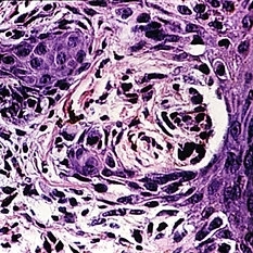
Histopathological examination of the retina showing no inflammation and a large multinucleated CMV infected cell. Sections of the choroid shows no inflammation, the choroid capillaries and normal choroidal melanocytes.
Photographer: Miguel N. Burnier, McGill University Health Center-McGill University Ocular Pathology & Translational Research Laboratory
Imaging device: Zeiss
Condition/keywords: choroid, cytomegalovirus (CMV), histopathology, multinucleated giant cells, retina
-
 Optic Nerve Melanocytoma
May 4 2025 by KANWALJEET HARJOT MADAN, M.S. (Ophthalmology), FAICO (Vitreous - Retina)
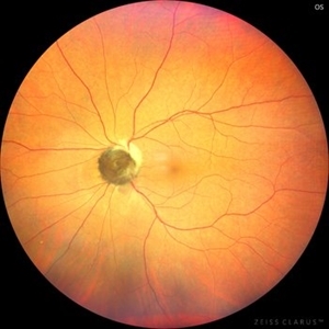
This is a fundus picture of a young 42-year male who visited for a routine eye exam. His BCVA was 20/20 in both eyes. Anterior segment examination was normal. His left eye showed grey-black pigmentation at the infero-nasal margin of the optic disc. Fundus of the right eye was normal. The patient was diagnosed to have optic disc melanocytoma on multimodal imaging and was advised regular follow-up. Optic nerve melanocytoma is typically a benign tumor made up of melanocytes and melanin. It can grow, but rarely transforms into a malignancy. Patients with Optic Nerve Melanocytoma should be periodically examined for evidence of growth, loss of visual field and optic nerve compression.
Photographer: Dr. Kanwaljeet Harjot Madan, Thind Eye Hospital, Jalandhar City (Punjab) INDIA.
Imaging device: Zeiss Fundus Camera
Condition/keywords: melanocytoma, melanoma, optic nerve
-
 Optic Nerve Melanocytoma
Nov 20 2019 by McGill University Health Centre
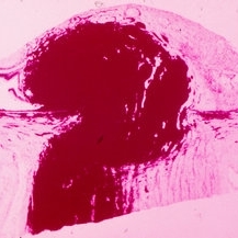
A 39-year-old female with a pigmented mass on the optic nerve. Differential diagnosis with peripapillary melanoma. Histopathology of a typical melanocytoma with large benign melanocytes which are heavily pigmented.
Condition/keywords: melanocytoma, optic nerve
-
 Slide 5-43
Feb 25 2019 by Lancaster Course in Ophthalmology
Nest of atypical melanocytes resembling a junctional nevus at the edge of a superficial spreading melanoma in a 70-year-old female. Note the inflammatory cells in the dermis.
Condition/keywords: junctional nevus, melanocytes, melanoma
 Loading…