Search results (146 results)
-
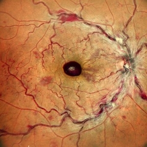
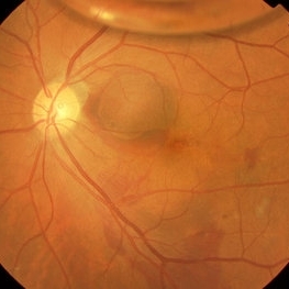
 Aggressive Posterior Retinopathy of Prematurity with Macular Hemorrhage
Oct 9 2012 by Audina M. Berrocal, MD FASRS
APROP with multiple pre-retinal hemorrhages
Photographer: Ditte Hess CRA, BPEI
Imaging device: RETCAM
Condition/keywords: macular hemorrhage, retinopathy of prematurity (ROP)
-
 Massive Submacular Hemorrhage
Sep 28 2012 by Joseph M. Civantos, MD
82-year-old gentleman who developed this massive submacular hemorrhage 3 days after his 16th Lucentis injection. Visual acuity dropped from 20/80 to LP.
Condition/keywords: subretinal hemorrhage
-
 Optic Nerve Head Avulsion
Sep 24 2024 by Gustavo Uriel Fonseca Aguirre
A 14-year-old male with a history of blunt ocular trauma in the right eye presented partial avulsion of the optic nerve head and submacular hemorrhage that was managed with neumatic displacement.
Photographer: Gustavo U. Fonseca Aguirre, Fundación Hospital Nuestra Señora de la Luz, Ciudad de México
Condition/keywords: optic nerve head avulsion
-
 Post Subretinal tpa , viterectomy and gas
May 6 2022 by Shobhit Chawla, M.S.
SUBMACULAR HAEMORRHAGE IN A 38YEAR OLD LADY PATIENT CAUSE POLYP BLEED IN PCV. Following viterectomy , subretinal tpa . gas and aflibercept injection. 7 day post operative image.
Photographer: Shobhit Chawla
Imaging device: Zeiss Clarus 500
Condition/keywords: aflibercept, intravitreal gas bubble, submacular hemorrhage, tissue plasminogen activator (tPA), vitrectomy
-
 Preretinal Hemorrhage
May 6 2017 by Mitzy E Torres Soriano, MD
Fundus photograph of a 36-year-old-woman with a preretinal subhyaloid hemorrhage (valsalva retinopathy).
Photographer: Mitzy Torres Soriano
Condition/keywords: macular hemorrhage, premacular hemorrhage, preretinal hemorrhage, subhyaloid hemorrhage, valsalva retinopathy
-
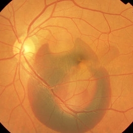
 Aggressive Posterior Retinopathy of Prematurity with Macular Hemorrhage
Oct 9 2012 by Audina M. Berrocal, MD FASRS
Aggressive posterior Type 1 ROP
Photographer: Ditte Hess CRA, BPEI
Imaging device: RETCAM
Condition/keywords: aggressive posterior retinopathy of prematurity (APROP), macular hemorrhage, retinopathy of prematurity (ROP)
-
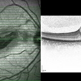
 Central Retinal Vein Occlusion with Foveal Hemorrhage
Apr 17 2025 by Malvika Singh
Fundus photograph of a 41 year-old, male, with a central retinal vein occlusion and a foveal sub-internal limiting membrane hemorrhage.
Photographer: Dr Malvika Singh, Retina Foundation, Ahmedabad, India
Imaging device: Mirante SLO/OCT
Condition/keywords: central retinal vein occlusion (CRVO), macular hemorrhage
-
 Chronical Submacular Hemorrhage in the Setting of Neovascular AMD
Mar 23 2015 by Rita Couceiro, MD, MS
An 80-year-old male, with a history of hypertension and high cholesterol, complained of acute and painless vision loss in his left eye (OS) in the previous 5 months. On observation best corrected visual acuity in OS was hand motion. A dense vitreous opacity in OS precluded fundus examination. Ocular ultrasound revealed vitreous hemorrhage and thickening of the macular area. The patient was submitted to pars plana vitrectomy, which disclosed a large submacular hemorrhage with chronical features and disciform scarring in the setting of neovascular AMD.
Imaging device: Intraoperative fundus photograph
Condition/keywords: neovascular age-related macular degeneration (AMD), submacular hemorrhage, wet age-related macular degeneration (wet AMD)
-
 Myopic Degeneration, Macular Hemorrhage
Sep 10 2014 by Mehul A Shah
A 50-year-old male patient presented with complaint of sudden loss of vision.
Photographer: Drashti Netralaya,Dahod
Imaging device: FF 450
Condition/keywords: myopic degeneration
-
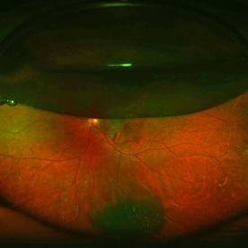 Pneumatic Displacement of a Massive Submacular Hemorrhage
Aug 3 2013 by Yusuke Oshima, MD, PhD
Pneumatic displacement of massive submacular hemorrhage with C3F8 gas.
Condition/keywords: gas pneumatic displacement, polypoidal choroidal vasculopathy (PCV), submacular hemorrhage, subretinal hemorrhage
-
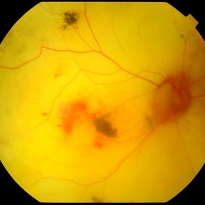
 Premacular Hemorrhage in Acute Myeloid Leukemia
Feb 10 2016 by Mallika Goyal, MD
Fundus photograph of a 45-year-old male with acute myeloid leukemia and thrombocytopenia shows premacular & retinal haemorrhage.
Photographer: Mallika Goyal, MD, Apollo Health City, Jubilee Hills, Hyderabad, India
Condition/keywords: premacular hemorrhage
-
 Sub Macular Fibrosis in the Setting of Old Sub Macular Hemorrhage
Feb 15 2024 by Sayena . Jabbehdari, MD, MPH, MBA
Pseudo color fundus photo of 78 years old male with history of sub macular hemorrhage in the setting of wet age-related macular degeneration. You can appreciate the tear shaped appearance of blood due to gravity. The OCT of the macula depicts the huge (>950mm) sub retinal fibrosis.
Photographer: Sayena Jabbehdari MD MPH , University of Arkansas in Little Rock
Imaging device: Clarus
Condition/keywords: macular neovascular disease, retina, submacular hemorrhage, Wet age related macular degeneration
-
 Submacular Hemorrhage PCV
May 6 2022 by Shobhit Chawla, M.S.
Submacular hemorrhage in a 38 years old female patient cause polyp bleed in PCV.
Photographer: Shobhit Chawla
Imaging device: Zeiss Clarus 500
Condition/keywords: polypoidal choroidal vasculopathy (PCV), submacular hemorrhage
-
 Submacular Hemorrhage After Pneumatic Displacement
Mar 21 2013 by Yusuke Oshima, MD, PhD
Fundus photograph demonstrates an effective displacement of the subretinal hemorrhage from the fovea.
Photographer: Yusuke Takada, Osaka University Graduate School of Medicine
Condition/keywords: submacular hemorrhage
-
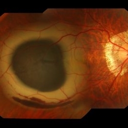
 Submacular Hemorrhage Before Treatment
Mar 21 2013 by Yusuke Oshima, MD, PhD
Fundus photograph of an 83-year-old man with a submacular hemorrhage due to polypoidal choroidal vasculopathy.
Photographer: Yusuke Takada, Osaka University Graduate School of Medicine
Condition/keywords: submacular hemorrhage
-
 t-PA with SF6 Gas for Submacular Hemorrhage
Aug 13 2019 by ANUBHAV GOYAL, DNB, FVRS
t-PA with SF6 gas for fresh submacular hemorrhage.
Photographer: ANUBHAV GOYAL, GIRIDHAR EYE INSTITUTE, COCHIN, KERALA , INDIA 682020
Imaging device: OPTOS FUNDUS CAMERA
Condition/keywords: submacular hemorrhage, sulphur hexafluoride gas, tissue plasminogen activator (tPA)
-
 Terson's Syndrome
Jan 7 2015 by H. Michael Lambert, MD
Color photograph LE, premacular boat shaped hemorrhage due to Terson's syndrome.
Condition/keywords: premacular hemorrhage
-
 Submacular Hemorrhage
Mar 12 2016 by Sjakon G Tahija, MD
This a fundus photograph of a high myope who presented with a submacular hemorrhage.
Photographer: Avris Siahaan, Klinik Mata Nusantara, Jakarta, Indonesia
Condition/keywords: high myopia, spontaneous submacular hemorrhage
-
 Submacular Hemorrhage
Apr 24 2018 by Pauline T Merrill, MD, FASRS
Fundus photo of left eye of a 65-year-old AMD patient who presented with sudden drop of vision from 20/30 to CF due to a large submacular hemorrhage, 7 months following her last Eylea injection. She underwent immediate injection of C3F8 in the office, with little effect. 10 days later vitrectomy with subretinal tPA and air-fluid exchange was performed, with successful displacement of the hemorrhage.
Photographer: Ermelinda Diaz, Illinois Retina Associates, Chicago, Illinois
Imaging device: Topcon 50DX
Condition/keywords: neovascular age-related macular degeneration (AMD), submacular hemorrhage
-
 Atrophic Pigment Epithelium
Jul 19 2019 by JEFFERSON R SOUSA, Tecg.º (Biomedical Systems Technology)
Patient 16-years-old, visual acuity with light perception. In retinal evaluation presented total atrophy retinal pigment epithelium and total papilla excavation. Mobilization of pigments and presence of macular hemorrhage.
Photographer: JEFFERSON R SOUSA - Study Center and Ophthalmological Research Dr. Andre M V Gomes, Institute Dr. Suel Abujamra São Paulo-Brazil
Imaging device: Topcon TRC-50 DX, Imaginet 4.0, angle de 35 graus. Flash 12w-s
Condition/keywords: atrophic pigment epithelium
-
 Blunt Ocular Trauma Due to Firework Injury
Jun 9 2020 by Brittany Rota
Ultra- widefield pseudocolor image of an 18-year-old male with blunt ocular trauma in the right eye due to a firework injury. The patient presented with commotio retinae (sclopteria), an acute vitreous hemorrhage, choroidal rupture, and a subretinal hemorrhage. The referring physician performed surgery on the lateral rectus muscle which was macerated but not severed, and several orbital fibrous foreign bodies were removed from the posterior orbit. The globe was intact. There is no evidence of retinal tear in the region of sclopetaria; however, there is complete necrosis of the temporal peripheral choroid and retina. The vitreous hemorrhage was slowly clearing on his exam 6-9-2020. The patient is developing subretinal fibrosis. The physician is concerned about the choroidal rupture that is visible through the submacular hemorrhage. There is one rupture that appears to course directly under the fovea. The physician states that if this is the case, his vision most likely will be 20/200 or worse. His vision was hand motion in all fields except nasally, which he was unable to see hand motion at his visit on 6-9-2020.
Photographer: Brittany Rota
Imaging device: Optos California
Condition/keywords: blunt trauma, choroidal rupture, commotio retinae, fibrosis, firework injury, fundus photograph, hand motion, necrotizing retina, Optos, pseudocolor, subretinal hemorrhage, vitreous hemorrhage
-
 Leukemia1
Mar 16 2013 by Roy Schwartz, MD
At presentation, spectral domain OCT shows intraretinal and sub-ILM hemorrhage as well as thickening of the RNFL in the area of the cotton wool spot.
Photographer: Galit Yair-Pur
Condition/keywords: acute leukemia, macular hemorrhage
-
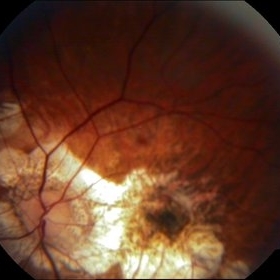
 Macular Degeneration with Large Submacular Hemorrhage
Oct 10 2012 by Joseph M. Civantos, MD
Macular degeneration with large submacular hemorrhage.
Condition/keywords: submacular hemorrhage
-
 Submacular Hemorrhage
Feb 28 2018 by Theodore Leng, MD, MS, FASRS
75-year-old woman with submacular hemorrhage secondary to wet AMD.
Condition/keywords: submacular hemorrhage
-
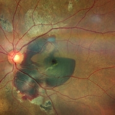
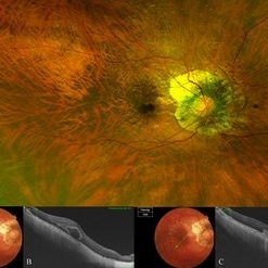 Bow-Tie Macular Hemorrhage With Cyst- Atypical Presentation of Myopic Choroidal Neovascularization
Mar 26 2021 by RUSHIK PATEL
The image of right eye of 51-year-old lady with high myopia show " Bow-Tie" macular hemorrhage (A). Optical coherence tomography (B) scan passing through hemorrhage showed intraretinal cystic lesion. During the course of intravitreal anti-VEGF injection treatment, the lesion converted into typical myopic choroidal neovascularization (C).
Photographer: Rushik Patel, Netralaya Super Speciality Eye Hospital
Condition/keywords: cyst, macular hemorrhage, myopic choroidal neovascularization (CNV)
 Loading…