Search results (64 results)
-
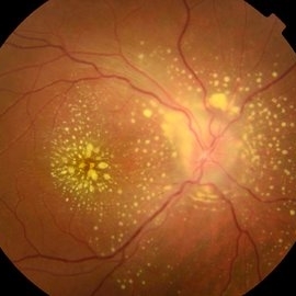
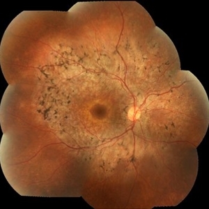
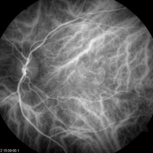
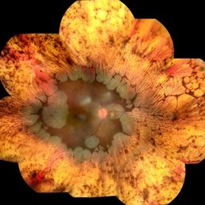 Gyrate Atrophy
Oct 30 2020 by JEFFERSON R SOUSA, Tecg.º (Biomedical Systems Technology)
Female patient, 28-year-old, with low vision in both eyes since childhood. In routine examination, important changes were observed with atrophic, symmetrical and bilateral aspects with apparently preservation of the central retina.
Condition/keywords: gyrate atrophy
-
 Arterial Occlusion
Jul 25 2019 by JEFFERSON R SOUSA, Tecg.º (Biomedical Systems Technology)
Male patient 16-years-old, was admitted to the clinic with low vision failure. On evaluation, signs of arterial occlusion in the right eye were observed. The imaging exams in the clinical evaluation showed important changes in the blood flow of one of the carotid arteries (partial obstruction), probably atherosclerotic carotid disease.
Photographer: JEFFERSON R SOUSA - Study Center and Ophthalmological Research Dr. Andre M V Gomes, Institute Dr. Suel Abujamra São Paulo-Brazil
Imaging device: Topcon TRC-50 DX, Imaginet 5.0, angle de 50 graus. Flash 36
Condition/keywords: arterial occlusion
-
 Choroideremia
Sep 21 2022 by Zach Seim
Ultra-widefield fundus photo of a 74 year old male presenting with severe vision loss beginning at age 55. Patient sought a second opinion with our office and was diagnosed with Choroideremia. Patient denies hearing loss, heart problems, balance issues, polydactyly, kidney problems, and dental problems. Patient reports that nobody in the family had blindness. Choroideremia is an X-linked chorioretinal dystrophy characterized by the diffuse, progressive degeneration of the retinal pigment epithelium (RPE), photoreceptors and choriocapillaris. It is caused by a mutation in the CHM gene.
Photographer: Zach Seim
Imaging device: Optos California
Condition/keywords: choroideremia, hereditary choroidal atrophy, hereditary retinal dystrophy, left eye, light perception, low vision, Optos, pseudocolor, ultra-wide field imaging
-
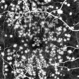
 Drusen
Mar 29 2018 by JEFFERSON R SOUSA, Tecg.º (Biomedical Systems Technology)
Male patient 27-years-old, with complaint of low vision in both eyes. The fundoscopic evaluation found the presence of drusen topography in the posterior pole with foveal. Fluorescein angiography shows the typical pattern of hyperfluorescence of drusen in the first minute of angiography.
Photographer: JEFFERSON R SOUSA - Study Center and Ophthalmological Research Dr. Andre M V Gomes, Institute Dr. Suel Abujamra São Paulo-Brazil
Imaging device: Topcon TRC-50 DX, Imaginet 5.0, angle de 50 graus. Flash 150.
Condition/keywords: colloidal drusen, drusen
-
 DUSN (Diffuse Unilateral Subacute Neuroretinitis)
Sep 2 2016 by JEFFERSON R SOUSA, Tecg.º (Biomedical Systems Technology)
Patient female, 15-year-old, he entered the clinic with complaint of low vision, visual acuity without correction was 20/60 in the right eye and 20/30 in the left eye. In the ocular exam of retinografia, there was change in the epithelium macular pigment and a small larva juxtafoveal above.
Photographer: JEFFERSON R SOUSA - Study Center and Ophthalmological Research Dr. Andre M V Gomes, Institute Dr. Suel Abujamra São Paulo-Brazil
Imaging device: Topcon TRC-50 Dx - Angulation of field photo of 35 Degrees, flash 36, Digital system Imaginet
Condition/keywords: diffuse unilateral subacute neuroretinitis (DUSN), larva, uveitis
-
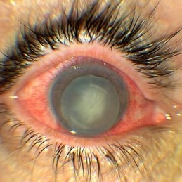 Luxated lens to anterior segment
Sep 7 2022 by JEFFERSON R SOUSA, Tecg.º (Biomedical Systems Technology)
Patient 61 years old, Female, subta low vision after blunt trauma. In the anterior segment photograph, the presence of a lens in the anterior chamber is observed. In the previous follow-up OCT, the disorganization of this follow-up is clear. Above all, the documentation of these cases is essential for future decisions. This patient was urgently referred for a surgical procedure, mainly to control the intraocular pressure, which was at 60 IOP.
Photographer: JEFFERSON ROCHA DE SOUSA - Retinal Department at Instituto Dr. Suel Abujamra Sao Paulo-Brazil.
Imaging device: Clarus 700 - Zeiss,
Condition/keywords: lens luxation, Luxated lens to anterior segment, subluxation of lens
-
 Mixed Occlusion of Artery and Vein
Jan 6 2021 by Renata Garcia Franco, Md
Male with a history of smoking, sudden low vision of the right eye, retinal neovascularization and inferior preretinal hemorrhage.
Photographer: Fatima Hernandez, Instituto de la Retina del Bajio SC
Imaging device: Zeiss
Condition/keywords: arterial occlusion
-
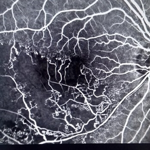
 Retinoschisis
Mar 28 2021 by JEFFERSON R SOUSA, Tecg.º (Biomedical Systems Technology)
A 14-year-old male patient was admitted for visual assessment. Visual acuity s/c in the right eye and 20/80 in the left eye. According to family members, he reported low vision since childhood. He had already undergone photocoagulation treatment at another service for which he had a diagnostic hypothesis of Coats' disease. Laboratory tests were requested (HIV, TOXO, TOXOCARIASIS, ACE, VDRL, PPD). In the evaluation, there was significant exudation in the posterior pole, some vascular irregularities in the right eye. In the left eye, there is retinoschisis affecting the entire posterior pole and the nasal region to the optic disc, macula with a characteristic chariot-wheel appearance, well exemplified by OCT-A (Structrure Deep: IPL - 25, OPL - 25).
Photographer: JEFFERSON R SOUSA - Study Center and Ophthalmological Research Dr. Andre M V Gomes, Institute Dr. Suel Abujamra São Paulo-Brazil
Imaging device: Optical coherence tomography system Optical Coherence Tomography system OCT CIRRUS 5000, Line Protocol, HD 21 line. Cirrus 5000 does not do a wide-angle tomographic image. (Structrure Deep: IPL - 25, OPL - 25).
Condition/keywords: Coats' disease, retinoschisis
-
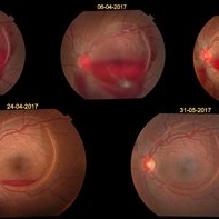 valsalva retinopathy
Feb 6 2018 by JEFFERSON R SOUSA, Tecg.º (Biomedical Systems Technology)
A 28-year-old female patient, at the 28th week of gestation, presented low vision. Retinal mapping and retinography examination revealed extensive subhaloidal haemorrhage suggestive of RETINOPATHY VALSALVA in pregnancy. In periodic follow-up, spontaneous reabsorption of the hemorrhage was observed.
Photographer: JEFFERSON R SOUSA - Study Center and Ophthalmological Research Dr. Andre M V Gomes, Institute Dr. Suel Abujamra São Paulo-Brazil
Imaging device: Fundus camera Topcon TRC-50 DX, Imaginet, 50 degree field. Flash 50
Condition/keywords: valsalva retinopathy
-
 Uveitis Posterior
Jul 19 2019 by JEFFERSON R SOUSA, Tecg.º (Biomedical Systems Technology)
A 23-year-old male patient attended the clinic with low vision of the right eye. In the evaluation it presented important fundoscopical alterations like retinal exudations in the posterior pole and nasal retina, aspects of macular star. It was proven that it was a posterior uveitis.
Photographer: JEFFERSON R SOUSA - Study Center and Ophthalmological Research Dr. Andre M V Gomes, Institute Dr. Suel Abujamra São Paulo-Brazil
Imaging device: Topcon TRC-50 DX, Imaginet 4.0, angle de 50 graus. Flash 50w-s
Condition/keywords: uveitis
-
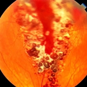
 Retinal Detachment
Feb 17 2018 by JEFFERSON R SOUSA, Tecg.º (Biomedical Systems Technology)
A 42-year-old patient complained of low vision in the left eye. In retinal mapping and background color photography, extensive retinal detachment was observed.
Photographer: JEFFERSON R SOUSA - Study Center and Ophthalmological Research Dr. Andre M V Gomes, Institute Dr. Suel Abujamra São Paulo-Brazil
Imaging device: Fundus camera Topcon TRC-50 DX, Imaginet 5.0, angle de 50 graus. Flash 36 / Mosaic with 10 images.
-
 Retinal Detachment
Feb 8 2018 by JEFFERSON R SOUSA, Tecg.º (Biomedical Systems Technology)
The male patient attended the clinic with low vision. In the retinal and retinal mapping examination, important fudoscopical alterations were observed. Full retinal detachment with Giant rupture in upper temporal arch.
Photographer: JEFFERSON R SOUSA - Study Center and Ophthalmological Research Dr. Andre M V Gomes, Institute Dr. Suel Abujamra São Paulo-Brazil
Imaging device: Fundus camera Topcon TRC-50 DX, Imaginet 5.0, campo de 50 graus. Flash 36 / Mosaic with 16 images.
Condition/keywords: retinal in rupture
-
 Coloboma
Jan 23 2018 by JEFFERSON R SOUSA, Tecg.º (Biomedical Systems Technology)
Male patient, 22 years old, with low vision since infancy. In retinal and retinal mapping examinations, important alterations were observed in the formation of retinochoroidal structures suggestive of coloboma.
Photographer: JEFFERSON R SOUSA - Study Center and Ophthalmological Research Dr. Andre M V Gomes, Dr. Suel Abujamra Institute São Paulo-Brazil
Imaging device: Acquisition of the image in the Camera background Topcon TRC-50 Dx - IA, Keystone field photo of 50 Degrees. Composition automatic of Imaginet with manual adjustment
Condition/keywords: coloboma, coloboma of choroid
-
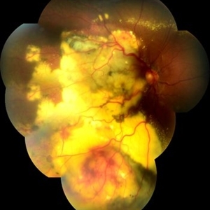
 Syphilis Neuroretinopathy
Apr 2 2018 by JEFFERSON R SOUSA, Tecg.º (Biomedical Systems Technology)
Female patient, 21-years-old, with complaint of low vision in the right eye for 3 years. According to information from the patient's history, at the time she noticed the low vision, it also coincided with a picture of a strong urinary infection as well as episodes of constant tonsillitis. Yes, the patient did not seek medical attention and self-medicated with antibiotics. In ophthalmologic evaluation, as well as examinations of color retinography and ocular fundus autofluorescence, important pigmentary alterations were observed following vascular arches with pigment mobilization in osteoclasts (aspect of a unilateral pigmentary retinitis secondary to the inflammatory process). Which suggested inflammatory process sequelae. Through the laboratory tests, he had positive (+) confirmation for SYPHILIS NEURORETINOPATHY .
Photographer: JEFFERSON R SOUSA - Study Center and Ophthalmological Research Dr. Andre M V Gomes, Institute Dr. Suel Abujamra São Paulo-Brazil
Imaging device: Fundus camera Topcon TRC-50 DX, Imaginet 5.0, angle de 50 graus. Flash 36 / Mosaic with 10 images.
Condition/keywords: neurosyphilitic optic atrophy, retinitis pigmentosa, syphilis, syphilis neuroretinopathy
-
 Drusen
Mar 29 2018 by JEFFERSON R SOUSA, Tecg.º (Biomedical Systems Technology)
Male patient, 27-years-old, with complaint of low vision in both eyes. The fundoscopic evaluation was found the presence of drusen topography in the posterior pole with foveal. Fluorescein angiography shows the typical pattern of hyperfluorescence of drusen in the first minute of angiography.
Photographer: JEFFERSON R SOUSA - Study Center and Ophthalmological Research Dr. Andre M V Gomes, Institute Dr. Suel Abujamra São Paulo-Brazil
Imaging device: Topcon TRC-50 DX, Imaginet 5.0, angle de 20 graus. Flash 150.
Condition/keywords: colloidal drusen, drusen
-
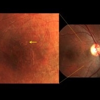
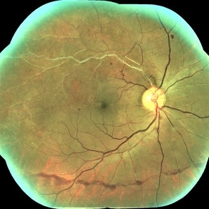
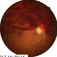 Hemi-Central Retinal Venous Occlusion
Apr 17 2018 by Ronald Silva
Fundus photograph of an 55-year-old man with low vision acuity for 2 weeks, and was observed hemi-central retinal venous oclusion right eye.
Photographer: Ronald Rocha da Silva, HCOE, Feira de Santana-BA
Condition/keywords: central retinal vein occlusion (CRVO)
-
 Leber's Miliary Aneurysm
Feb 17 2018 by JEFFERSON R SOUSA, Tecg.º (Biomedical Systems Technology)
Male patient, 29 years old, with low vision in the right eye has 9 months. In the retinal mapping and color retinography examination, there were important fundoscopical alterations.
Photographer: JEFFERSON R SOUSA - Study Center and Ophthalmological Research Dr. Andre M V Gomes, Institute Dr. Suel Abujamra São Paulo-Brazil
Imaging device: Fundus camera Topcon TRC-50 DX, Imaginet 5.0, angle de 50 graus. Flash 36 / Mosaic with 11 images.
Condition/keywords: Leber's miliary aneurysm, lipid exudation, massive lipid exudation
-
 occlusion of retinal vein
Mar 15 2018 by JEFFERSON R SOUSA, Tecg.º (Biomedical Systems Technology)
Male patient, 55 years old, with low vision in the right eye. In the retinal mapping examination and color photography, important alterations were observed suggesting an occlusion of the inferior vein. It was later confirmed in fluorescein angiography.
Photographer: JEFFERSON R SOUSA - Study Center and Ophthalmological Research Dr. Andre M V Gomes, Institute Dr. Suel Abujamra São Paulo-Brazil
Imaging device: Heidelberg Engineering HRA - 2 or Spectralis Angiograph, 30 degrees.
Condition/keywords: occlusion of partial of retinal vein, occlusion of retinal vein, oclus
-
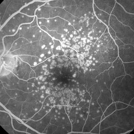
 Age-Related Macular Degeneration
Sep 12 2016 by JEFFERSON R SOUSA, Tecg.º (Biomedical Systems Technology)
Female patient, 57-years-old complaining of low vision, image with tortuosity in both eyes.
Photographer: JEFFERSON R SOUSA - Study Center and Ophthalmological Research Dr. Andre M V Gomes, Institute Dr. Suel Abujamra São Paulo-Brazil
Imaging device: OCT-Cirrus, HD 5 online with 3D cube. Color photography with Topcon TRC-50 Ex/OphthaVision
Condition/keywords: age-related macular degeneration (AMD)
-
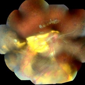
 Cavernous Hemangioma of the Retina
Sep 11 2016 by JEFFERSON R SOUSA, Tecg.º (Biomedical Systems Technology)
A female patient, 13 years of age, with complaint of low vision in her left eye, had esotropia in this eye. In the examination of fundoscopy and color photograph, we observed a pattern of multiple formations venous aneurysm with aspects of bunches of grapes in the nasal cavity above, which is characteristic of the cavernous hemangiomas of the retina.
Photographer: JEFFERSON R SOUSA - Study Center and Ophthalmological Research Dr. Andre M V Gomes, Institute Dr. Suel Abujamra São Paulo-Brazil
Imaging device: Topcon TRC-50VT, Film, Kodak Ektachrome 160 - ASA 100 / 35mm, field of 35 degrees. Flash 100.
Condition/keywords: cavernous hemangioma of the retina, tumor
-
 Choroidal Neovascular Membrane (CNVM)
Jan 23 2018 by JEFFERSON R SOUSA, Tecg.º (Biomedical Systems Technology)
Female patient, 56 years old with low vision in the left eye on Fluorescent Retinography with Green Indocyanine showed a pattern of subretinal neovascular formation, typical of the subetinal neovascular membrane.
Photographer: JEFFERSON R SOUSA - Study Center and Ophthalmological Research Dr. Andre M V Gomes, Dr. Suel Abujamra Institute São Paulo-Brazil
Imaging device: Acquisition of the image in the Camera background Zeiss - Visucam-500.
Condition/keywords: choroidal neovascular membrane (CNVM)
-
 Coats' Disease
Mar 28 2021 by JEFFERSON R SOUSA, Tecg.º (Biomedical Systems Technology)
A 14-year-old male patient was admitted for visual evaluation. Visual acuity s/c in the right eye and 20/80 in the left eye. According to family members, he reported low vision since childhood. He had already undergone treatment with photocoagulation in another service to which he had a diagnostic hypothesis of Coatas disease. Laboratory tests were requested (HIV, TOXO, TOXOCARIASIS, ECA, VDRL, PPD). In the evaluation it was observed important exudation in the posterior pole, some vascular irregularities in the right eye. In the left eye, there is retinoschisis affecting the entire posterior pole and the region nasal to the optic disc, macula with a characteristic aspect of a cartwheel. Well exemplified by OCT-A (Structrure Deep: IPL - 25, OPL - 25).
Photographer: JEFFERSON R SOUSA - Study Center and Ophthalmological Research Dr. Andre M V Gomes, Institute Dr. Suel Abujamra São Paulo-Brazil
Imaging device: Topcon TRC-50 DX, Imaginet 4.0, angle de 50 graus. Flash 50w-s
Condition/keywords: Coats' disease, retinoschisis
-
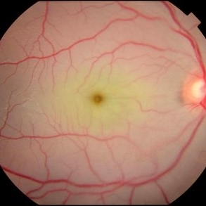
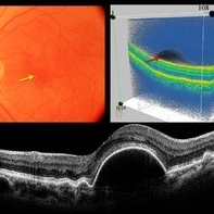
 Cone Dystrophy
Mar 29 2013 by Henry J. Kaplan, MD
Fundus photograph of a patient with low vision and hemeralopia and typical bull`s eye in cone dystrophy #2.
Condition/keywords: bull's eye maculopathy, cone dystrophy
-
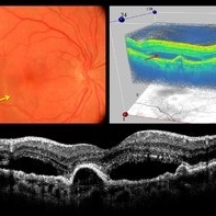
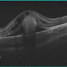 Cystoid Macular Edema (CME) in Vitelliform Macular Dystrophy (VMD)
Apr 22 2018 by Ronald Silva
Macula OCT of a 3-year-old boy with low vision and cystoid macular edema (CME) in vitelliform macular dystrophy (VMD) in right eye.
Photographer: Ronald Rocha da Silva, HCOE, Feira de Santana-BA
Condition/keywords: Best disease, cystoid macular edema (CME), vitelliform macular dystrophy
 Loading…