Search results (22 results)
-
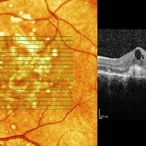
 Macular fold posterior microphthalmos OS
Apr 24 2022 by Mariam Cernichiaro-Espinosa, MD
Macular OCT of 6-year-old girl with posterior microphthalmos, showing macular fold and one intraretinal cyst OS.
Photographer: Mariam Cernichiaro-Espinosa, Asociación para Evitar la Ceguera, I.A.P. Mexico City, Mexico.
Imaging device: Zeiss Clarus
Condition/keywords: posterior microphthalmos
-
 Retinal Detachment
Nov 9 2012 by Norman Byer
This 18-year-old girl gave the history of having been hit in this eye three years before with a fist and of having retinal surgery nine months previously, which was temporarily successful. When the photograph was taken, she had a total left retinal detachment with a small nasal dialysis which had not been treated. She also had two prominent intraretinal cysts, one of which is shown here. The retina promptly reattached following further surgery and the next slide shows an interesting change in this cyst.
Condition/keywords: intraretinal cyst, small nasal dialysis
-
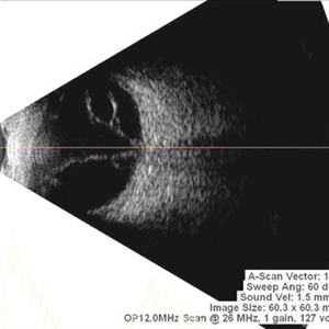
 Intraretinal Cysts in Chronic Retinal Detachment
Dec 8 2020 by Alice Kim
B-scan ultrasound showing multiple intraretinal cysts in the setting of chronic retinal detachment and proliferative vitreoretinopathy.
Condition/keywords: chronic retinal detachment, intraretinal cyst, proliferative vitreoretinopathy (PVR)
-
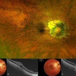 Bow-Tie Macular Hemorrhage With Cyst- Atypical Presentation of Myopic Choroidal Neovascularization
Mar 26 2021 by RUSHIK PATEL
The image of right eye of 51-year-old lady with high myopia show " Bow-Tie" macular hemorrhage (A). Optical coherence tomography (B) scan passing through hemorrhage showed intraretinal cystic lesion. During the course of intravitreal anti-VEGF injection treatment, the lesion converted into typical myopic choroidal neovascularization (C).
Photographer: Rushik Patel, Netralaya Super Speciality Eye Hospital
Condition/keywords: cyst, macular hemorrhage, myopic choroidal neovascularization (CNV)
-
 Diabetic Macular Edema
Feb 12 2025 by Kimberly Wakester
Horizontal OCT scan of a 63-year-old woman with diabetic macular edema in the right eye. When reviewing the scan, one of the intraretinal cyst (IRC) appears heart shaped. A fun scan to see just a few day's before Valentine's day.
Photographer: Kimberly Wakester, COA
Imaging device: Heidelberg
Condition/keywords: diabetic macular edema, intraretinal cyst
-
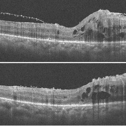
 Intraretinal cysts
Nov 15 2021 by Marcelo Zas, MD PhD
Left eye from a young patient with a chronic rhegmatogenous retinal detachment presenting intraretinal cysts.
Photographer: Zas Marcelo MD PhD
Condition/keywords: chronic retinal detachment, intraretinal cyst
-
 Intraretinal cysts
Nov 15 2021 by Marcelo Zas, MD PhD
Left eye from a young patient with a chronic rhegmatogenous retinal detachment presenting intraretinal cysts.
Photographer: Zas Marcelo MD PhD
Condition/keywords: chronic retinal detachment, intraretinal cyst
-
 Macular fold posterior microphthalmos OD
Apr 24 2022 by Mariam Cernichiaro-Espinosa, MD
Macular OCT of 6-year-old girl with posterior microphthalmos, showing macular fold and intraretinal cysts OD.
Photographer: Mariam Cernichiaro-Espinosa, Asociación para Evitar la Ceguera, I.A.P. Mexico City, Mexico.
Imaging device: Zeiss Clarus
Condition/keywords: posterior microphthalmos
-
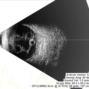
 Multiple Retinal Cysts Associated With Chronic Retinal Detachment
Sep 24 2018 by samarth mishra
Patient presented with a diminution of vision in left eye since few months. On B-scan ultrasonography multiple retinal cysts with a total retinal detachment were noted.
Photographer: Aditya Birla Sankara Nethralaya, West Bengal , Kolkata , India
Condition/keywords: B scan ultrasound, chronic retinal detachment, intraretinal cyst, retinal cyst
-
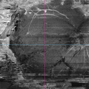 OCT en face of a 360 retinotomy for closed funnel combined retinal detachment
Jan 1 2023 by Malek Yassine, MD
Swept Source OCT en face at deep capillary plexus, shows foveal and parafoveal intraretinal cysts corresponding to macular edema under silicon oil
Imaging device: Topcon Triton DRI-OCT
Condition/keywords: combined retinal detachment, OCT EN FACE
-
 Optic Nerve Pit / Serous Detachment
Feb 20 2013 by From the Collections of Thomas M. Aaberg, MD and Thomas M. Aaberg Jr., MD
No history; FA; serous detachment intraretinal cyst; lamellar hole.
Condition/keywords: intraretinal cyst, optic nerve pit
-
 Optic Nerve Pit / Serous Detachment
Feb 20 2013 by From the Collections of Thomas M. Aaberg, MD and Thomas M. Aaberg Jr., MD
No history; FA; serous detachment intraretinal cyst; lamellar hole.
Condition/keywords: intraretinal cyst, optic nerve pit
-
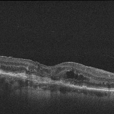
 Post Retinal Reattachment Surgery Epiretinal Membrane
Sep 14 2021 by Ogugua Ndubuisi Okonkwo, MD, FRCS (Edin), FASRS
Postoperative optical coherence tomography (OCT) of the right eye in a 65-year-old male who had retinal reattachment surgery for a macular hole retinal detachment. This OCT scan shows epiretinal membrane and intraretinal cystic fluid spaces.
Photographer: Oreoluwa Olabode , Eye Foundation Hospital, Lagos.
Imaging device: Optovue Avanti RTVue.
Condition/keywords: epiretinal membrane (ERM), macular hole retinal detachment, Retinal Reattachment surgery
-
 Punctate Inner Choroidopathy Complicated with CNV
Jun 5 2013 by Henry J. Kaplan, MD
OCT of the same patient demonstrates CNV complex with intraretinal cystoid edema #3.
Photographer: Angela Andersson
Imaging device: HRA II
Condition/keywords: choroidal neovascularization (CNV), punctate inner choroidopathy (PIC)
-
 Retinal Detachment
Nov 9 2012 by Norman Byer
This eye of a 25-year-old man has a retinal detachment of about six year’s duration. This photograph shows an intraretinal cyst, which is a secondary result of the longstanding detachment.
Condition/keywords: intraretinal cyst
-
 Retinal Detachment
Nov 9 2012 by Norman Byer
This is the same lesion as in the previous photograph shown 13 days after surgery. Not only is the retina reattached, but the cyst has now completely disappeared even though no treatment of any kind was applied in the vicinity of the cyst. Long detached retinas tend to develop intraretinal cysts, and these tend to disappear following reattachment of the retina even without direct treatment to the cysts.
Condition/keywords: intraretinal cyst, re-attached retinal detachment (RRD)
-
 Rhegmatogenous Retinal Detachment With Retinal Dialysis and Intraretinal Cyst
Mar 18 2020 by Giridhar Anantharaman, MS
Optos ultra-widefield retinal imaging of the left eye of a 30-year-old lady with rhegmatogenous retinal detachment with inferotemporal retinal dialysis and a large intraretinal cyst.
Photographer: Rakesh PR, Giridhar Eye Institute, Kerala, India
Imaging device: Optos UWF Daytona plus
Condition/keywords: intraretinal cyst, retinal dialysis
-
 RPE rip macular OCT
Dec 23 2012 by Alex P. Hunyor, MD
80-year-old female with subfoveal occult CNV and large extrafoveal PED which underwent spontaneous RPE rip. OCT shows subfoveal CNV and intraretinal cystic edema
Condition/keywords: pigment epithelial detachment (PED), retinal pigment epithelium (RPE) tear
-
 Total Retinal Detachment
Dec 10 2012 by Yale L. Fisher, MD
Retinal detachment secondary to PVR, patient is NLP. Yellow arrow indicates intraretinal cyst due to longstanding detachment.
Condition/keywords: video
 Loading…