Search results (2824 results)
-
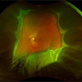
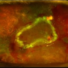
 Coats' Disease
Apr 27 2018 by Brenda Fallas
3-year-old boy with unilateral Coats' Disease fundus photo.
Photographer: Brenda Fallas, Bascom Palmer Eye Institute, Miami, FL
Imaging device: Retcam III 130 degree lens
Condition/keywords: Coats' disease, color fundus photograph, retinal telangiectasia
-
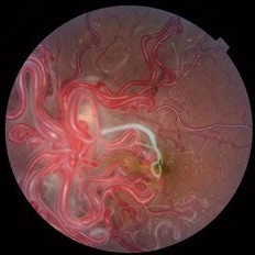 Wyburn Mason
Jun 23 2018 by Caesar K. Luo, MD, FASRS
Fundus photograph of a 16-year-old female patient with poor vision in one eye demonstrates racemose angiomatosis as seen in Wyburn-Mason Syndrome.
Photographer: Joseph Trabucco, Progressive Vision Institute, Allentown, PA
Condition/keywords: Wyburn-Mason
-
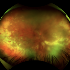
 Tractional Retinal Detachment
Dec 4 2019 by Janet Brazil
Fundus photograph of a 32-year-old female with severe end-stage diabetic tractional retinal detachment.
Photographer: Janet Atkinson, Eye Associates of New Mexico, Albuquerque, NM
Imaging device: Topcon TRC- 50EX
Condition/keywords: diabetes, proliferative diabetic retinopathy (PDR), tractional retinal detachment
-
 Ocular Manifestation of Acute Leukemia
Sep 8 2012 by Hamid Ahmadieh, MD
Color fundus photograph of a 26-year-old man with acute leukemia.
Photographer: Hamid Ahmadieh, MD, Ophthalmic Research Center, Labbafinejad Medical Center, Shahid Beheshti University of Medical Sciences , Tehran
Imaging device: Topcon Fundus Camera
Condition/keywords: acute leukemia, white centered retinal hemorrhage (Roth Spot)
-
 Giant Retinal Tear
Feb 20 2024 by Soobien Lee
Optos color fundus photograph of a 40-year-old caucasian male who is a UFC fighter with a total retinal detachment in his right eye secondary to a giant retinal tear from 10 o'clock to 2 o'clock.
Photographer: Trinity Wolf, Elman Retina Group
Imaging device: Optos Ultra-Widefield Imaging
Condition/keywords: giant retinal tear, optos, Retinal Detachment, Retinal tear with detachment, trauma
-
 Acute Posterior Multifocal Placoid Pigment Epitheliopathy
Feb 20 2024 by Soobien Lee
Optos color fundus photograph of a 20-year-old caucasian female with viral prodrome and vision loss OS>OD secondary to Acute Posterior Multifocal Placoid Pigment Epitheliopathy (APPME). Imaging of her left eye shows multiple bilateral creamy yellow-white placoid lesions at the level of RPE and choroid throughout the posterior pole.
Photographer: Ashley Metzger, Elman Retina Group
Imaging device: Optos Ultra-Widefield Imaging
Condition/keywords: acute posterior multifocal placoid pigment epitheliopathy (APMPPE), bacilliary layer detachment, Optos, uveitis, white dot syndrome
-
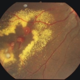
 Disseminated Retinitis and Retinochoroiditis, Metastatic
May 16 2017 by Karen Panzegrau
Fundus photograph of 44-year-old male with plasmacytoma infiltation of the choroid confirmed by biopsy, associated with disseminated retinitis, and retinochoroiditis. Vision is LP. Patient treated with intravitreal methotrexate
Photographer: Karen Panzegrau
Imaging device: Optos
Condition/keywords: metastatic lesion, methotrexate, Optos, plasmacytoma, retinitis, retinochoroiditis, unilateral exudative retinal detachment
-
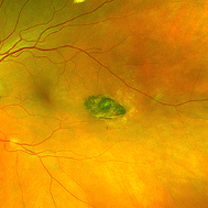 Torpedo Maculopathy
Feb 20 2024 by Soobien Lee
Optos color fundus photograph of a 35-year-old asymptomatic female with no ocular or medical history with stable and chronic appearing torpedo-shaped macula lesion in the left eye.
Photographer: Peter Sotirakos, Elman Retina Group
Imaging device: Optos Ultra-Widefield Imaging
Condition/keywords: macula, Optos, torpedo maculopathy
-
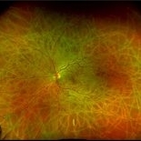
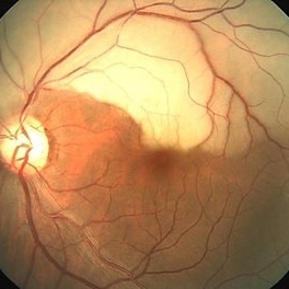
 Proliferative Diabetic Retinopathy
Sep 17 2012 by Michael P. Kelly, FOPS
Retinal fundus photograph of a patient with PDR and NVD.
Photographer: Michael P. Kelly, FOPS Director, Duke Eye Labs, Duke University Hospital, Duke Eye Center
Imaging device: Topcon
Condition/keywords: neovascularization of the disc (NVD)
-
 Tumor of Retina (Retinocytoma)
Jan 9 2019 by Janet Brazil
Fundus photograph of a 13-year-old male with a bilateral tumor of the retina, with positive RB gene mutation.
Photographer: Janet Atkinson, Eye Associates of New Mexico
Imaging device: TOPCON TRC-50EX
Condition/keywords: RB gene mutation, tumor
-
 Choroidal Melanoma with Exudative Retinal Detachment
Mar 2 2023 by Aditya S Kelkar, MS, FRCS, FASRS,FRCOphth
Color fundus photograph of the left eye of a 45 year old male showing choroidal melanoma with exudative retinal detachment.
Photographer: Dr. Pranali Surawase, National Institute of Ophthalmology, Pune, India.
Imaging device: Zeiss Clarus 500
Condition/keywords: choroidal mass, exudative retinal detachment, Retinal detachment
-
 Posterior Ophthalmomyiasis Interna
Sep 20 2021 by Haley Tamanosky
Fundus photograph of 36-year-old woman with Posterior Ophthalmomyiasis Interna and moving larva.
Photographer: Haley Tamanosky
Condition/keywords: linear track marks, Posterior Ophthalmomyiasis Interna
-
 Retinal Arterio-Venous Malformations
Apr 7 2017 by Deepak Bhojwani, MS
Multimodal imaging of a 16-year-old boy with retinal arterio-venous malformations(AVM). He also had cerebral AVM's on MRI-contrast studies suggesting Wyburn-Mason syndrome.
Photographer: DEEPAK BHOJWANI, RAGHUDEEP EYE HOSPITAL, AHMEDABAD.
Imaging device: Zeiss VISUCAM
Condition/keywords: color fundus photograph, FA early phase, optical coherence tomography (OCT), Wyburn-Mason
-
 Retinal Detachment with PVR (s/ SPR, PPV, MPV, 360 Retinectomy, PFO, PI, FAx, SO)
Aug 22 2019 by Merrick Avila
Ultra-wide field pseudocolor fundus photograph of a 64-year-old female with a treated retinal detachment with proliferative vitreoretinopathy. Patient has a history of complex retinal detachments that have been treated multiple times. On exam 8-22-19, there were large macular holes with LP vision. There was a long discussion about guarded nature of her condition and goals or trial for repair including globe sparing prevention of phthisis.
Photographer: Merrick Avila
Imaging device: Optos
Condition/keywords: diabetic retinopathy, hemorrhage, Optos, proliferative vitreoretinopathy (PVR), retinectomy, silicone oil
-
 Acute Retinal Necrosis (ARN)
Jul 3 2025 by Heitor Nogueira
Fundus photograph of an 63-year-old woman who reported unilateral visual acuity loss for 10 days associated with ocular pain. He presented conjunctival hyperemia with temporal and nasal nodular scleritis, anterior chamber reaction 2+/4+, Koeppe nodules, granulomatous PKs, vitreitis 2+/4+, multiple areas of vasculitis in the arcades and periphery, associated with hemorrhages and necrotizing retinitis in the temporal, inferior and nasal periphery. Positive serology for Herpes Virus
Photographer: Heitor Nogueira, Penido Burnier Institute, Campinas, São Paulo, Brazil
Imaging device: Optos Daytona
Condition/keywords: ARN complications, Herpes, progressive outer retinal necrosis (PORN), Uveitis
-
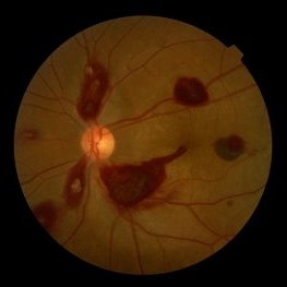
 Bilateral Lebers Miliary Aneurysm in a Female
Sep 5 2017 by Ogugua Ndubuisi Okonkwo, MD, FRCS (Edin), FASRS
Fundus photograph of the active left eye of a 26-year-old female with bilateral LMA. Shows severe exudation in the nasal retina by leaking aneurysms.
Condition/keywords: aneurysm
-
 Branch Retinal Artery Occlusion With Calcium Embolus at the Disc - Fundus Photo
Apr 7 2018 by Rameez N Hussain, MD
Acute branch retinal artery occlusion with a calcium embolus at the disc with retinal whitening in the area of retinal edema.
Photographer: DR RAMEEZ N HUSSAIN
Imaging device: zeiss
Condition/keywords: branch retinal artery occlusion (BRAO), embolus, fundus photograph, retinal edema
-
 Central Retinal Artery Occlusion With Cilioretinal Sparing
Apr 4 2018 by Soumya Venkatesh
Fundus photograph of a 23-year-old gentleman presenting with sudden loss of vision 2 days prior to presentation. He underwent all relevant investigations and found to have APLA positive. He also had dengue serology positive. On follow up, his retinal edema reduced unmasking the underlying hemorrhages( flame shaped).
Photographer: Soumya Harapanahalli Venkatesh, JSS university, Karnataka, India
Condition/keywords: central retinal artery occlusion (CRAO), cherry red spot, cilioretinal sparing, retinal ischemia
-
 Choroidal Detachment
Jan 17 2022 by Logan ryzenga
Left ultra-wide field photograph of an 81-year old female with a choroidal detachment affecting her left eye. Patient had a stent placed November, 2021 and following the procedure she complains of variable blurred vision and severe constricted visual fields. She presented at our office with flashes a month prior but without pain or floaters.
Photographer: Logan Ryzenga
Imaging device: Optos California
Condition/keywords: choroidal detachment, fundus photograph, left eye, Optos, pseudocolor, superior retina, ultra-wide field imaging
-
 Choroidal Fracture
Oct 27 2024 by César Adrián Gómez Valdivia, MD
Fundus photograph of a traumatic choroidal fracture & extra-macular sub-retinal hemorrhage.
Photographer: @eyemissu2
Imaging device: TOPCON TRC-50DX
Condition/keywords: Choroidal Fracture
-
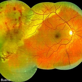 Commotio Post-Blunt Trauma
Apr 3 2018 by Paulo Bueno
Fundus photograph of an 35-year-old man with commotio retinae after indoor soccer ball blunt trauma.
Photographer: Paulo Bueno, Taubaté, Brazil.
Imaging device: Zeiss Visucam
Condition/keywords: blunt trauma, commotio retinae
-
 Cuticular and soft drusen
Jun 14 2021 by Gerardo Garcia-Aguirre, MD
Fundus photograph (left) and Retro mode infrared image (right) of an eye with soft and cuticular drusen. Drusen are highlighted and better visualized with retro mode imaging.
Photographer: Gerardo Garcia-Aguirre
Imaging device: Nidek Mirante
Condition/keywords: drusen, dry age-related macular degeneration (dry AMD)
-
 Disciform Scar
Aug 18 2020 by Aditya S Kelkar, MS, FRCS, FASRS,FRCOphth
Left eye fundus photograph of 75-year-old male, showing large disciform scar post subretinal bleeding secondary to idiopathic polypoidal choroidal vasculopathy
Photographer: Dr.Mounika Bolisetty
Imaging device: CLARUS 500
Condition/keywords: disciform scar, idiopathic polypoidal choroidal vasculopathy
-
 Hemangioma Capilar Retina
Apr 9 2023 by Gustavo Aguirre-Suarez
Fundus photograph composition of a Retinal Capilar Hemangioma
Photographer: Dr. Gustavo Aguirre-Suarez
Imaging device: Visucam 500
Condition/keywords: hemangioma, Von Hippel-Lindau
-
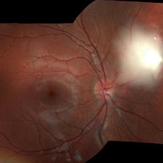
 Optic Nerve Head Drusen With Idiopathic CNV
Feb 17 2017 by Kristen Wagner
22-year-old female fundus photograph of a right eye with Optic Nerve Drusen with Idiopathic CNV.
Photographer: Kristen Wagner, COT, OSC Ophthalmic Photographer, Tennessee Retina, Nashville TN
Condition/keywords: choroidal neovascularization (CNV), drusen of optic disc, optic disc drusen
 Loading…