Search results (195 results)
-
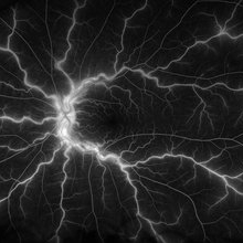
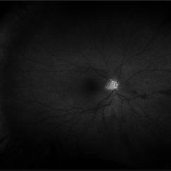
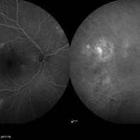
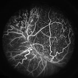 Coats' Disease FA
Apr 27 2018 by Brenda Fallas
3-year-old boy with unilateral Coats' Disease FA photo.
Photographer: Brenda Fallas, Bascom Palmer Eye Institute, Miami, FL
Imaging device: Retcam III 130 degree lens
Condition/keywords: Coats' disease, FA early phase, fluorescein angiogram (FA), retinal telangiectasia
-
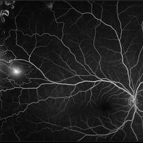
 Venous Loop
Feb 20 2024 by Soobien Lee
A 77-year-old male with a history of bilateral optic neuropathy from bilateral optic nerve sheath meningiomas S/P radiation/proton-beam therapies. Presented with radiation retinopathy OS and a known venous loop OS.
Photographer: Gavin Bragdon, Elman Retina Group
Imaging device: Optos Ultra-Widefield Fluorescein Angiography
Condition/keywords: fluorescein angiogram (FA), Optos, radiation retinopathy, retinal vascular disease, venous loop
-
 Acute Posterior Multifocal Placoid Pigment Epitheliopathy
Feb 20 2024 by Soobien Lee
Fluorescein angiogram of a 20-year-old caucasian female with viral prodrome and vision loss OS>OD secondary to Acute Posterior Multifocal Placoid Pigment Epitheliopathy (APPME). Early blockage with late hyperfluorescent leakage can be seen on fluorescein angiography of the left eye.
Photographer: Ashley Metzger, Elman Retina Group
Imaging device: Optos Ultra-Widefield Fluorescein Angiography
Condition/keywords: acute posterior multifocal placoid pigment epitheliopathy (APMPPE), bacilliary layer detachment, FA, FA early phase, fluorescein angiogram (FA), Optos, uveitis, white dot syndrome
-
 Acute Posterior Multifocal Placoid Pigment Epitheliopathy
Feb 20 2024 by Soobien Lee
Fluorescein angiogram of a 20-year-old caucasian female with viral prodrome and vision loss OS>OD secondary to Acute Posterior Multifocal Placoid Pigment Epitheliopathy (APPME). Early blockage with late hyperfluorescent leakage can be seen on fluorescein angiography of the left eye.
Photographer: Ashley Metzger, Elman Retina Group
Imaging device: Optos Ultra-Widefield Fluorescein Angiography
Condition/keywords: acute posterior multifocal placoid pigment epitheliopathy (APMPPE), bacilliary layer detachment, FA, FA late phase, FA late phase leakage, fluorescein angiogram (FA), Optos, uveitis, white dot syndrome
-
 Radiation Retinopathy; BRVO with Macular Edema
Apr 26 2023 by Denica Rodriguez
Ultra-wide field fluorescein angiography of a 61 year old male with radiation retinopathy following brachytherapy for choroidal melanoma of his left eye. Following treatment, patient developed a branch retinal vein occlusion both ischemic and non-ischemic. Anti-VEGF injections were recommended. The fine needle biopsy showed a class 2 uveal melanoma. Patient also has diabetic retinopathy affecting both eyes. Patient's vision at the time the image was taken was Dcc 20/80-1.
Photographer: Denica Rodriguez COA, ST
Imaging device: Optos California
Condition/keywords: branch retinal vein occlusion (BRVO), Choroidal melanoma, diabetic retinopathy, FA, fluorescein angiogram (FA), I-125 brachytherapy, macular edema, melanoma, Optos, radiation retinopathy, Retina, ultra-wide field imaging
-
 Rod/Cone dystrophy
Aug 20 2021 by Jeffrey Barker
Fluorescein Angiogram Early frame (29 seconds)
Photographer: Jeffrey P. Barker, B.S. Retina Vitreous Surgeons of C.N.Y.
Condition/keywords: cone dystrophy, fluorescein angiogram (FA)
-
 Branch Retinal Artery Occlusion With Calcium Embolus at the Disc - Fundus Fluorescence Angiogram (FA)
Apr 7 2018 by Rameez N Hussain, MD
Acute branch retinal artery occlusion with a calcium embolus at the disc which is hyperfluorescent in FA.
Photographer: DR RAMEEZ N HUSSAIN
Imaging device: Zeiss
Condition/keywords: branch retinal artery occlusion (BRAO), embolus, fluorescein angiogram (FA), retinal edema
-
 Central Retinal Vein Occlusion
Jan 21 2022 by Olivia Rainey
Ultra-widefield fluorescein angiogram of a 23-year-old female with a Central Retinal Vein Occlusion affecting her left eye. The patient presented on 12/22/2021 cc20/40-2 vision in the left eye. The patient reported recent trauma of being hit with a fist on both sides of face followed by vision loss. The patient has history of Hashimoto's thyroid disease. The following labs have been ordered, PT, PTT, CBC, antithrombin III activity, protein C, protein S, Factor V Leiden mutation, Prothrombin (G20210A), lipid panel, HbA1c, quantiferon gold, RPR, and CXR.
Photographer: Olivia Rainey, OCT-C, COA
Imaging device: Optos California
Condition/keywords: central retinal vein occlusion (CRVO), disc leakage, fluorescein angiogram (FA), fluorescein leakage, left eye, non-ischemic central retinal vein occlusion (CRVO), Optos, trauma, ultra-wide field imaging
-
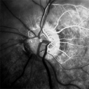
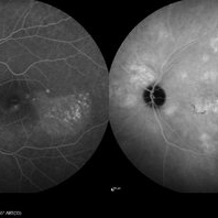
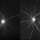
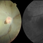 Ophthalmic Artery Occlusion in a 39-Year-Old with Rheumatoid Arthritis
Oct 6 2020 by Michael Izzo, MD
Left image: fundus photograph of a 39-year-old male with rheumatoid arthritis found to have ophthalmic artery occlusion depicting boxcar segmentation of blood in retinal vasculature and macular ischemia demonstrated by retinal whitening without cherry red fovea. Right image: early phase fluorescein angiography demonstrating patchy choroidal filling, arterial non-perfusion and optic nerve head leakage.
Photographer: Karen Rivera, COA; Washington National Eye Center
Condition/keywords: fluorescein angiogram (FA), ophthalmic artery occlusion, rheumatoid arthritis
-
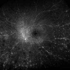
 Proliferative Diabetic Retinopathy
Aug 16 2022 by Donnie Willis
51 yo female. Uncontrolled Diabetes. Active PDR.
Photographer: Donnie Willis, Tennessee Retina
Imaging device: Optos
Condition/keywords: capillary dropouts, Diabetes, FA, fluorescein angiogram (FA), Optos, proliferative diabetic retinopathy (PDR), vitreomacular traction (VMT)
-
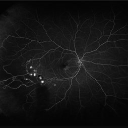
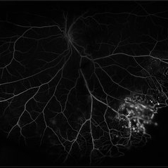
 Proliferative Sickle Cell Retinopathy
Feb 1 2023 by Olivia Rainey
Ultra-widefield fluorescein angiography of a 25-year old male with Proliferative Sickle Cell Retinopathy affecting his right eye. Patient stated that he was born with Sickle disease (SC), and has yearly eye exams. He noted no vision concerns over the last year but has typically experienced sickle attacks about 1-2 per year. The physician noted that the fluorescein obtained showed peripheral nonperfusion affecting the patient's nasal and temporal retina as well as neovascularization affecting his left eye more than his right. He recommended pan retinal photocoagulation in his left eye for his temporal and nasal retina, as as well as his right eye following.
Photographer: Olivia Rainey, OCT-C, COA
Imaging device: Optos California
Condition/keywords: early phase, fluorescein angiogram (FA), fluorescein leakage, neovascularization (NV), non-perfusion, proliferative retinopathy, right eye, sickle cell retinopathy, ultra-wide field imaging, ultra-widefield image
-
 Proliferative Sickle Cell Retinopathy
Jan 29 2021 by Olivia Rainey
Ultra-widefield fluorescein angiogram of a 24-year-old female with proliferative sickle cell retinopathy affecting her right eye. The physician's interpretation of the fluorescein shows seafan neovascularization superotemporally, AV anastomeses, and good peripheral laser. He performed scatter PRP OD on 12/2/2020 to nonperfusion in temporal far periphery. The patient's 12/2020 Hb electrophoresis came back showing Hb SC (rather than sickle cell trait). Patient was born at full term, but she reports that her mother used drugs while pregnant with the patient. The patient also mentioned that her niece has full sickle cell disease and her grandmother, mother, and sibling all have condition on the sickle cell spectrum.
Photographer: Olivia Rainey, OCT-C, COA
Imaging device: Optos California
Condition/keywords: fluorescein angiogram (FA), fluorescein leakage, neovascularization (NV), neovascularization elsewhere (NVE), Optos, sea fan, sickle cell retinopathy
-
 Retinal Hemorrhages
Mar 10 2021 by Kachelle Brown
Ultra widefield Fluorescein Angiography of a 48-year-old female with retinal hemorrhages affecting her right eye. Physician suspect sickle cell due to family history, and has ordered labs to rule out.
Photographer: Kachelle Brown
Imaging device: Optos California
Condition/keywords: fluorescein angiogram (FA), fluorescein leakage, Optos, retinal hemorrhage, sickle cell retinopathy, ultra-wide field imaging
-
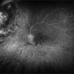
 VKH Syndrome
Jun 12 2025 by Virginia Gebhart
Fluorescein angiogram of 22 year old male with VKH syndrome. Significant cell in AC and vitreous, multiple punched-out CR scars in periphery, mild vascular leakage. Pt referred to rheumatology for immunomodulatory treatment.
Photographer: Virginia Gebhart, Retina Consultants of Carolina
Imaging device: Optos California
Condition/keywords: FA, fluorescein angiogram (FA), multifocal choroiditis, panuveitis, VKH, Vogt-Koyanagi-Harada
-
 Acute Zonal Occult Outer Retinopathy (AZOOR) FA, Fluorescein Angiography, Peripheral Vasculitis
Jan 19 2022 by James B. Soque, CRA, OCT-C, COA, FOPS
Acute Zonal Occult Outer Retinopathy (AZOOR). Peripheral Vasculitis OD. Fluorescein angiography showing vasculitis in the far right periphery 8-10 o'clock. 46-year-old white male, VA CC 20/16, 20/12.5, has had recurrent vasculitis for 11 years. No treatment.
Photographer: James Soque, CRA, OCT-C, COA, FOPS, Island Retina, Shirley, NY
Imaging device: Optos California
Condition/keywords: acute zonal occult outer retinopathy (AZOOR), FA early phase, fluorescein angiogram (FA), Peripheral Vasculitis, ultra-wide field imaging
-
 Central Retinal Artery Occlusion
May 25 2017 by Olivia Rainey
Ultra-wide field fluorescein angiography, taken at 42 seconds, of an 73-year-old female with a central retinal artery occlusion in her right eye.
Photographer: Olivia Rainey
Imaging device: Optos California
Condition/keywords: central retinal artery occlusion (CRAO), early phase, fluorescein angiogram (FA), ischemia, non-perfusion, Optos, ultra-wide field imaging
-
 Central Retinal Vein Occlusion with Retinal Neovascularization
Jan 19 2022 by Olivia Rainey
Ultra-widefield fluorescein angiogram of a 56-year-old male with a Central Retinal Vein Occlusion with Retinal Neovascularization affecting his left eye. The patient presented on 1/19/2022 with scNLP vision in the left eye. The patient has good PRP, however areas of ischemia still remain untreated by laser. He also has severe neovascular glaucoma contributing to his poor vision.
Photographer: Olivia Rainey, OCT-C, COA
Imaging device: Optos California
Condition/keywords: central retinal vein occlusion (CRVO), FA early phase, fluorescein angiogram (FA), hemorrhage, ischemic CRVO, left eye, neovascular glaucoma, Optos, pan-retinal photocoagulation (PRP), retinal ischemia, retinal neovascularization, ultra-wide field imaging
-
 Central Retinal Vein Occlusion with Severe Retinal Ischemia
Jan 19 2022 by Olivia Rainey
Ultra-widefield fluorescein angiogram of a 56-year-old male with a Central Retinal Vein Occlusion with Severe Retinal Ischemia affecting his right eye. The patient presented on 1/19/2022, sc20/20-2 vision in the right eye. The patient has had a good response to Eylea with complete resolution of edema. The physician is considering PRP to ischemic periphery in the future and given the degree of ischemia in both eyes, she recommends that the patient's PCP check carotid Doppler US.
Photographer: Olivia Rainey, OCT-C, COA
Imaging device: Optos California
Condition/keywords: central retinal vein occlusion (CRVO), FA late phase, fluorescein angiogram (FA), ischemic CRVO, Optos, retinal ischemia, ultra-wide field imaging
-
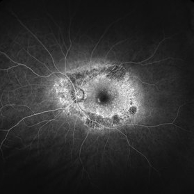
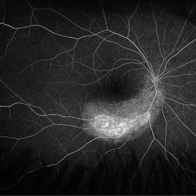
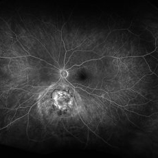
 Central Serous Chorioretinopathy
Jan 25 2022 by Olivia Rainey
Late phase widefield fluorescein angiography of a 60-year-old male with Central Serous Chorioretinopathy. Chronic history of CSR followed with observation without treatment prior to presenting at our office. The physician noted subfoveal subretinal fluid with recent visual decline. FA shows multifocal leakage and ICG shows hypercyanescence. OCTA, ICG, and FA consistent with CSR, and without concern for CNVM thus will observe without anti-VEGF at this time. PDT therapy recommended.
Photographer: Olivia Rainey, OCT-C, COA
Imaging device: Heidelberg Spectralis
Condition/keywords: 55-degrees, central serous chorioretinopathy (CSCR), central serous retinopathy (CSR), chronic central serous chorioretinopathy (CSCR), fluorescein angiogram (FA), heidelberg spectralis, indocyanine green (ICG) angiography, left eye
-
 Central Serous Chorioretinopathy
Jan 25 2022 by Olivia Rainey
Late phase widefield fluorescein angiography of a 60-year-old male with Central Serous Chorioretinopathy. Chronic history of CSR followed with observation without treatment prior to presenting at our office. The physician noted significant findings on exam and imaging with multifocal areas of inactive and active changes OD. FA shows superotemporal macular leakage, subtle inferonasal macular leakage and staining as well as multifocal hypercyanescence on ICG. Fortunately foveal sparing and thus observation is recommended at this time OD.
Photographer: Olivia Rainey, OCT-C, COA
Imaging device: Heidelberg Spectralis
Condition/keywords: 55-degrees, central serous chorioretinopathy (CSCR), central serous retinopathy (CSR), chronic central serous chorioretinopathy (CSCR), fluorescein angiogram (FA), fluorescein leakage, heidelberg spectralis, indocyanine green (ICG) angiography, late phase
-
 Choroidal Melanoma 3 Ways
Jan 16 2025 by Virginia Gebhart
RGB/FA/ICG of 76 year old female with a new choroidal melanoma. Pt scheduled for plaque radiation. BCVA 20/400
Photographer: Virginia Gebhart, Retina Consultants of Carolina
Imaging device: Optos California
Condition/keywords: fluorescein angiogram (FA), indocyanine green (ICG) angiography, OPTOS CALIFORNIA RGB
-
 Choroidal Melanoma FA
Nov 14 2023 by Virginia Gebhart
Fluorescein angiogram of 69 year old male with small lesion consistent with choroidal melanoma. Small pigmented elevated choroidal lesion just below ON with drusen, RPE changes and trace questionable OP present in the left eye. Extensive imaging and ultrasound was performed for further evaluation and documentation.
Photographer: Virginia Gebhart
Imaging device: Optos
Condition/keywords: fluorescein angiogram (FA), Fluorescein angiography
-
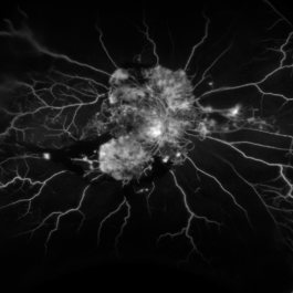
 Coats' Disease
Jul 16 2019 by Kim Barrett
Ultra-wide field fluorescein angiogram of a 23-year-old male with Coats' disease, presented with distorted vision affecting his left eye. He reported seeing flashes and floaters since January of 2019, but the flashes had resolved. He was treated with Intravitreal Preservative Free Triamcinolone in the office and scheduled for PRP laser in the near future.
Photographer: Kim Barrett
Imaging device: Optos
Condition/keywords: Coats' disease, fluorescein angiogram (FA), fluorescein leakage, inferior retina, ischemia, left eye, Optos, ultra-wide field imaging
-
 CRVO
CRVO
Jan 21 2021 by Jamin S. Brown, MD
Fluorescein Image of a central retinal vein occlusion of the right eye.
Photographer: Stefanie Palmer CRA, Retina Vitreous Surgeons of CNY
Condition/keywords: central retinal vein occlusion (CRVO), fluorescein angiogram (FA)
-
 Cystoid Macular Degeneration
Feb 1 2023 by Kachelle Brown
Fluorescein Angiogram of a 56 year old woman with bilateral Cystoid Macular Degeneration. Patient vision was 20/60 OU.
Photographer: Kachelle Brown OMA, Retina Specialist of Michigan
Condition/keywords: cystoid macular degeneration, cystoid macular edema (CME), FA late phase, fluorescein angiogram (FA)
 Loading…