Search results (78 results)
-
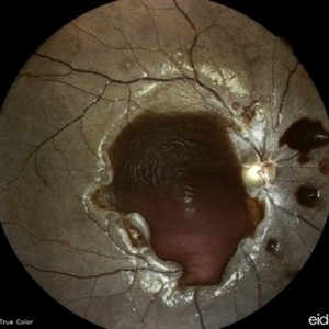
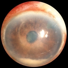
 Amelanotic Choroidal Melanoma
May 18 2020 by McGill University Health Centre
The enucleation specimen in (A) shows an amelanotic, mushroom-shaped tumor arising from the choroid. Microhemorrhages are present within the tumor and also surround the tumor base (arrow). True retinal detachment is present (arrowhead). The subretinal fluid is mixed: clear (1), hemorrhagic (2), and fibrinoid (3).
Condition/keywords: enucleation, mushroom-shaped
-
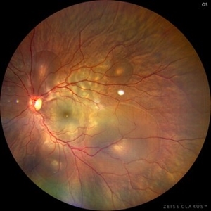 Central Serous Chorioretinopathy in Pregnancy (OS)
Apr 28 2024 by Vishal Agrawal, MD, FRCS,FACS,FASRS
30-year female with sudden loss of vision came for examination. She was in her first trimester of pregnancy. Examination revealed bilateral bullous NSD with subretinal fibrin s/o CSR.
Photographer: Dr Ayushi
Imaging device: Clarus 700
Condition/keywords: Central Serous Chorioretinopathy (CSR), neurosensory detachment of retina, pregnancy
-
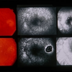
 Central Serous Retinopathy with Fibrin
Oct 13 2012 by Edwin H. Ryan, MD
Recurrent central serous with fibrin in a 54-year-old man.
Condition/keywords: central serous chorioretinopathy (CSCR), subretinal fluid
-
 Anemic Retinopathy Related Retinal Hemorrhages
Nov 5 2019 by Chinmayi Vyas
Anemic retinopathy related retinal hemorrhages in a 24 years old male with Hb of 4.2gm/ dl. The manifestations of anemic retinopathy are nonspecific and may closely simulate hypertensive or diabetic retina. Retinal changes in anemia are cotton wool spots, venous tortuosity, and hemorrhages which may be present at all levels of the retina and choroid. All retinal hemorrhages can occur when Hb falls below 8 g/100 ml or if the platelet count falls below 50,000/cumm. The combination of severe anemia and thrombocytopenia is likely to produce retinal hemorrhages. The Roth’s spots or white centre hemorrhages are typically associated with bacterial endocarditis , anemia and other systemic conditions. The white center is suspected to represents focal ischemia, inflammatory or infectious infiltrate, fibrin or accumulation of neoplasticism cells.
Photographer: Dr Chinmayi Vyas
Condition/keywords: retinal hemorrhage
-
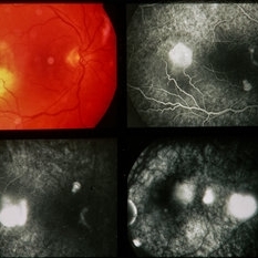
 Central Serous Chorioretinopathy
Apr 19 2017 by Gustavo Barreto de Melo, MD, PhD, FASRS
Fundus photograph of a 32-year-old pregnant woman with a serous detachment and subretinal fibrin deposit surrounding the fovea.
Photographer: Denyson Silva, Sergipe Eye Hospital
Condition/keywords: central serous chorioretinopathy (CSCR), pregnancy
-
 Central Serous Retinopathy with Fibrin
Oct 13 2012 by Edwin H. Ryan, MD
EDI scan of choroid in patient with CSC
Condition/keywords: central serous chorioretinopathy (CSCR), choroidal thickening, fluorescein leakage
-
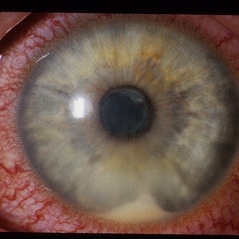 HLA-B27 Associated Uveitis
Jun 4 2014 by Henry J. Kaplan, MD
Severe anterior uveitis with fibrinous reaction and hypopyon formation related to HLA-B27. Notice the membrane on the lens surface.
Condition/keywords: acute anterior uveitis, HLA-B27, hypopyon
-
 Anemic Retinopathy Related Retinal Hemorrhages
Nov 5 2019 by Chinmayi Vyas
Anemic retinopathy related retinal hemorrhages in a 24 years old male with Hb of 4.2gm/ dl. The manifestations of anemic retinopathy are nonspecific and may closely simulate hypertensive or diabetic retina. Retinal changes in anemia are cotton wool spots, venous tortuosity, and hemorrhages which may be present at all levels of the retina and choroid. All retinal hemorrhages can occur when Hb falls below 8 g/100 ml or if the platelet count falls below 50,000/cumm. The combination of severe anemia and thrombocytopenia is likely to produce retinal hemorrhages. The Roth’s spots or white centre hemorrhages are typically associated with bacterial endocarditis , anemia and other systemic conditions. The white center is suspected to represents focal ischemia, inflammatory or infectious infiltrate, fibrin or accumulation of neoplasticism cells.
Photographer: Dr Chinmayi Vyas, Nethradhama superspeciality eye hospital , banglore, india
Imaging device: Eidon fundus imaging
Condition/keywords: anaemic retinopathy
-
 Anterior Capsular Opacity
Feb 8 2018 by Claire Kiernan, MD
Slit lamp photograph of a 39-year-old female following uncomplicated cataract surgery shown here with dense fibrinous changes of the anterior capsule. This patient underwent Nd:YAG laser anterior capsulotomy with clearing of her visual axis.
Photographer: Steve Crow, University of Tennessee Hamilton Eye Institute, Memphis, TN
Condition/keywords: anterior capsule opacification, cataract extraction, cataract surgery
-
 Bleb-related Endophthalmitis Slide 2
Oct 22 2012 by Ronald C. Gentile, MD
Anterior chamber has a hypopyon with fibrin.
Photographer: The New York Eye & Ear Infirmary Department of Medical Imaging
Condition/keywords: Bleb-related endophthalmitis
-
 Branch Retinal Artery Occlusion (BRAO) - Embolic
Mar 27 2019 by Gary R. Cook, MD, FACS
Superonasal BRAO with fibrin-platelet embolus OD.
Imaging device: Topcon VT-50
Condition/keywords: branch retinal artery occlusion (BRAO), embolus
-
---thumb.jpg/image-square;max$300,300.ImageHandler) Central Serous Chorioretinopathy 1
Central Serous Chorioretinopathy 1
Mar 18 2013 by Maurice F. Rabb
Woman with a 3 month history of reduced vision, and her fundi appeared as if she had a severe form of central serous chorioretinopathy, including subretinal febrin deposition, serous pigment epithelial detachments, patchy zones of pigment epithelial atrophy, and dependent, bullous detachments bilaterally. There are also multifocal areas of orange subretinal deposits, some in the form of an irregular sequence or change. These looked like Elschnig spots and Siegrist lines, consistent with choroidal ischemia that could account for the exudative detachments as well.
Condition/keywords: bullous detachments bilaterally, central serous chorioretinopathy (CSCR), choroidal ischemia, dependent, orange subretinal deposits, patchy zones of pigment epithelial atrophy, reduced vision, serous pigment epithelial detachment, Siegrist Streaks, subretinal fibrin deposition
-
---thumb.jpg/image-square;max$300,300.ImageHandler) Central Serous Chorioretinopathy 2
Central Serous Chorioretinopathy 2
Mar 18 2013 by Maurice F. Rabb
Woman with a 3 month history of reduced vision, and her fundi appeared as if she had a severe form of central serous chorioretinopathy, including subretinal febrin deposition, serous pigment epithelial detachments, patchy zones of pigment epithelial atrophy, and dependent, bullous detachments bilaterally. There are also multifocal areas of orange subretinal deposits, some in the form of an irregular sequence or change. These looked like Elschnig spots and Siegrist lines, consistent with choroidal ischemia that could account for the exudative detachments as well.
Condition/keywords: bullous detachments bilaterally, central serous chorioretinopathy (CSCR), choroidal ischemia, dependent, orange subretinal deposits, patchy zones of pigment epithelial atrophy, reduced vision, serous pigment epithelial detachment, Siegrist Streaks, subretinal fibrin deposition
-
---thumb.jpg/image-square;max$300,300.ImageHandler) Central Serous Chorioretinopathy 3
Central Serous Chorioretinopathy 3
Mar 18 2013 by Maurice F. Rabb
Woman with a 3 month history of reduced vision, and her fundi appeared as if she had a severe form of central serous chorioretinopathy, including subretinal febrin deposition, serous pigment epithelial detachments, patchy zones of pigment epithelial atrophy, and dependent, bullous detachments bilaterally. There are also multifocal areas of orange subretinal deposits, some in the form of an irregular sequence or change. These looked like Elschnig spots and Siegrist lines, consistent with choroidal ischemia that could account for the exudative detachments as well.
Condition/keywords: bullous detachments bilaterally, central serous chorioretinopathy (CSCR), choroidal ischemia, dependent, orange subretinal deposits, patchy zones of pigment epithelial atrophy, reduced vision, serous pigment epithelial detachment, subretinal fibrin deposition
-
---thumb.jpg/image-square;max$300,300.ImageHandler) Central Serous Chorioretinopathy 4
Central Serous Chorioretinopathy 4
Mar 18 2013 by Maurice F. Rabb
Woman with a 3 month history of reduced vision, and her fundi appeared as if she had a severe form of central serous chorioretinopathy, including subretinal febrin deposition, serous pigment epithelial detachments, patchy zones of pigment epithelial atrophy, and dependent, bullous detachments bilaterally. There are also multifocal areas of orange subretinal deposits, some in the form of an irregular sequence or change. These looked like Elschnig spots and Siegrist lines, consistent with choroidal ischemia that could account for the exudative detachments as well.
Condition/keywords: bullous detachments bilaterally, central serous chorioretinopathy (CSCR), choroidal ischemia, dependent, orange subretinal deposits, patchy zones of pigment epithelial atrophy, reduced vision, serous pigment epithelial detachment, Siegrist Streaks, subretinal fibrin deposition
-
---thumb.jpg/image-square;max$300,300.ImageHandler) Central Serous Chorioretinopathy 5
Central Serous Chorioretinopathy 5
Mar 18 2013 by Maurice F. Rabb
Woman with a 3 month history of reduced vision, and her fundi appeared as if she had a severe form of central serous chorioretinopathy, including subretinal febrin deposition, serous pigment epithelial detachments, patchy zones of pigment epithelial atrophy, and dependent, bullous detachments bilaterally. There are also multifocal areas of orange subretinal deposits, some in the form of an irregular sequence or change. These looked like Elschnig spots and Siegrist lines, consistent with choroidal ischemia that could account for the exudative detachments as well.
Condition/keywords: bullous detachments bilaterally, central serous chorioretinopathy (CSCR), dependent, orange subretinal deposits, patchy zones of pigment epithelial atrophy, reduced vision, serous pigment epithelial detachment, Siegrist Streaks, subretinal fibrin deposition
-
---thumb.jpg/image-square;max$300,300.ImageHandler) Central Serous Chorioretinopathy 6
Central Serous Chorioretinopathy 6
Mar 18 2013 by Maurice F. Rabb
Woman with a 3 month history of reduced vision, and her fundi appeared as if she had a severe form of central serous chorioretinopathy, including subretinal febrin deposition, serous pigment epithelial detachments, patchy zones of pigment epithelial atrophy, and dependent, bullous detachments bilaterally. There are also multifocal areas of orange subretinal deposits, some in the form of an irregular sequence or change. These looked like Elschnig spots and Siegrist lines, consistent with choroidal ischemia that could account for the exudative detachments as well.
Condition/keywords: bullous detachments bilaterally, central serous chorioretinopathy (CSCR), choroidal ischemia, dependent, orange subretinal deposits, patchy zones of pigment epithelial atrophy, reduced vision, serous pigment epithelial detachment, Siegrist Streaks, subretinal fibrin deposition
-
---thumb.jpg/image-square;max$300,300.ImageHandler) Central Serous Chorioretinopathy 7
Central Serous Chorioretinopathy 7
Mar 18 2013 by Maurice F. Rabb
Woman with a 3 month history of reduced vision, and her fundi appeared as if she had a severe form of central serous chorioretinopathy, including subretinal febrin deposition, serous pigment epithelial detachments, patchy zones of pigment epithelial atrophy, and dependent, bullous detachments bilaterally. There are also multifocal areas of orange subretinal deposits, some in the form of an irregular sequence or change. These looked like Elschnig spots and Siegrist lines, consistent with choroidal ischemia that could account for the exudative detachments as well.
Condition/keywords: bullous detachments bilaterally, central serous chorioretinopathy (CSCR), choroidal ischemia, dependent, orange subretinal deposits, reduced vision, serous pigment epithelial detachment, Siegrist Streaks, subretinal fibrin deposition
-
---thumb.jpg/image-square;max$300,300.ImageHandler) Central Serous Chorioretinopathy 8
Central Serous Chorioretinopathy 8
Mar 18 2013 by Maurice F. Rabb
Woman with a 3 month history of reduced vision, and her fundi appeared as if she had a severe form of central serous chorioretinopathy, including subretinal febrin deposition, serous pigment epithelial detachments, patchy zones of pigment epithelial atrophy, and dependent, bullous detachments bilaterally. There are also multifocal areas of orange subretinal deposits, some in the form of an irregular sequence or change. These looked like Elschnig spots and Siegrist lines, consistent with choroidal ischemia that could account for the exudative detachments as well.
Condition/keywords: bullous detachments bilaterally, central serous chorioretinopathy (CSCR), choroidal ischemia, dependent, orange subretinal deposits, patchy zones of pigment epithelial atrophy, reduced vision, serous pigment epithelial detachment, Siegrist Streaks, subretinal fibrin deposition
-
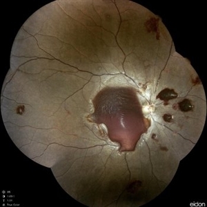
 Central Serous Chorioretinopathy in Pregnancy (OD)
Apr 28 2024 by Vishal Agrawal, MD, FRCS,FACS,FASRS
30-year female with sudden loss of vision came for examination. She was in her first trimester of pregnancy. Examination revealed bilateral bullous NSD with subretinal fibrin s/o CSR.
Photographer: Dr Ayushi
Imaging device: Clarus 700
Condition/keywords: Central Serous Chorioretinopathy (CSR), neurosensory detachment of retina, pregnancy
-
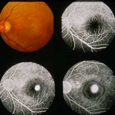
 Central serous chorioretinopathy with subretinal fibrin
Apr 4 2022 by T. P . VIGNESH, MBBS,MS
Fundus photo of left eye of a 37 year old female patient, who presented with defective vision of 1 month duration ,at 24 weeks of pregnancy, revealing Central serous chorioretinopathy with dense subretinal fibrin.
Photographer: Bharathi Singaravel
Imaging device: Zeiss Clarus
Condition/keywords: central serous chorioretinopathy (CSCR)
-
 Central Serous Chorioretinopathy With Subretinal Fibrin
May 16 2024 by T. P . VIGNESH, MBBS,MS
SD-OCT of the right eye of a 45 year old man revealing Central serous chorioretinopathy with subretinal fibrin .
Photographer: Sivanath
Imaging device: Heidelberg Spectralis
Condition/keywords: central serous chorioretinopathy (CSCR)
-
 Central Serous Retinopathy
Mar 17 2014 by Howard Schatz, MD
54-year-old male. CSR (FIBRIN).
Condition/keywords: central serous retinopathy (CSR)
-
 Central Serous Retinopathy
Mar 17 2014 by Howard Schatz, MD
54-year-old male. CSR (FIBRIN).
Condition/keywords: central serous retinopathy (CSR)
-
 Central Serous Retinopathy
Mar 17 2014 by Howard Schatz, MD
38-year-old male. CSR (FIBRIN). LE 20/40.
Condition/keywords: central serous retinopathy (CSR)
 Loading…