Search results (78 results)
-
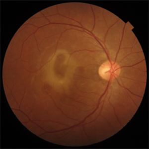
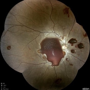
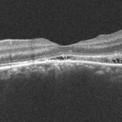
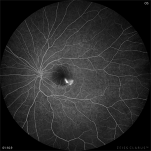 CSR with Fibrin-FFA
Jan 29 2025 by Vishal Agrawal, MD, FRCS,FACS,FASRS
A 31-year-old female was referred with a diagnosis of subretinal cysticercosis. BCVA was 20/200 OS. OCT showed a large subfoveal bacillary layer detachment (BALAD) without any scolex. FFA revealed a smoke-stack appearance. A final diagnosis of CSR with Fibrin was made and was managed conservatively. BCVA at final visit was 20/20.
Photographer: Dr Ayushi Gupta
Imaging device: Clarus 700
Condition/keywords: central serous chorioretinopathy (CSCR)
-
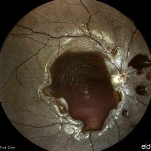
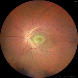 CSR with Fibrin
Jan 28 2025 by Vishal Agrawal, MD, FRCS,FACS,FASRS
A 31-year-old female was referred with a diagnosis of subretinal cysticercosis. BCVA was 20/200 OS. OCT showed a large subfoveal bacillary layer detachment (BALAD) without any scolex. FFA revealed a smoke-stack appearance. A final diagnosis of CSR with Fibrin was made and was managed conservatively. BCVA at final visit was 20/20.
Photographer: Dr Ayushi Gupta
Imaging device: Clarus 700
Condition/keywords: central serous chorioretinopathy (CSCR)
-
 Central Serous Chorioretinopathy With Subretinal Fibrin
May 16 2024 by T. P . VIGNESH, MBBS,MS
SD-OCT of the right eye of a 45 year old man revealing Central serous chorioretinopathy with subretinal fibrin .
Photographer: Sivanath
Imaging device: Heidelberg Spectralis
Condition/keywords: central serous chorioretinopathy (CSCR)
-
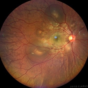 Central Serous Chorioretinopathy in Pregnancy (OD)
Apr 28 2024 by Vishal Agrawal, MD, FRCS,FACS,FASRS
30-year female with sudden loss of vision came for examination. She was in her first trimester of pregnancy. Examination revealed bilateral bullous NSD with subretinal fibrin s/o CSR.
Photographer: Dr Ayushi
Imaging device: Clarus 700
Condition/keywords: Central Serous Chorioretinopathy (CSR), neurosensory detachment of retina, pregnancy
-
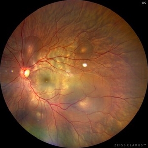 Central Serous Chorioretinopathy in Pregnancy (OS)
Apr 28 2024 by Vishal Agrawal, MD, FRCS,FACS,FASRS
30-year female with sudden loss of vision came for examination. She was in her first trimester of pregnancy. Examination revealed bilateral bullous NSD with subretinal fibrin s/o CSR.
Photographer: Dr Ayushi
Imaging device: Clarus 700
Condition/keywords: Central Serous Chorioretinopathy (CSR), neurosensory detachment of retina, pregnancy
-
 CSCR with Sub-Retinal Fibrin
Feb 6 2024 by Thirumalesh Mochi Basavaraj, MD
43 year old gentleman with a chronic central serous chorioretinopathy with sub retinal fibrin deposition.
Photographer: Puttaswamy
Condition/keywords: central serous chorioretinopathy (CSCR), subretinal fibrin deposition
-
 Endophthalmitis one day after tap and injection.
Nov 19 2022 by Gareth Lema, MD, PhD
A fibrin clot, consolidating one day after tap and injection for post-op endophthalmitis by Staph. aureus.
Photographer: Gareth Lema, MD, PhD, New York Eye and Ear of Mount Sinai
Imaging device: Cell phone with a macro lens and muscle light for illumination.
Condition/keywords: endophthalmitis
-
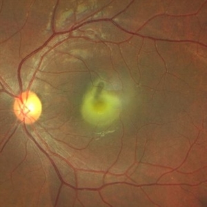 Central serous chorioretinopathy with subretinal fibrin
Apr 4 2022 by T. P . VIGNESH, MBBS,MS
Fundus photo of left eye of a 37 year old female patient, who presented with defective vision of 1 month duration ,at 24 weeks of pregnancy, revealing Central serous chorioretinopathy with dense subretinal fibrin.
Photographer: Bharathi Singaravel
Imaging device: Zeiss Clarus
Condition/keywords: central serous chorioretinopathy (CSCR)
-
Macular Hematoma Secondary Valsalva Maneuver
Oct 14 2021 by Islam bechakh
A 32-year-old man, who has presented for 02 months, a macular hematoma secondary to a Valsalva maneuver. He benefited from an attempt to open the hematoma with a Yag laser, but to no avail. We operated on and performed a 23G vitrectomy with posterior vitreous detachment, and discovered an epiretinal membrane which separated the hematoma from the posterior hyaloid. After removal of this membrane and aspiration of red blood cells and fibrin, the macula regained a normal appearance with good functional recovery.
Photographer: Islam Bechakh
Condition/keywords: epiretinal membrane (ERM), ERM, Macular hematoma, Valsalva maneuver
-
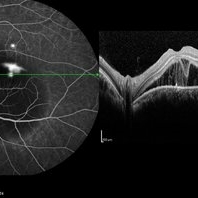 Fibrin Dipping Sign in OCT in Smokestack CSCR Leak
Aug 27 2020 by KRISHNENDU NANDI, MS
Image of a 42-year-old male showing smokestack CSCR leak in DFA with subretinal fibrin generates a dipping morphological pattern on OCT.
Photographer: Dr. Krishnendu Nandi
Condition/keywords: central serous chorioretinopathy (CSCR), fibrin
-
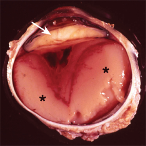
 Amelanotic Choroidal Melanoma
May 18 2020 by McGill University Health Centre
The enucleation specimen in (A) shows an amelanotic, mushroom-shaped tumor arising from the choroid. Microhemorrhages are present within the tumor and also surround the tumor base (arrow). True retinal detachment is present (arrowhead). The subretinal fluid is mixed: clear (1), hemorrhagic (2), and fibrinoid (3).
Condition/keywords: enucleation, mushroom-shaped
-
 Diffuse Uveitis
May 18 2020 by McGill University Health Centre
In this enucleation specimen, the uveal tract is completely replaced by purulent material (*). There is a subretinal hemorrhage underlying the detached retina and fibrous material in the vitreous cavity (arrowhead). The lens is cataractous and is surrounded by fibrinoid membranes (arrow).
Condition/keywords: uveitis
-
 Anemic Retinopathy Related Retinal Hemorrhages
Nov 5 2019 by Chinmayi Vyas
Anemic retinopathy related retinal hemorrhages in a 24 years old male with Hb of 4.2gm/ dl. The manifestations of anemic retinopathy are nonspecific and may closely simulate hypertensive or diabetic retina. Retinal changes in anemia are cotton wool spots, venous tortuosity, and hemorrhages which may be present at all levels of the retina and choroid. All retinal hemorrhages can occur when Hb falls below 8 g/100 ml or if the platelet count falls below 50,000/cumm. The combination of severe anemia and thrombocytopenia is likely to produce retinal hemorrhages. The Roth’s spots or white centre hemorrhages are typically associated with bacterial endocarditis , anemia and other systemic conditions. The white center is suspected to represents focal ischemia, inflammatory or infectious infiltrate, fibrin or accumulation of neoplasticism cells.
Photographer: Dr Chinmayi Vyas, Nethradhama superspeciality eye hospital , banglore, india
Imaging device: Eidon fundus imaging
Condition/keywords: anaemic retinopathy
-
 Anemic Retinopathy Related Retinal Hemorrhages
Nov 5 2019 by Chinmayi Vyas
Anemic retinopathy related retinal hemorrhages in a 24 years old male with Hb of 4.2gm/ dl. The manifestations of anemic retinopathy are nonspecific and may closely simulate hypertensive or diabetic retina. Retinal changes in anemia are cotton wool spots, venous tortuosity, and hemorrhages which may be present at all levels of the retina and choroid. All retinal hemorrhages can occur when Hb falls below 8 g/100 ml or if the platelet count falls below 50,000/cumm. The combination of severe anemia and thrombocytopenia is likely to produce retinal hemorrhages. The Roth’s spots or white centre hemorrhages are typically associated with bacterial endocarditis , anemia and other systemic conditions. The white center is suspected to represents focal ischemia, inflammatory or infectious infiltrate, fibrin or accumulation of neoplasticism cells.
Photographer: Dr Chinmayi Vyas
Condition/keywords: retinal hemorrhage
-
 Branch Retinal Artery Occlusion (BRAO) - Embolic
Mar 27 2019 by Gary R. Cook, MD, FACS
Superonasal BRAO with fibrin-platelet embolus OD.
Imaging device: Topcon VT-50
Condition/keywords: branch retinal artery occlusion (BRAO), embolus
-
 Slide 9-19
Feb 26 2019 by Lancaster Course in Ophthalmology
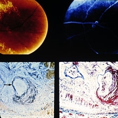
Retinal arterial macroaneurysm. A ring of retinal exudate partially surrounds the macroaneurysm (upper left), which is more clearly delineated by fluorescein (upper right). The retinal arteriole is greatly dilated, and the stain for elastic tissue shows a localized area of disruption and loss of the internal elastic membrane (arrow). The surrounding retina is thickened by edema and some hemorrhage. The ectatic area of the vessel wall is greatly thickened by the accumulation of a laminated fibrinous material. (Courtesy of Alan Friedman, M.D.)
Condition/keywords: retinal arterial macroaneurysm, retinal exudates
-
 Slide 9-18
Feb 26 2019 by Lancaster Course in Ophthalmology
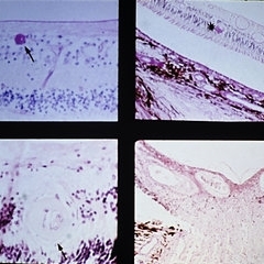
Malignant hypertension with retinal arterioles that are thickened and have fibrinoid necrosis (arrows). Retinal exudates (asterisk) and papilledema are also present. Papilledema is evidenced by fullness of the optic nerve head and peripapillary crowding of the retina (lower right).
Condition/keywords: fibrinoid, malignant hypertension, papilledema, retinal arteriole, retinal exudates
-
 Slide 7-59
Feb 25 2019 by Lancaster Course in Ophthalmology
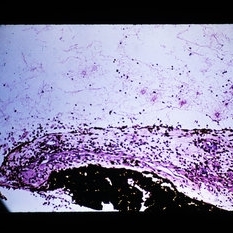
Fibrinoid necrosis of the sclera with palisading of granulomatous inflammation at the edge of the necrosis.
Condition/keywords: fibrinoid, sclera
-
 Slide 2-16
Feb 19 2019 by Lancaster Course in Ophthalmology
Organization of fibrinous exudate from iris, causing posterior synechiae and pupillary membrane.
Condition/keywords: fibrinous exudate, membranes, posterior synechiae
-
 Slide 2-14
Feb 19 2019 by Lancaster Course in Ophthalmology
Papillitis indicated by the disc edema, perivascular infiltrate, and overlying fibrinous exudate in fungal endophthalmitis.
Condition/keywords: edema, fibrinous exudate, fungal endophthalmitis, papillitis
-
 Slide 1-29
Feb 19 2019 by Lancaster Course in Ophthalmology
Smudgy, "fibrinoid" necrosis of collagen in the sclera of a patient with rheumatoid scleritis. Some abscess formation is seen above, rimmed by a granulomatous reaction. (H&E stain)
Condition/keywords: fibrinoid, sclera, scleritis
-
 Slide 1-2
Feb 14 2019 by Lancaster Course in Ophthalmology
Pink network of fibrin strands lying over the iris in recurrent iritis ("fibrin- o s exudate"). (H&E stain)
Condition/keywords: fibrin, iris, iritis
-
 Platelet Fibrin Emboli Fisher
May 3 2018 by Alexandr Stepanov
Platelet fibrin emboli Fisher.
Photographer: Alexandr Stepanov MD, PhD, FEBO, Faculty Hospital Hradec Kralove, Czech Republic
Condition/keywords: platelet fibrin emboli
-
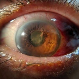
 Anterior Capsular Opacity
Feb 8 2018 by Claire Kiernan, MD
Slit lamp photograph of a 39-year-old female following uncomplicated cataract surgery shown here with dense fibrinous changes of the anterior capsule. This patient underwent Nd:YAG laser anterior capsulotomy with clearing of her visual axis.
Photographer: Steve Crow, University of Tennessee Hamilton Eye Institute, Memphis, TN
Condition/keywords: anterior capsule opacification, cataract extraction, cataract surgery
-
 OCT Showing Subretinal Hyperreflective Material in a Patient w[With CSCR
May 5 2017 by Gustavo Barreto de Melo, MD, PhD, FASRS
32-year-old pregnant woman with a serous detachment and subretinal fibrin deposit surrounding the fovea.
Photographer: Denyson Silva
Condition/keywords: central serous chorioretinopathy (CSCR), pregnancy
 Loading…