Search results (171 results)
-
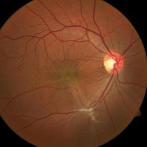
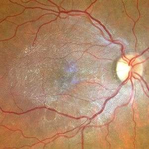
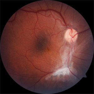
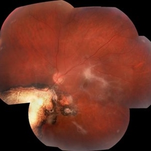
 Epiretinal Membrane/Macular Pucker With Combined Hamartoma of Retina and RPE
Jul 8 2015 by Emmanuel Chang, MD PhD FACS FASRS
10-year-old with history of progressive severe distortion in the left eye over the past year.
Photographer: Retina and Vitreous of Texas
Imaging device: Heidelberg Autofluorescence
Condition/keywords: combined hamartoma, epiretinal membrane (ERM), retinal pigment epithelium (RPE) hamartoma
-
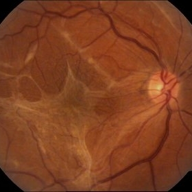
 Pucker
Oct 8 2012 by David R. Chow, MD, FRCS(C)
Condition/keywords: epiretinal membrane (ERM)
-
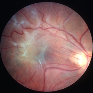
 Epiretinal Membrane
Oct 15 2012 by Sharon Fekrat, MD FACS FASRS
Fundus photograph of an epiretinal membrane
Photographer: Michael P. Kelly, FOPS, Director, Duke Eye Labs, Duke University Eye Center, Durham, NC
Condition/keywords: epiretinal membrane (ERM)
-
 Epiretinal Membrane
Sep 14 2012 by Michael P. Kelly, FOPS
Epiretinal membrane imaged using a high magnification retinal fundus camera and red free illumination.
Photographer: Michael P. Kelly, FOPS, Director, Duke Eye Center Labs, Duke Universtiy Hospital
Condition/keywords: epiretinal membrane (ERM), high magnification, monochromatism, red-free
-
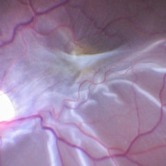 ERM With Retinal Detachment
May 25 2017 by Manish Nagpal, MD, FRCS (UK), FASRS
Per operative photo prior to ERM removal in a case of retinal detachment with ERM.
Photographer: MANISH NAGPAL
Imaging device: SONY 3 CHIP HD CAMERA
Condition/keywords: epiretinal membrane (ERM), internal limiting membrane (ILM) peeling
-
 OCT Image of Epiretinal Membrane
Aug 29 2017 by Carolyn Daley
OCT photograph of a 64-year-old women with an epiretinal membrane in the right eye. Patient has not noticed any decline in vision so surgery was not recommended at this time.
Photographer: Carolyn Daley
Imaging device: Heidelberg Spectralis
Condition/keywords: epiretinal membrane (ERM), optical coherence tomography (OCT)
-
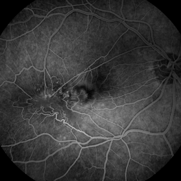
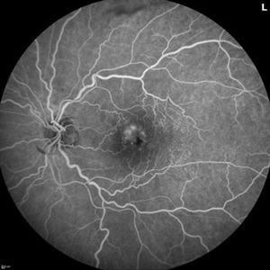 Branch Retinal Vein Occlusion with Multifactorial Macular Edema and Epiretinal Membrane
Oct 3 2024 by Logan ryzenga
Fluorescein angiogram of a 62 year old woman with cystoid macular edema from concurrent Epiretinal Membrane and Branch Retinal Vein occlusion. She has an extensive history of anti-VEGF injections with stable but unresolved macular edema. Following angiography, it was determined that an epiretinal membrane peel would be indicated in an attempt to achieve resolution of macular edema.
Photographer: Logan Ryzenga
Imaging device: Heidelberg Spectralis
Condition/keywords: 55-degrees, branch retinal vein occlusion (BRVO), cystoid macular edema (CME), epiretinal membrane (ERM), Fluorescein angiography, heidelberg spectralis, hyperfluorescence, leakage, left eye, OS, wide angle imaging
-
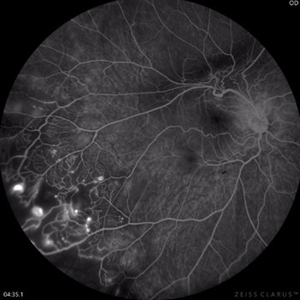 Coats Disease Fluorescein Angiography
Sep 2 2022 by FLOR ANGELICA JACOME GUTIERREZ
Fluorescein angiography of a patient with Coats disease where we found telangiectatic vessels, aneurysms, peripheral capillary nonperfusion and perivascular leak.
Photographer: Dr.Guillermo Salcedo Villanueva
Imaging device: Zeiss CLARUS 700 (FA)
Condition/keywords: Coats' disease, epiretinal membrane (ERM)
-
 Epiretinal Membrane
Sep 6 2021 by Ricardo Leitão Guerra
65-year-old woman with an asymptomatic ERM (BCVA=20/20).
Imaging device: Zeiss Clarus 700
Condition/keywords: epiretinal membrane (ERM)
-
 Epiretinal Membrane
May 10 2017 by Nichole Lewis
Epiretinal membrane.
Photographer: Nichole Lewis
Condition/keywords: epiretinal membrane (ERM)
-
 Epiretinal Membrane
Oct 11 2012 by Michael P. Kelly, FOPS
This is a patient with idiopathic panuveitis who developed a visually significant epiretinal membrane. Pars plana vitrectomy with membrane peeling was performed to remove the epiretinal proliferation. I recommend magnifying the image to see the exquisite detail centrally.
Photographer: Michael P. Kelly, FOPS Director, Duke Eye Center Labs, Duke Universtiy Hospital
Imaging device: Zeiss 450Plus
Condition/keywords: epiretinal membrane (ERM), panuveitis
-
 Epiretinal membrane - Fundus photograph
Feb 5 2014 by Gerardo Garcia-Aguirre, MD
Fundus photograph of a 62 year old female with metamorphopsia and decreased visual acuity. A stage 2 epiretinal membrane is observed, causing distortion of the retinal vasculature.
Photographer: Gerardo Garcia-Aguirre, MD
Condition/keywords: epiretinal membrane (ERM)
-
 ERM
ERM
Nov 26 2020 by Priya Rasipuram Chandrasekaran, MBBS, DO, DNB, FRCS
A 58-year-old female presented with distortion of images 1 month following cataract surgery in the right eye and fundus examination showed epiretinal membrane extending from the disc to the macula and OCT macula showing epiretinal membrane with disorganization of the foveal architecture.
Condition/keywords: epiretinal membrane (ERM)
-
 Full Thickness Macular Hole With ERM
Feb 26 2014 by Sharon Fekrat, MD FACS FASRS
Middle aged woman with a full thickness macular hole in the left eye associated with an epiretinal membrane.
Photographer: Michael P Kelly, Ophthalmic Photographer, Duke Eye Imaging, Duke Eye Center
Condition/keywords: epiretinal membrane (ERM), macular hole
-
 Macular Pucker
Mar 29 2013 by Henry J. Kaplan, MD
Large epiretinal membrane with straightened vessels in the papillomacular bundle and distorsion of retinal vessels.
Condition/keywords: epiretinal membrane (ERM), macular pucker
-
 Macular Traction Related to Toxoplasma Chorioretinitis
Jan 7 2021 by Lucas Zago Ribeiro, MD
Fundus image of a 50-year-old woman with macular traction and epiretinal membrane after toxoplasma chorioretinitis.
Photographer: Lucas Zago Ribeiro, UNIFESP / EPM, Brazil
Condition/keywords: epiretinal membrane (ERM), toxoplasmosis
-
 Partial Optic Disc Avulsion with Optic Disc Pit
Jul 1 2018 by John S. King, MD
16-year-old with acute loss of vision after blunt finger injury to eye while playing football. This photo is three weeks post-injury. Vision HM. Retinal striae with subhyaloid heme. Decreased retinal whitening. Peripapillary heme clearing, and temporal optic disc avulsion with optic disc pit can be seen.
Photographer: Maisee Yang
Imaging device: Topcon
Condition/keywords: epiretinal membrane (ERM), optic nerve head avulsion, optic nerve pit, traumatic optic neuropathy
-
 Per Operative Photo Post ILM Removal
May 25 2017 by Manish Nagpal, MD, FRCS (UK), FASRS
Per operative photo immediately following ILM removal.
Photographer: manish nagpal
Imaging device: SONY HD SURGICAL MICROSCOPE CAMERA
Condition/keywords: dye, epiretinal membrane (ERM), internal limiting membrane (ILM) peeling, staining
-
 Pre-Macular Fibrosis
Jul 26 2014 by Avris Romario Diparaja Siahaan
Red Free Image of an 88-year-old-woman with pre-macular fibrosis in her left eye. Her left eye was pseudophakia.
Photographer: Avris Romario Diparaja Siahaan, Klinik Mata Nusantara
Imaging device: Heidelberg Spectralis
Condition/keywords: epiretinal membrane (ERM), red-free
-
 Spiral ERM
Dec 20 2019 by Anfisa Ayalon, MD
OCT of a 60 -year-old man with epiretinal membrane. Note a spiral-like edge.
Photographer: Anfisa Ayalon,MD., Meir Medical Center, Kfar Saba, Israel.
Condition/keywords: epiretinal membrane (ERM), optical coherence tomography (OCT), rare
-
 Stained ILM Post ERM Removal-1-018
May 25 2017 by Manish Nagpal, MD, FRCS (UK), FASRS
Per operative photo of stained ILM status post ERM removal in a case of RD with ERM.
Photographer: MANISH NAGPAL
Imaging device: SONY 3 CHIP HD CAMERA
Condition/keywords: epiretinal membrane (ERM), internal limiting membrane (ILM) peeling, staining
-
 Status Post ERM Removal
May 25 2017 by Manish Nagpal, MD, FRCS (UK), FASRS
Per operative photo of ERM removal in a case of retinal detachment with ERM.
Photographer: MANISH NAGPAL
Imaging device: SONY 3 CHIP HD CAMERA
Condition/keywords: epiretinal membrane (ERM), internal limiting membrane (ILM) peeling
-
 Thickening of the Posterior Hyaloid
Dec 12 2020 by Anyssa Montenegro
Color fundus photograph of the right eye of a 36-year-old man showing thickening of the posterior hyaloid associated with an epiretinal membrane due to ocular toxoplasmosis.
Photographer: Anyssa Montenegro, Centro Brasileiro da Visão, Brasília-DF, Brazil
Condition/keywords: epiretinal membrane (ERM), ocular toxoplasmosis, thickening of the posterior hyaloid
-
 Toxoplasmosis Associated Epiretinal Membrane
Oct 27 2016 by Gabriel Costa Andrade, PhD
Fundus photograph of a 26-year-old woman with a chorioretinal scar due to toxoplasmosis and secondary epiretinal membrane.
Photographer: Gabriel Andrade, Federal University of São Paulo, São Paulo, Brazil
Condition/keywords: epiretinal membrane (ERM), toxoplasmosis
-
 Epiretinal Membrane
Jun 1 2018 by vitor borges porfirio pereira pereira
Epiretinal membrane by multicolor
Photographer: Marcos Avila, CBV Hospital de Olhos, Brasilia
Condition/keywords: epiretinal membrane (ERM)
 Loading…