Search results (121 results)
-
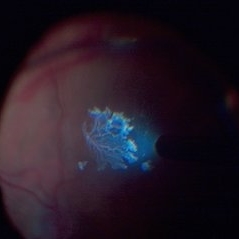 Detached NVE During PVD induction
Apr 27 2018 by Michael J. Koss, MD, PhD, MBA
A 73-year-old woman with macular pucker underwent a pars plana vitrectomy with membrane peeling. Additionally the patient suffers from diabetic retinopathy after being diagnosed with type 2 diabetes mellitus sixteen years ago. Prior to the procedure she was treated with a series of intravitreal Bevacizumab-injections due to diabetic macular edema. There was no history of a proliferative DRP. During the vitrectomy a branch of an obliterated NVE spontaneously detached and floated freely in the vitreous. The 3D shot was captured via Alcon’s NGENUITY® 3D Visualization System in form of photograph and video providing an outstandingly detailed image of the branched NVE.
Photographer: Michael Koss, Augenzentrum Nymphenburger Hoefe
Imaging device: Alcon’s NGENUITY® 3D Visualization System
Condition/keywords: diabetes, diabetic retinopathy, neovascularization elsewhere (NVE), pars plana vitrectomy (PPV), PVD induction
-
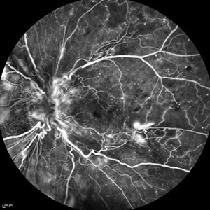
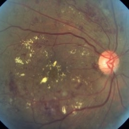
 Diabetic Macular Edema, Proliferative Diabetic Retinopathy, Neovascularization Elsewhere, DME, PDR, NVE
Apr 1 2013 by James B. Soque, CRA, OCT-C, COA, FOPS
39-year-old white female and long standing diabetis, c/o new peripheral symptoms of left eye. FA OS reveals diabetic macular edema, microaneurysms, and neovasculaization elsewhere. Fluorescein Angogram, Early Phase, 50 Deg, 2x Mag.
Photographer: James B Soque, CRA, COA
Imaging device: Topcon TRC 50DX with MERGE software, OIS 10.6.45
Condition/keywords: diabetic macular edema, neovascularization (NV), proliferative diabetic retinopathy (PDR)
-
---thumb.jpg/image-square;max$300,300.ImageHandler) Diabetic Retinopathy Hard Exudates OD
Diabetic Retinopathy Hard Exudates OD
Jun 30 2013 by Rogerio N Shinsato, MD, PhD
Fundus photograph with diabetic retinopathy.
Condition/keywords: diabetic macular edema, foveal hard exudates
-
 Diabetic Retinopathy Hard Exudates OS
Jun 30 2013 by Rogerio N Shinsato, MD, PhD
Fundus photograph with diabetic retinopathy.
Condition/keywords: diabetic macular edema, foveal hard exudates
-
 Diabetic Retinopathy Hard Exudates OS
Jun 30 2013 by Rogerio N Shinsato, MD, PhD
Fundus photograph with diabetic retinopathy.
Condition/keywords: diabetic macular edema, foveal hard exudates
-
 Ozurdex Sarcophagus in DME
Apr 17 2017 by Manish Nagpal, MD, FRCS (UK), FASRS
50-year-old male treated with Ozurdex implant for DME came for a follow up after 4 months and we could see the sarcophagus of the implant dangling in vitreous in front of the macula.
Photographer: POOJA BAROT
Condition/keywords: diabetic macular edema, Ozurdex implant, sarcophagus
-
 Ozurdex Sarcophagus in DME
Apr 17 2017 by Manish Nagpal, MD, FRCS (UK), FASRS
50-year-old male treated with Ozurdex implant for DME came for a follow up after 4 months and we could see the sarcophagus of the implant dangling in vitreous in front of the macula in this montage view.
Photographer: pooja barot
Condition/keywords: diabetic macular edema, Ozurdex implant, sarcophagus
-
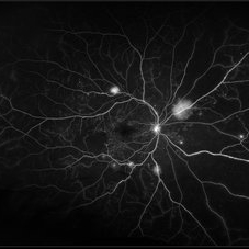
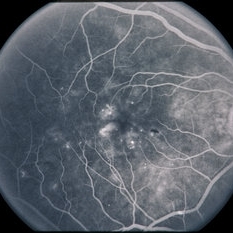
 Proliferative Diabetic Retinopathy
Mar 16 2015 by Matt Poe, COA
IVFA of 53-year-old male with Proliferative Diabetic Retinopathy, Diabetic macular edema, and a tractional retinal detachment.
Photographer: Matt Poe, COA. Northwest Arkansas Retina Associates, Springdale, AR.
Imaging device: Heidelberg HRA
Condition/keywords: neovascularization (NV), proliferative diabetic retinopathy (PDR)
-
 Proliferative Diabetic Retinopathy
Jan 29 2021 by Olivia Rainey
Ultra-widefield fluorescein angiogram of a 65-year-old male with proliferative diabetic retinopathy affecting his right eye. The patient's diabetic retinopathy has progressed significantly since he was last seen in 2014. It was recommended to begin antiVEGF to control DME followed by laser treatment OU.
Photographer: Olivia Rainey, OCT-C, COA
Imaging device: Optos California
Condition/keywords: anti-VEGF, diabetes, diabetic macular edema, neovascularization (NV), neovascularization elsewhere (NVE), non-perfusion, Optos, proliferative diabetic retinopathy (PDR), ultra-wide field imaging
-
 Serous Retinal Detachment in Advanced Proliferative Diabetic Retinopathy
Feb 15 2024 by Annaka Gooding
Ultra-Wide fundus photograph of a 29 year old female with a Serous Retinal Detachment in Advanced PDR. Patient present to clinic with LP vision following PPV and fill in PRP. Physician recommended oral prednisone treatment and to reassess at their following visit.
Photographer: Annaka Gooding, CPO
Imaging device: Optos California RGB
Condition/keywords: Diabetes, diabetic macular edema, fundus photography, OPTOS CALIFORNIA, pan-retinal photocoagulation (PRP), pars plana vitrectomy (PPV), proliferative diabetic retinopathy (PDR), serous retinal detachment, ultra-wide field imaging
-
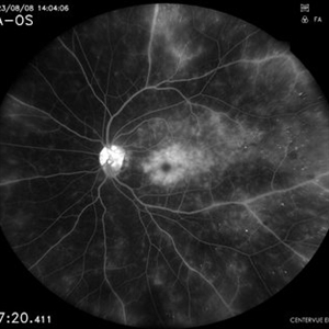
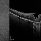
 Vitreomacular traction
Jun 23 2022 by T. P . VIGNESH, MBBS,MS
SD-OCT of RE reveals Vitreomacular traction resembling bow and arrow and diabetic macular edema with intraretinal hard exudates in a 60 year old female patient with Moderate NPDR .
Imaging device: Heidelberg Spectralis
Condition/keywords: vitreomacular traction (VMT)
-
 CIRCINATE RETINOPATHY
Oct 19 2022 by Akansha Sharma
COLOUR FUNDUS PHOTOGRAPH OF A 51 YEAR OLD MALE WITH DIABETIC MACULOPATHY
Photographer: Dr. Akansha Sharma-Retina Foundation, Ahmedabad
Condition/keywords: circinate retinopathy, diabetic macular edema, diabetic maculopathy
-
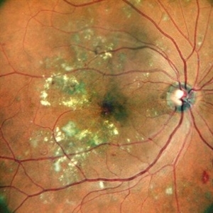
 Ischaemic Diabetic Maculopathy
Dec 18 2012 by Mallika Goyal, MD
Right eye of a 57-year-old diabetic gentleman with diabetic macular edema. OCT shows macular thickening and fluid, and fluorescein shows foveal leak with parafoveal non-perfusion. In view of the parafoveal non-perfusion, anti-VEGF therapy may worsen the situation in this eye.
Photographer: Mallika Goyal, MD, Apollo Health City, Hyderabad, India
Condition/keywords: ischaemic diabetic maculopathy
-
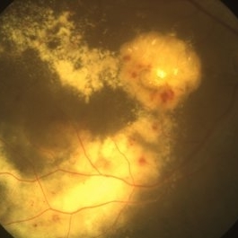 Abundant Hard Exudates - Diabetic Macular Edema
Oct 3 2013 by Gerardo Garcia-Aguirre, MD
Abundant hard exudates - diabetic macular edema.
Condition/keywords: diabetic macular edema
-
 Angiographic Diabetic Macular Edema in a Case of Proliferative Diabetic Retinopathy
Apr 9 2024 by Akansha Sharma
Fundus fluorescein angiographic image of 62 year old male demonstrating angiographic diabetic macular edema in a case of proliferative diabetic retinopathy.
Photographer: Dr. Akansha Sharma, Bharati Eye Hospital
Condition/keywords: clinically significant macular edema (CSME), diabetic blindness, diabetic macular edema, proliferative diabetic retinopathy (PDR)
-
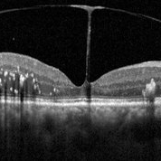
 Bullseye Maculopathy
Jan 22 2024 by Kali Jend
Optical coherence tomography of a 73-year-old female with Bullseye Macular Changes affecting her left eye. Patient reports having a family history of this condition and denies prior Plaquenil or Elmiron use. Compared to previous imaging, the patient's condition progressed in the left eye from 2020 to 2023. Patient has a history of fluctuating Diabetic Macular Edema and a current Epiretinal Membrane as well. Patient's vision was Ncc20/60 at the time the image was taken.
Photographer: Kali Jend
Imaging device: Heidelberg Spectralis
Condition/keywords: bullseye maculopathy, epiretinal membrane (ERM), heidelberg spectralis, left eye, macular pucker, OCT, optical coherence tomography (OCT)
-
 Clinically Significant Macular Edema
Apr 23 2015 by Mehul A Shah
Patient presented with complaints of diminished vision ou.
Photographer: Mehul Shah
Imaging device: Zeiss FF450 Plus
Condition/keywords: diabetic macular edema
-
 CME DME After CE
Aug 27 2014 by Susanna S. Park, MD, PhD
Macular OCT of a 62-year-old diabetic woman with severe vision loss 2 weeks after cataract surgery due to severe worsening of macular edema. Exam also showed new proliferative diabetic retinopathy.
Photographer: chandra
Condition/keywords: diabetic macular edema, optical coherence tomography (OCT)
-
 Combined Tractional and Rhegmatogenous Retinal Detachment
Jan 30 2023 by Olivia Rainey
Ultra-widefield fluorescein angiography of a combined tractional and rhegmatogenous retinal detachment repair affecting the left eye. The retina is attached following silicone oil placement during most recent surgery. The patient was seeing CF at the time the image was taken.
Photographer: Olivia Rainey, OCT-C, COA
Imaging device: Optos California
Condition/keywords: diabetes, diabetic macular edema, diabetic retinopathy, hyperfluorescence, right eye, scleral buckle, silicone oil, tractional retinal detachment, ultra-wide field imaging, ultra-widefield image
-
 CRAO with PDR and DME
Oct 27 2014 by Mallika Goyal, MD
Right fundus of a 66-year-old diabetic tobacco chewing male who presented with sudden vision loss to HMCF 12 days back shows Lasered PDR with severe diabetic macular edema with retinal infarct from CRAO (accounting for the sudden recent vision loss).
Photographer: Mallika Goyal, MD, Apollo Health City, Jubilee Hills, Hyderabad-500033
Condition/keywords: central retinal artery occlusion (CRAO), proliferative diabetic retinopathy (PDR)
-
 Diabetic Macular Edema
Oct 12 2012 by Gregg T. Kokame, MD, MMM, FASRS
Diabetic macular edema
Photographer: Jaclyn Pisano, Retina Consultants of Hawaii
Imaging device: Zeiss FF-450 plus
Condition/keywords: diabetic macular edema
-
---thumb.JPG/image-square;max$300,300.ImageHandler) Diabetic Macular Edema
Diabetic Macular Edema
Oct 26 2012 by Mallika Goyal, MD
Fundus photograph of left eye of 55-year-old diabetic and hypertensive gentleman with normal serum lipids showing abundant foveal hard exudates.
Condition/keywords: diabetic macular edema
-
---thumb.JPG/image-square;max$300,300.ImageHandler) diabetic macular edema
diabetic macular edema
Oct 26 2012 by Mallika Goyal, MD
Fundus photograph of left eye of 58-year-old diabetic gentleman with normal serum lipids showing foveal hard exudates.
Condition/keywords: foveal hard exudates
-
 Diabetic Macular Edema
Feb 7 2014 by David Callanan, MD
56-year-old patient with diabetic macular edema.
Condition/keywords: diabetic macular edema
-
 Diabetic Macular Edema
Feb 7 2014 by David Callanan, MD
56-year-old patient with diabetic macular edema.
Condition/keywords: diabetic macular edema
 Loading…