Search results (466 results)
-
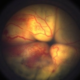
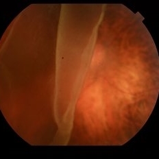
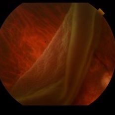
 Vortex Vein Varix - supine
Dec 19 2012 by Eric A. Postel, MD
Color photograph of a vortex vein varix in a patient in the supine position
Condition/keywords: vortex vein
-
 Coats' Disease
Apr 30 2020 by Giselle DeOliveira
Color Photograph of 20-month-old male infant.
Photographer: Giselle DeOliveira, University of Miami, Bascom Palmer Eye Institute
Imaging device: Retcam III
Condition/keywords: Coats' disease
-
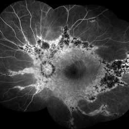
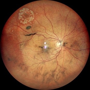
 Coats' Disease
Feb 25 2021 by Niloofar Piri, MD
Collage color photo and FA image of the same patient with Coats' Disease demonstrating telangiectatic aneurysmal lesions in the temporal periphery, associated with hard exudate deposition posteriorly. FA (AV phase) demonstrating hyperfluorescent aneurysmal lesions as well as peripheral capillary non perfusion. Note the posterior hypofluorescence where the hard exudates are located.
Condition/keywords: Coats' disease, congenital retinal telangiectasis, retinal telangiectasia
-
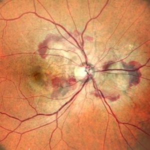
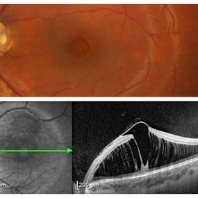 Optic Disc Pit with Maculopathy
Feb 25 2021 by Niloofar Piri, MD
Color fundus photograph and SD OCT of a 6-year-old patient with optic disc pit associated with large retinoschisis involving the entire macula. SD OCT demonstrating large retinoschisis with ILM draping centrally giving it the appearance of pseudohole on the corresponding central area of color photo. Vision 20/80
Photographer: Lisa Breeding, St Louis University
Condition/keywords: maculopathy, optic disc
-
 Prepapillary Vascular Loop
Mar 11 2020 by Asdrubal F Moreno, MD
Fundus color photograph of a 80-year-old woman with a unilateral congenital prepapillary vascular loop and hypertensive retinopathy, focus on the vascular loop end for perception.
Photographer: Asdrubal Moreno, Fundacion AVAO, Universidad de Los Andes, Venezuela
Imaging device: Zeiss Visucam 500
Condition/keywords: congenital prepapillary vascular loop, peripapillary
-
 A Large Break at the Posterior Pole With RD With PVR (S/p Old Blunt Trauma)
Jan 16 2025 by Anand Temkar
Right eye central fundus color photo of a 10 year old kid who noticed diminution of vision in right eye since a month. We can see the large break at the posterior pole with rolled up margins associated with retinal detachment and PVR changes.
Photographer: Dr.Anand Temkar- Retina Foundation, Ahmedabad
Imaging device: Mirante
Condition/keywords: Posterior pole break, proliferative vitreoretinopathy (PVR), Retinal Detachment
-
 A Large Break at the Posterior Pole With RD With PVR (S/p Old Blunt Trauma)
Jan 16 2025 by Anand Temkar
Right eye widefield fundus color photo of a 10 year old kid who noticed diminution of vision in right eye since a month. We can see the large break at the posterior pole with rolled up margins associated with retinal detachment and PVR changes.
Photographer: Dr.Anand Temkar- Retina Foundation, Ahmedabad
Imaging device: Mirante
Condition/keywords: posterior pole break, proliferative vitreoretinopathy (PVR), Retinal Detachment
-
 Bullous Retinal Detachment
Jul 9 2021 by Anton Orlin, MD
This is a color photograph of a right eye with a superior, bullous, macula-splitting retinal detachment. These features place the patient at a higher risk for macular fold formation postoperatively. To prevent fold formation, a surgeon should attempt for more complete subretinal fluid drainage during repair. This can be done with the use of perfluorocarbon liquid or by making a drainage retinotomy.
Condition/keywords: macular fold
-
 Coat's Disease
Jan 14 2025 by Kimberly Wakester
Fundus photographs of an 7-year-old boy with Coat's Disease in the right eye. There is subfoveal lipid end scarring in the macula and "light bulb" type telangiectasias temporally noted on exam and shown in Optos color photos. FA findings show anastomoses, capillary dropout, and "light bulb" type telangiectasias temporally with mild late leakage. Patient will be monitored at this time and have repeat imaging in 4 months.
Photographer: Kimberly Wakester, COA
Imaging device: Optos California
Condition/keywords: Coat's disease
-
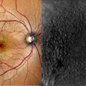 Color Photo and Retro Mode of Cuticular Drusens
Aug 30 2020 by Sham Talati, DOMS
A patient With cuticular drusens in both eyes.
Photographer: Dr. Sham Talati,Retina Foundation,Ahmedabad
Imaging device: Nidek Mirante
Condition/keywords: cuticular drusen
-
 Degeneration Paravenous
Sep 20 2016 by JEFFERSON R SOUSA, Tecg.º (Biomedical Systems Technology)
Female patient, 32-years-old, Asian, appeared at the clinic with a history of glaucoma. 20/20 visual acuity in both eyes. Examination of color photography, pigmentary changes were observed following the vascular arcades only in the left eye. Suggestive of paravenous degeneration.
Photographer: JEFFERSON R SOUSA - Study Center and Ophthalmological Research Dr. Andre M V Gomes, Institute Dr. Suel Abujamra São Paulo-Brazil
Imaging device: Zeiss / VisuCam-500 - Angulation of field photo of 45 Degrees, flash 24.
Condition/keywords: degeneration paravenous
-
 Dislocated Lens, Posterior OD
Jan 26 2024 by Corey Grant
OPTOS California photo presents a 71 year old male patient with a dislocated lens, posterior in the right eye. Presented on 1/26/24 with posteriorly dislocated SN60WF with a Soemmerring ring. Associated retinal hemorrhage within retinoschisis as well. This will result in a PPV/IOL exchange/SFIOL/STK for the right eye.
Photographer: Corey Grant, Ophthalmic Imager, Retina Specialist of Michigan
Imaging device: OPTOS California
Condition/keywords: color photo, IOL, OD, Optos, OPTOS CALIFORNIA, pars plana vitrectomy (PPV), retina
-
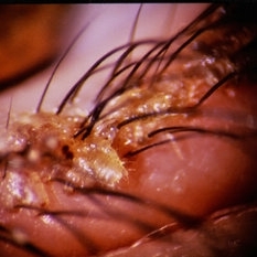 Lice in Lashes
-
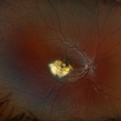
 Macular Pucker With Myelinated Nerve Fiber Layer
Nov 1 2018 by Kevin J. Blinder, MD, FASRS
Multi-color photo of macular pucker with myelinated nerve fiber layer.
Photographer: Jarrod Wehmeier
Imaging device: Heidelberg Spectralis
Condition/keywords: macular pucker
-
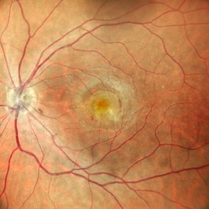
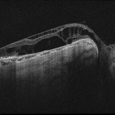 Optic Nerve Pit OD - OCT
Aug 6 2018 by Hosam Attia, MD
65-year-old white male, presented for a second opinion for possible cataract extraction OD. BCVA: OD: 20/70 OS: 20/60 WRx: OD: -3.75 +1.50 x 5 OS: -1.75 +1.50 x 178 SLE: +2 NS OD>OS DFE: OD: Nasal macular GA, connected by milder track of RPE changes to an optic nerve pit OD (no fluid seen clinically) OS: enlarged C/D w/ no pits, macular RPE change w/ No heme, CME/ SRF OCT: OD: Peri-papillary cystoid changes & outer retinal atrophy (corresponding to the area of GA on the pseudocolor photo) w/ No SRF (mimicking PP CNVM), connected to the optic disc pit by shallow sinus/ tract. OS: Drusenoid RPE changes, No cystoid changes/ SRF
Imaging device: Zeiss Cirrus -5000
Condition/keywords: congenital optic nerve pit
-
 Retinal Detachment
May 15 2018 by Morgan Benton
Ultra-wide field pseudocolor image of a 54-year-old male with a retinal detachment affecting his left eye after trauma. Patient was only able to see hand motion.
Photographer: Morgan Benton
Imaging device: Optos
Condition/keywords: color photo, left eye, Optos, ultra-wide field imaging
-
 Retinal Detachment
Oct 25 2018 by Graciela Nahuelquín Ríos
A 45-year-old patient reported a blow to the right eye 1 month ago, and a week ago he presented with low visual acuity. In retinal mapping and background color photography retinal detachment with giant rupture in temporal arch.
Photographer: Lic. TM. Graciela Nahuelquín Ríos
Imaging device: TRC-50DX - Topcon
-
 Retinal Hole
Feb 11 2024 by Anjana Mirajkar, MS Ophthalmology
A color photo of RE of a 50 year old male showing lasered retinal hole superiorly with vitreous degeneration.
Photographer: Dr. Anjana Mirajkar -Retina Foundation, Ahmedabad
Imaging device: Mirante-Nidek
Condition/keywords: full thickness retinal hole
-
 Right Eye Color Photo With Hemorrhages in Case of CNVM With Angioid Streaks
Nov 29 2024 by Anand Temkar
A 45 year old male came with chief complaint of blurring vision in right eyes since past 4 days. His vision is 6/12 in right eye and 6/9 in left eye. His vision was 14 mmHg in right eye and 16 mmHg in left eye. He was diagnosed with Angioid Streaks in both eyes about a year ago, then he developed choroidal neovascularization in his left eye 8 months ago, for which he received AntiVEGF injections x 3. Left eye is a stable eye now. Patient presented with right eye choroidal neovascularization in a case of Angioid Streaks on recent follow up. We have advised him right eye AntiVEGF injections x 3. In this image, the right eye color photo shows bleed from CNVM in case of angioid streaks.
Photographer: Dr.Anand Temkar- Retina Foundation, Ahmedabad
Imaging device: Mirante
Condition/keywords: Angioid Streaks, choroidal neovascular membrane (CNVM)
-
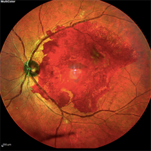 Subhyaloid Hemorrhage without Foveal Involvement Multicolor Photo
Jul 17 2020 by Sergio Groman-Lupa, MD, PhD
Subhyaloid hemorrhage without foveal involvement multicolor photo.
Photographer: Garcia Lelevier OD, Codet Vision Institute
Imaging device: Heidelberg Spectralis
Condition/keywords: subhyaloid hemorrhage
-
 Subretinal Neovascular Membrane
Oct 25 2023 by Vaidehi Sathaye
Fundus of a 45 year old female with SRNVM in the LE
Photographer: Dr. Vaidehi Sathaye
Imaging device: Mirante
Condition/keywords: color photo, SRNVM
-
 Tamoxifen
Jan 7 2015 by H. Michael Lambert, MD
Color photograph of advanced tamoxifen retinopathy with extensive crystalline deposits
Condition/keywords: tamoxifen retinopathy
-
 Terson's Syndrome
Jan 7 2015 by H. Michael Lambert, MD
Color photograph LE, premacular boat shaped hemorrhage due to Terson's syndrome.
Condition/keywords: premacular hemorrhage
-
 Toxoplasmosis Scar
Sep 26 2021 by Nivesh Gupta
Color photo of a 18-year-old male with Pigmented Parafoveal Scar
Photographer: DR. NIVESH GUPTA, RETINA FOUNDATION, AHMEDABAD
Imaging device: NIDEK MIRANTE
Condition/keywords: coloboma of macula, Parafoveal scar, Pigmented Parafoveal Scar, toxoplasmosis
 Loading…