Search results (143 results)
-
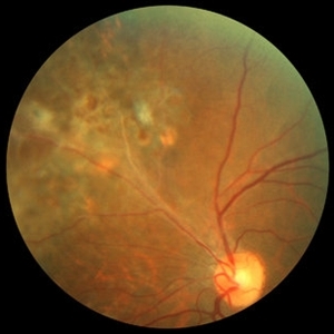
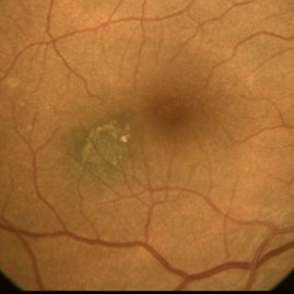 Choroidal Nevus Associated with Drusen
Jan 11 2021 by Gabriel Costa Andrade, PhD
Fundus photograph of an 53-year-old man with a macular melanotic nevus.
Photographer: Gabriel Andrade
Condition/keywords: choroidal nevus
-
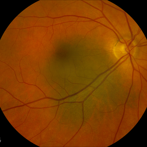
 Choroidal Nevus with Drusen
Oct 18 2012 by Suber S. Huang, MD, MBA, FASRS
65-year-old woman with choroidal nevus with drusen
Photographer: Irit Baum-Rawraway
Condition/keywords: choroidal nevus
-
 Choroidal nevus with halo
Aug 21 2022 by Niloofar Piri, MD
Fundus photograph of the right eye in a 43 yo patient demonstrating a flat choroidal nevsu with halo nasal to the optic nerve
Photographer: Andrew Polk, MD
Condition/keywords: Choroidal nevus, nevus with halo
-
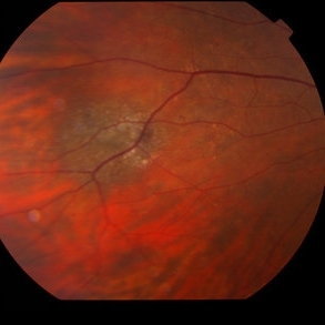
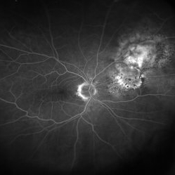
 choroidal nevus with polypoidal choroidal vasculopathy
Nov 20 2012 by Roy Schwartz, MD
Rare combination of a choroidal nevus complicated by polypoidal choroidal vasculopathy. The lesion is temporal to the fovea, and leakage of subretinal fluid almost reaching the fovea is demonstrated.
Photographer: Galit Yair-Pur
Condition/keywords: choroidal nevus, polypoidal choroidal vasculopathy (PCV)
-
 Choroidal Nevus with Subretinal Hemorrhage
Jan 29 2015 by Gordon Finnerty
47-year-old woman diagnosed with choroidal nevus with subretinal hemorrhage.
Photographer: Gordon Finnerty
Imaging device: Topcon TRC -50DX
Condition/keywords: choroidal nevus, subretinal hemorrhage
-
 Choroidal-Nevus
Feb 25 2023 by Aditya S Kelkar, MS, FRCS, FASRS,FRCOphth
Color fundus photograph of the left eye of a 55 year old male showing large choroidal nevus.
Photographer: Dr. Apoorva Jadhav, National Institute of Ophthalmology, Pune. India.
Imaging device: Zeiss Clarus 500
Condition/keywords: Choroidal nevus
-
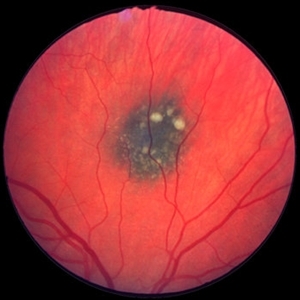
 Macular Nevus
Jan 20 2021 by Jamin S. Brown, MD
Macular Nevus as well as CSR.
Photographer: Stefanie Palmer CRA, Retina Vitreous Surgeons of CNY
Condition/keywords: choroidal nevus, macula lesion
-
 Torpedo Maculopathy 1
Feb 26 2014 by Raj K. Maturi, MD
Color fundus of a 47-year-old female with torpedo maculopathy- atypical choroidal nevus.
Photographer: Charlotte Harris COA Midwest Eye Institute Indianapolis, Indiana
Imaging device: TOPCON 50EX
Condition/keywords: choroidal nevus, fundus photograph, torpedo maculopathy
-
 Choroidal Melanoma
Jan 30 2019 by Karen Panzegrau
Ultra-wide field optos image of a 27-year-old male patient who presented with loss of vision for about 6-8 weeks. Previous choroidal nevus seen. Recommended annual monitoring. No exam for since 10/2014. Brachytherapy vs enucleation was discussed. Brachytherapy was decided as treatment. Full metastatic work up is being performed.
Photographer: Karen Panzegrau
Imaging device: Optos
Condition/keywords: choroidal nevus, exudative retinal detachment, malignant neoplasm of eye, Optos, ultra-wide field imaging
-
 Retinal Angiomas In VHL
Dec 24 2012 by Roy D. Brod, MD
Fundus photograph of 16 year old male with recent diagnosis of Von Hippel-Lindau disease showing typical appearance of a retinal angioma in superior mid periphery OD. Note unrelated choroidal nevus above superior arcade.
Photographer: Julia Walker
Condition/keywords: hemangioma, Von Hippel-Lindau
-
 Benign Choroidal Nevus
Dec 21 2012 by Gary S. Gutow, MD, MS
Photographer: Alecia Camp, CRA - Tennessee Retina, Nashville,TN
-
 Benign Choroidal Nevus
Dec 21 2012 by Gary S. Gutow, MD, MS
Photographer: Alecia Camp, CRA - Tennessee Retina, Nashville,TN
-
 Choroidal Nevus[002]
Dec 31 2012 by Raj K. Maturi, MD
Photographer: Tom Steele CRA Midwest Eye Institute Indianpolis, In
Imaging device: Optos
Condition/keywords: choroidal nevus
-
 Choroidal Nevus[004]
Dec 31 2012 by Raj K. Maturi, MD
Photographer: Tom, Steele CRA Midwest Eye Institute Indianapolis, In
Imaging device: Optos
Condition/keywords: Optos
-
 Choroidal naevus
Jan 11 2013 by Alex P. Hunyor, MD
Choroidal naevus with overlying drusen
Condition/keywords: benign pigmented lesions, choroidal nevus
-
 Amelenotic Choroidal Nevus
Oct 16 2012 by Jeffrey G. Gross, MD, FASRS
Amelenotic choroidal nevus.
-
 Benign Choroidal Nevus
Dec 21 2012 by Gary S. Gutow, MD, MS
Photographer: Nichole Lewis - Tennessee Retina, Nashville, TN
-
 Benign Choroidal Nevus
Dec 21 2012 by Gary S. Gutow, MD, MS
Photographer: Alecia Camp, CRA - Tennessee Retina, Nashville,TN
-
 Benign Choroidal Nevus
Dec 21 2012 by Gary S. Gutow, MD, MS
Photographer: Alecia Camp, CRA - Tennessee Retina, Nashville,TN
-
 Benign Choroidal Nevus
Dec 21 2012 by Peter L. Sonkin, MD
Photographer: Alecia Camp, CRA - Tennessee Retina, Nashville,TN
-
 Benign Choroidal Nevus
Dec 21 2012 by Carl C. Awh, MD, FASRS
Photographer: Alecia Camp, CRA - Tennessee Retina, Nashville,TN
-
 Benign Choroidal Nevus
Dec 21 2012 by Brandon G. Busbee, MD
Photographer: Alecia Camp, CRA - Tennessee Retina, Nashville,TN
-
 Benign Choroidal Nevus
Dec 21 2012 by Franco M. Recchia, MD
Photographer: Alecia Camp, CRA - Tennessee Retina, Nashville,TN
-
 Benign Nevus
Dec 6 2023 by Virginia Gebhart
71 year old female with benign choroidal nevus with fibrotic PED
Photographer: Virginia Gebhart
Imaging device: Topcon
Condition/keywords: choroidal nevus, fibrotic neovascularization, nevus
-
 Choroidal Mass
Jul 14 2013 by Jason S. Calhoun
Fundus photo shows yellowish choroidal mass in the left eye.
Photographer: Jason S. Calhoun, Department of Ophthalmology, Mayo Clinic Jacksonville, Florida
Imaging device: TOPCON TRC 50-EX
Condition/keywords: choroidal nevus, indolent choroidal mass
 Loading…