Search results (345 results)
-
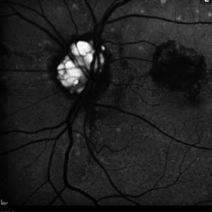
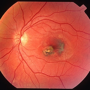
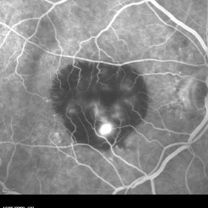
 Optic Nerve Head Drusen With Idiopathic CNV
Feb 17 2017 by Kristen Wagner
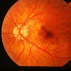
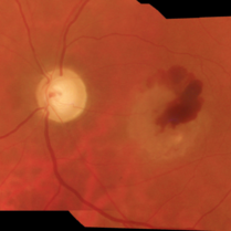
22-year-old female fundus photograph of a right eye with Optic Nerve Drusen with Idiopathic CNV.
Photographer: Kristen Wagner, COT, OSC Ophthalmic Photographer, Tennessee Retina, Nashville TN
Condition/keywords: choroidal neovascularization (CNV), drusen of optic disc, optic disc drusen
-
 Cat Eye Syndrome
Feb 11 2020 by Sophia El Hamichi, MD
A 3-year-old female with cat eye syndrome including iris, chorioretinal and optic nerve colobomas. Note the CNV temporally to the optic nerve coloboma (blue arrows)
Photographer: Giselle De Oliveira, Bascom Palmer Eye Institute, Miami
Imaging device: RetCam
Condition/keywords: cat eye syndrome, chorioretinal coloboma, choroidal neovascularization (CNV), coloboma, coloboma of optic disc, optic nerve coloboma
-
 Choroidal Neovascularization
May 27 2020 by Jamin S. Brown, MD
73-year-old female with CNV.
Photographer: Jeffrey Barker, Retina-Vitreous Surgeons of CNY
Condition/keywords: choroidal neovascularization (CNV)
-
---thumb.jpg/image-square;max$300,300.ImageHandler) Active Choroidal Neovascularization With Subretinal Hemorrhage
Active Choroidal Neovascularization With Subretinal Hemorrhage
Nov 25 2013 by Maurice F. Rabb
Active choroidal neovascularization with subretinal hemorrhage.
Condition/keywords: choroidal neovascularization (CNV), subretinal hemorrhage
-
 Choroidal Osteoma Plus CNV
Sep 2 2012 by Hamid Ahmadieh, MD
Color fundus photograph and OCT imaging of a 47-year-old man with a juxtafoveal CNV superimposed on a choroidal osteoma.
Photographer: Hamid Ahmadieh, Ophthalmic Research Center, Labbafinejad Medical Center
Imaging device: Topcon
Condition/keywords: choroidal neovascularization (CNV), choroidal osteoma, optical coherence tomography (OCT)
-
 CNV due to AMPPE
Oct 16 2012 by Ratimir Lazic, MD, PhD
Color fundus photography of a 58-year-old male. White dots with juxtafoveolar subretinal fluid can be seen. BCVA of that eye is 0.35.
Photographer: Marko Lukic, MD
Imaging device: Zeis Visucam Lite 2
Condition/keywords: acute posterior multifocal placoid pigment epitheliopathy (APMPPE), choroidal neovascularization (CNV)
-
 CNV due to AMPPE
Oct 16 2012 by Ratimir Lazic, MD, PhD
FAG of 58-year-old male. In early venous phase hyperflorescence of white dots (caused by window defect) can be seen. Leakage of dye in juxtafoveolar region.
Photographer: Marko Lukic, MD
Imaging device: Zeis Visucam Lite 2
Condition/keywords: acute posterior multifocal placoid pigment epitheliopathy (APMPPE), choroidal neovascularization (CNV)
-
 Idiopathic Choroidal Neovascularization
Mar 2 2023 by Corey Grant
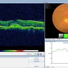
Optical coherence tomography and ultra-wide field fundus photograph of a 51 year old male with idiopathic choroidal neovascularization affecting his right eye. The patient had no symptoms at the time of the appointment and his vision was Dcc20/20-2 OU. The physcian stated that there wasn't active exudation on the exam or ocular imaging and based on the clinical findings, he has recommended we defer any treatments.
Photographer: Corey Grant
Imaging device: Heidelberg Spectralis, OPTOS California
Condition/keywords: choroidal neovascularization (CNV), CNVM, fundus photograph, OCT, optical coherence tomography (OCT), Optos, Right Eye, ultra-wide field imaging
-
 Idiopathic Peripapillary CNV
Jan 4 2024 by Virginia Gebhart
13 year old female with inactive CNV. Increased pigment 360 at 1 year follow up. No inflammation or SRF, pt remains asymptomatic
Photographer: Virginia Gebhart
Imaging device: Optos California
Condition/keywords: choroidal neovascularization (CNV), peripapillary choroidal neovascularization (PPCNVM)
-
 Juxtafoveal Choroidal Neovascularization Secondary to Choroidal Rupture
Aug 30 2012 by Young Hee Yoon, MD, PhD
Fluorescence Angiography (FA) image of a 14-year-old boy with a history of blunt trauma to his left eye 9 months ago. Best-corrected visual acuity remained at 20/30.
Photographer: Heon Eui Hong, Asan Medical Center
Imaging device: HHeidelberg HRA II/ version 1.7.0.0
Condition/keywords: choroidal rupture, juxtafoveal choroidal neovascularization (CNV)
-
 Macular Degeneration with Extensive Geographic Atrophy
Jan 26 2022 by Olivia Rainey
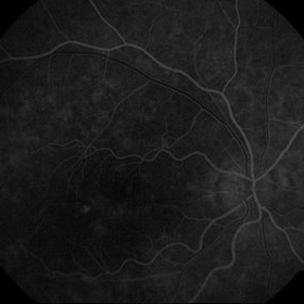
Heidelberg Spectralis fluorescein angiography of a 94-year-old woman with Macular Degeneration affecting both eyes. The FA reveals transmission defects consistent with RPE changes and geographic atrophy of RPE of both eyes, as well as window defects consistent with peripheral scarring in the right eye. The patient's vision was Dcc20/70 in both eyes at the visit the images were taken.
Photographer: Olivia Rainey, OCT-C, COA
Imaging device: Heidelberg Spectralis
Condition/keywords: 30-degrees, choroidal neovascularization (CNV), dry age-related macular degeneration (dry AMD), early phase, fluorescein angiogram (FA), geographic atrophy, heidelberg spectralis, macular degeneration, neovascular age-related macular degeneration (AMD)
-
 Myopic CNV
May 2 2013 by Henry J. Kaplan, MD
Choroidal neovascularization with hemorrhage in a highly myopic patient.
Condition/keywords: myopic choroidal neovascularization (CNV)
-
 Pigment Epithelial Detachment late FA with small occult CNV
Jul 6 2012 by Tarek S. Hassan, MD, FASRS
72-year-old man with VA loss and metamorphopsia of 2 months duration. PED found, testing done to rule out CNV. Very suspicious for CNV in superonasal fovea/parafovea.
Condition/keywords: choroidal neovascularization (CNV), pigment epithelial detachment (PED)
-
 Presumed Ocular Histoplasmosis Syndrome with Choroidal Neovascularization
Aug 24 2012 by John S. King, MD
22 sec - early somewhat lacy appearance c discrete border
Photographer: Kristin Konecki, OcuSight Eye Care Center, Rochester, NY
Condition/keywords: choroidal neovascularization (CNV), presumed ocular histoplasmosis syndrome (POHS)
-
 Punctate Inner Choroidopathy Complicated with CNV
Jun 5 2013 by Henry J. Kaplan, MD
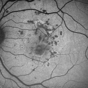
Fundus autofluorescence of the same patient demonstrates multiple hypoautofluorescent spots compatible with the clinical lesions #2.
Photographer: Angela Andersson
Imaging device: HRA II
Condition/keywords: choroidal neovascularization (CNV), punctate inner choroidopathy (PIC)
-
 Punctate Inner Choroidopathy with CNV Treated with Bevacizumab # 4 of 7
Feb 28 2013 by Gregory R. Blaha, MD, PhD
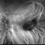
Mid-phase fluorescein angiogram in a 31-year-old female with vision loss from a choroidal neovascular membrane (CNV) from punctate inner choroidopathy.
Photographer: Gerard Gauthier, Spindel Eye Assoc., Derry, NH
Imaging device: Zeiss FF 450 Plus
Condition/keywords: bevacizumab, choroidal neovascularization (CNV), punctate inner choroidopathy (PIC)
-
 Toxoplasmic
May 27 2021 by Gabriel Costa Andrade, PhD
Fundus photograph of an 52-year-old woman with glaucoma and choroidal neovascularization due to toxoplasmosis.
Photographer: Gabriel Andrade
Condition/keywords: choroidal neovascularization (CNV), toxoplasmosis
-
 Choroidal Hemorrhage, Subretinal Hemorrhage
Dec 18 2017 by Nichole Lewis
Choroidal hemorrhage, Subretinal Hemorrhage, wet macular degeneration,
Photographer: Nichole Lewis
Condition/keywords: choroidal hemorrhage, choroidal neovascularization (CNV), exudative age-related macular degeneration, subretinal hemorrhage, wet age-related macular degeneration (wet AMD)
-
 Angioid Streaks With Associated Disc Drusen and CNV
Sep 21 2018 by Sarah Oelrich
Angioid streaks with associated disc drusen and CNV.
Photographer: Sarah Oelrich CRA COT, Southeastern Retina Associates Knoxville Tn
Condition/keywords: angioid streaks, autofluorescence imaging, choroidal neovascularization (CNV), disc drusen, infrared image
-
 ICG: Choroidal Aspergilloma With Secondary Choroidal Neovascularization and Exudative Retinal Detachment
Mar 21 2019 by Scott D Walter, MD, MSc, FASRS
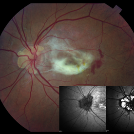
Multimodal imaging of a transplant patient with disseminated Aspergillosis and vision loss in her left eye.
Condition/keywords: choroidal neovascular membrane (CNVM), choroidal neovascularization (CNV), exudative detachment, focal chorioretinitis, fungal endophthalmitis, granulomatous choroiditis
-
 Optic Nerve Head Drusen
Feb 9 2018 by Olivia Rainey
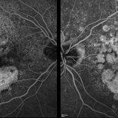
Fundus autofluorescence of a 49-year-old female with optic nerve head drusen affecting her left eye. The patient has pseudoxanthoma elasticum with choroidal neovascularization and has been receiving anti-VEGF treatment for many years.
Photographer: Olivia Rainey
Imaging device: Heidelberg Spectralis
Condition/keywords: 30 degrees, anti-VEGF, choroidal neovascularization (CNV), fundus autofluorescence (FAF), Heidelburg Spectralis, left eye, optic disc, optic nerve drusen, pseudoxanthoma elasticum (PXE)
-
 Punctate Inner Choroidopathy with CNV Treated with Bevacizumab # 6 of 7
Feb 28 2013 by Gregory R. Blaha, MD, PhD
Fundus photo following treatment with bevacizumab in a 31-year-old female with vision loss from a choroidal neovascular membrane (CNV) from punctate inner choroidopathy. The vision improved and was stable following a single injection.
Photographer: Gerard Gauthier, Spindel Eye Assoc., Derry, NH
Imaging device: Zeiss FF 450 Plus
Condition/keywords: bevacizumab, choroidal neovascularization (CNV), punctate inner choroidopathy (PIC)
-
 Secondary Choroidal Neovascularization Due to Toxoplasmosis
Feb 25 2013 by Henry J. Kaplan, MD
Left eye: secondary choroidal neovascularization and subretinal hemorrhage in a patient with old macular scar of toxoplasma.
Condition/keywords: choroidal neovascularization (CNV), toxoplasmosis, toxoplasmosis chorioretinitis
-
 Bruch’s membrane rupture
Jan 11 2013 by Hyung-Woo Kwak, MD
An area of Bruch’s membrane rupture involving the fovea is seen on indocyanine green angiography: late phase (right).
Photographer: Misook Lee, Kyung Hee Univsersity Hospital, Seoul
Imaging device: Zeiss f 450 plus
Condition/keywords: Bruch's membrane, myopic choroidal neovascularization (CNV)
-
 Choroidal Neovascularization with Subretinal Hemorrhage
Oct 11 2012 by Gabriela Lopezcarasa Hernandez, MD
70-year-old male with sudden decrease in visual acuity of right eye
Photographer: Azucena Rios, Macula Retina Consultores Mexico
Imaging device: Heidelberg Spectralis
Condition/keywords: choroidal neovascularization (CNV), subretinal hemorrhage
 Loading…