Search results (104 results)
-
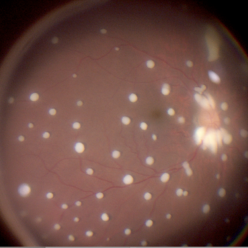 Seedlings of Fungal Endophthalmitis
Mar 14 2025 by SHILPI H NARNAWARE, ICO ( Retina) , FAICO ( Vitreo-Retina)
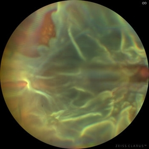
57 year diabetic female , was treated as a case of recurrent vitreous post cataract surgery. Patient was posted for vitrectomy 3 months post cataract surgery. Intra-operatively, multiple yellowish colonies were seen all over the posterior pole were seen, which were later found to be Aspergillus colonies.
Photographer: Shilpi Narnaware, Sarakshi Netralaya , Nagpur, Maharashtra , India
Imaging device: Ngenuity
Condition/keywords: endophthalmitis, fungal
-
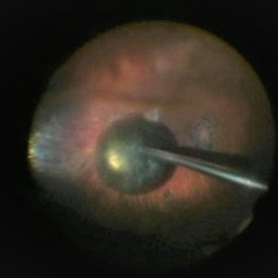 PPV retained cataract
Apr 19 2023 by Denica Rodriguez
A 46-year-old male with hypermature dense cataract. Patient got a piece of metal in his eye when he was 5 years old and was not able to see since. Patient was having cataract surgery and phacodonesis was present. The lens dropped to the back of the eye. Patient had to have another surgery to do vitrectomy. The lens removal was done with a fragmatome handpiece.
Photographer: Denica Rodriguez COA, ST
Imaging device: Zeiss Microscope with resight
Condition/keywords: cataract, dropped nucleus, fragmatome, lens capsule, ocular trauma, pars plana vitrectomy (PPV), retained lens fragments, Retina, retina surgery, traumatic cataract
-
 Prominent Long Ciliary Nerve
Jan 25 2022 by Kachelle Brown
Ultra-wide field photograph of a 48-year-old female with a prominent long ciliary nerve. Patient presented asymptomatic, and was referred for a macula on retinal detachment. Patient was diagnosed with high myopia and a posterior vitreous detachment, and the physician discussed increased risk of floaters, myopic degeneration and retinal detachment associated with high myopia. -24.00 prior to cataract surgery OU per patient.
Photographer: Kachelle Brown
Imaging device: Optos California
Condition/keywords: fundus photograph, high myopia, long ciliary nerve, optos, right eye, ultra-widefield image
-
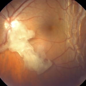 Retained Lens Fragment
Mar 2 2014 by Homayoun Tabandeh, MD, FASRS
Retained lens fragment, choroidal detachment, and serous retinal detachment post cataract surgery
Condition/keywords: retained lens fragments
-
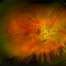
 Chronic Open Funnel Retinal Detachment With Horse Shoe Tear
Feb 7 2024 by Harsh Vardhan Singh, MS
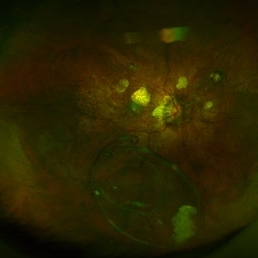
67 year old male with history of cataract surgery 1 year presented with old chronic retinal detachment with open funnel configuration with multiple breaks.
Photographer: Harsh Vardhan Singh
Imaging device: Clarus 700
Condition/keywords: chronic retinal detachment, Retinal Detachment, Retinal Detachment with multiple breaks
-
 Dislocated Capsular Tension Ring in Vitreous Cavity
Dec 21 2019 by Pablo Baquero Ospina, MD
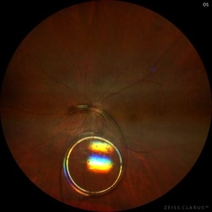
Fundus photograph of an 52-year-old woman with pseudoexfoliation glaucoma and previous cataract surgery with capsular tension ring. 5 years later she refers floaters.
Photographer: Pablo Baquero, Asociacion Para Evitar la Ceguera en Mexico, Mexico city
Imaging device: Optos/Daytona
Condition/keywords: fundus photograph, pseudoexfoliation glaucoma
-
 Dislocated Intraocular Lens (IOL)
Aug 2 2019 by JEFFERSON R SOUSA, Tecg.º (Biomedical Systems Technology)
A 53-year-old male patient suffered blunt trauma 15 days after cataract surgery. Note total dislocation of the intraocular lens. No glass reaction.
Photographer: JEFFERSON R SOUSA - Study Center and Ophthalmological Research Dr. Andre M V Gomes, Institute Dr. Suel Abujamra São Paulo-Brazil
Imaging device: Topcon TRC-50 DX, Imaginet 4.0, angle de 50 graus. Flash 18w-s
Condition/keywords: dislocated intraocular lens (IOL)
-
 Dislocated IOL
May 15 2018 by Morgan Benton
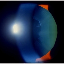
Ultra-wide field pseudocolor image of a 68-year-old male with a dislocated IOL after cataract surgery in the left eye. Patient was only able to count fingers at one foot and could pinhole to 20/60.
Photographer: Morgan Benton
Imaging device: Optos
Condition/keywords: color fundus photograph, dislocated intraocular lens (IOL), left eye, Optos, ultra-wide field imaging
-
 Dislocated IOL
Sep 20 2025 by JORGE SOBERANES
Fundus photograph of a 65-year-old man with a history of cataract surgery one year ago and bad vision since that.
Photographer: Dr. Jorge Soberanes, APEC, Universidad Nacional Autónoma México
Condition/keywords: dislocated lens, intraocular lens dislocation
-
 Endophthalmitis
Apr 9 2014 by Aleksandra V. Rachitskaya, MD, FASRS
Slit lamp photo of a patient with endophthalmitis after cataract surgery. An infectious infiltrate is noted next to the clear corneal incision.
Photographer: Bascom Palmer Eye Institute
Condition/keywords: cataract surgery, endophthalmitis
-
 ERM
ERM
Nov 26 2020 by Priya Rasipuram Chandrasekaran, MBBS, DO, DNB, FRCS
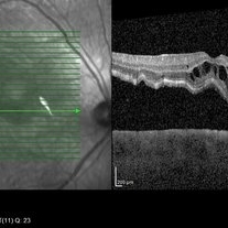
A 58-year-old female presented with distortion of images 1 month following cataract surgery in the right eye and fundus examination showed epiretinal membrane extending from the disc to the macula and OCT macula showing epiretinal membrane with disorganization of the foveal architecture.
Condition/keywords: epiretinal membrane (ERM)
-
 Macula Off Retinal Detachment
Jan 2 2018 by Carolyn Daley
55-year-old with macula off retinal detachment post cataract surgery.
Photographer: Carolyn Daley, Retina Specialists of Michigan
Imaging device: Heidelberg Spectralis
Condition/keywords: Heidelburg Spectralis, optical coherence tomography (OCT)
-
 Monochrome-frame
Nov 21 2023 by Nassim Alejandro Abreu Arbaje, MD
Frame grab from a 68 year male, who underwent cataract surgery and vitrectomy with ilm peel because of PDR. In the picture we can see the enhanced contrast we can get while doing an ILM peel with a monochromatic digital filter
Photographer: Nassim Abreu, Hospital Dr. Elías Santana
Imaging device: NGenuity 3D system
Condition/keywords: enhaced contrast, Macular surgery, monochrome
-
 Rhegmatogenous retinal detachment with dislocated IOL in a Morning Glory anomaly
Jul 27 2023 by Gustavo Aguirre-Suarez
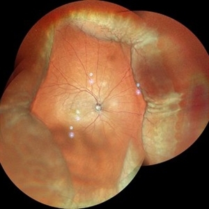
Fundus photograph of a 13-year-old male with a history of congenital cataract surgery in his right eye in 2019. The patient presents with sudden visual loss. Upon examination, a dislocated IOL is observed in the posterior segment, accompanied by a rhegmatogenous retinal detachment featuring peripheral retinal tears and horseshoe breaks. Additionally, a morning glory disc anomaly is also present in this patient.
Photographer: Gustavo Aguirre-Suarez
Imaging device: Mirante, NIDEK
Condition/keywords: dislocated posterior chamber intraocular lens (PCIOL), Morning Glory Anomaly, rhegmatogenous retinal detachment
-
 Scleral Buckling IOL Drop
Aug 6 2023 by Dr.Sheetal Divate
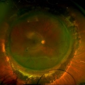
A 27 year old female with an old history of trauma and operated with scleral buckling and cataract surgery in the past came recently with complaints of DOV . Findings noted where IOL drop, inferior retinal detachment and old scleral buckle indent.
Photographer: Dr.Sheetal Divate
Imaging device: Optos Advance
Condition/keywords: dislocated intraocular lens (IOL), Retinal Detachment, scleral buckle
-
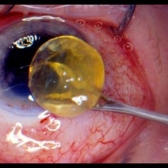
 Subconjuntival IOL After Blunt Trauma
Jun 27 2018 by Gabriel Costa Andrade, PhD
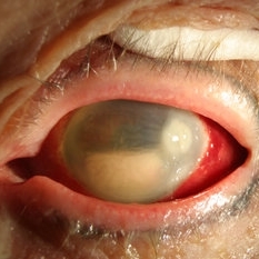
A 73-year-old male patient was referred to our ophthalmic emergency department with complaints of redness, pain, and diminution of vision in his left eye, after fall from height. The patient underwent small incision cataract surgery with polymethylmethacrylate (PMMA) IOL implantation in both the eyes 15 years back through superior sclerocorneal incision under local anesthesia. His best-corrected visual acuity was perception of light in the left eye. Ophthalmic examination using slit lamp biomicroscopy of the left eye revealed diffuse subconjunctival hemorrhage with no conjunctival laceration and inferior bulbar conjunctiva showed traumatic pseudophacocele with a sign “golden half ring,” suggesting the presence of PCIOL in subconjunctival space.There was total hyphema obscuring the view of rest of the ocular structures in his left eye.
Photographer: Gabriel Andrade, RETINA CLINIC, São Paulo, BRAZIL
Condition/keywords: dislocated intraocular lens (IOL), trauma
-
 Autofluorescence of Ocular Hypotony
May 29 2013 by Zofia Anna Nawrocka (vel Michalewska), MD, PhD
Autofluorescence image of a 75-year-old patient with hypotony, 2 weeks after trauma, 2 years after extracapsular cataract surgery.
Photographer: Zofia Michalewska, Ophthalmic Clinic "Jasne Blonia
Imaging device: Spectralis
Condition/keywords: hypotony
-
 Pseudoexfoliation
Feb 24 2018 by JEFFERSON R SOUSA, Tecg.º (Biomedical Systems Technology)
53-year-old patient, male, AV 20/60. It has a central pseudoexfoliation. Certainly you will have indication of cataract surgery.
Photographer: JEFFERSON R SOUSA - Study Center and Ophthalmological Research Dr. Andre M V Gomes, Institute Dr. Suel Abujamra São Paulo-Brazil
Condition/keywords: crystalline lens, crystals, pseudoexfoliation glaucoma, pseudoexfoliation of lens capsule, pseudoexfoliation syndrome
-
 360 Degrees Choroidal Detachment
Sep 22 2024 by Anand Temkar
A 52 year old male came with chief complaints of diminution of vision in RE since past 15 days. He gave history of ( RE ) cataract surgery + IOL about 2 months ago. His vision was 6/9 in RE and PL +ve, PR inaccurate in LE. His IOP was 10 mm of Hg in RE and 20 mm of Hg in LE.
Photographer: Dr.Anand Temkar- Retina Foundation, Ahmedabad
Imaging device: Mirante
Condition/keywords: choroidal detachment, choroidals, serous choroidal detachment
-
 Anterior Capsular Contraction Syndrome
Feb 8 2018 by Claire Kiernan, MD
Slit lamp photo of a female with underlying Retinitis Pigmentosa Zonulopathy and new anterior capsular contraction syndrome following cataract surgery.
Photographer: Steve Crow, University of Tennessee Hamilton Eye Institute, Memphis, TN
Condition/keywords: anterior capsule opacification, cataract surgery, zonules
-
 Anterior Capsular Opacity
Feb 8 2018 by Claire Kiernan, MD
Slit lamp photograph of a 39-year-old female following uncomplicated cataract surgery shown here with dense fibrinous changes of the anterior capsule. This patient underwent Nd:YAG laser anterior capsulotomy with clearing of her visual axis.
Photographer: Steve Crow, University of Tennessee Hamilton Eye Institute, Memphis, TN
Condition/keywords: anterior capsule opacification, cataract extraction, cataract surgery
-
 AV Anastomosis
Sep 19 2017 by Purva Patwari
54-year-old female post cataract surgery.
Photographer: Dr Purva Patwari, Patwari Retina Center,Ahmedabad
Imaging device: Zeiss visu 500
Condition/keywords: arteriovenous anastomosis
-
 Bullous Keratopathy
Jan 4 2025 by Mosab Salah
Corneal Slit photograph of an 84-year-old man underwent uneventful cataract surgery 1 year ago elsewhere, with a multiple fluid filled Bullae, not responding on conservative management and planned for KP.
Photographer: Abu-Ismail, Luai MD, The Islamic Hospital, Amman, Jordan
Imaging device: smartphone photography through SLB
Condition/keywords: bullous keratopathy, corneal edema
-
 Capsular Bag Dislocation into the Anterior Chamber
Jan 3 2020 by Manuel Ángel Alcántara Delgado, MD
Slit lamp photograph of a 65-year-old woman with previous history of complicated cataract surgery.
Photographer: Manuel Ángel Alcántara Delgado, CMN SXXI, Mexico City
Condition/keywords: anterior chamber, anterior dislocation of lens, anterior segment, cataract surgery, dropped capsular IOL bag complex
-
 Cataract
Feb 24 2018 by JEFFERSON R SOUSA, Tecg.º (Biomedical Systems Technology)
81-year-old patient, male, in surgical procedure (Facectomy - FEC). Removal of the fully opacified lens is observed.
Photographer: JEFFERSON R SOUSA - Study Center and Ophthalmological Research Dr. Andre M V Gomes, Institute Dr. Suel Abujamra São Paulo-Brazil
Imaging device: Canon / Lens Sigma 35mm F / 1.4 Dg Hsm. without flash user and keeping the limits of safety of the surgeon.
Condition/keywords: cataract surgery
 Loading…